Abstract
Many pathology laboratories are implementing digital pathology systems. The image resolution and scanning (digitization) magnification can vary greatly between these digital pathology systems. In addition, when digital images are compared with viewing images using a microscope, the cellular features can vary in size. This article highlights differences in magnification and resolution between the conventional microscopes and the digital pathology systems. As more pathologists adopt digital pathology, it is important that they understand these differences and how they ultimately translate into what the pathologist can see and how this may impact their overall viewing experience.
Keywords: Digital, magnification, pathology, resolution, whole slide image
INTRODUCTION
Digital pathology holds great promise for the future of anatomic pathology and many pathology laboratories today are starting to adopt this technology for a variety of applications, including telepathology (teleconsultation), quantitative image analysis and digitization of lab workflow.[1,2] Slide scanning systems are one component of the technology that will enable the digitization of the analog glass workflow to a digital workflow.
As more pathologists adopt digital pathology, one key question that is often asked by new users is “why is the image size not the same as my microscope?” More specifically, when a glass slide is manually viewed with a conventional light microscope using a ×20 objective and then viewed as a digital image that was created using the same ×20 objective lens, the perceived size of the image may not be the same.
Many pathologists are surprised to see that the images acquired with the same objective lens scale so differently when compared with the monitor, which is a common phenomenon in all digital pathology imaging systems. The reason this occurs is because the size of a whole slide image (WSI) is dependent on (1) the objective lens magnification being employed by the scanner, (2) size and number of individual pixels within the digital camera's sensor and (3) size and amount of individual pixels within the monitor. The images shown in Figure 1 illustrate this point. Figure 2 shows an image that was scanned on two different WSI systems. Both scanners used a ×20 objective lens to acquire the digital image. The images from the vendor 1, captured with a 5.5 micron pixel sensor, appear on the screen ×1.7 larger than the image on the right, captured with a 10 micron pixel size sensor. The image from vendor 1's scanner contains more information within the image due to its pixel resolution. Keeping the objective lens constant and varying the pixel size in the sensor will yield an image of different perceived size when viewed at full digital resolution. Depending on the digital camera and numerical aperture (NA) of the objective lenses within the WSI system, the digital resolution of the image can vary greatly. In this example, magnification is dictated by the microscope objective and should not be confused with scale.
Figure 1.

Images of an enterobius parasite in an appendix are shown using a similar ×20 objective, but with different sensor pixel sizes. Images a and c are sampled with a 5.5 micron pixel size, whereas b and d were sampled with a 10 micron pixel size. The apparent magnification differs by ×1.7, the ratio of the pixel sizes
Figure 2.

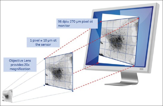
Graphic depicting the added boost in magnification that comes from the workstation monitor
MAGNIFICATION IS NOT RESOLUTION AND OPTICAL RESOLUTION IS NOT DIGITAL RESOLUTION
Another important consideration is the difference between magnification and resolution. Before this can be appreciated, one must understand the differences between digital resolution and optical resolution. Ultimately all definitions of resolution describe the minimum distance two objects can be separated by and still are distinguished as separate features. For example, using any viewing system, if the smallest distance between two objects is 1 micron before they get “blurred” together and appear as one object, we would state that the resolution is 1 micron (i.e., the system can resolve two objects that are 1 micron apart or a single object that is 1 micron in size). For a purely optical system, which has no sensor, the resolving power is dictated by a complex interplay of light and glass optics, which is often described in microscopy as the Rayleigh resolution limit (R).[3] We can calculate R as follows:
 provided that we know the color of light (λ is around 0.53 microns for the green light) and the property of the glass optics known as the NA.[4] The NA describes the number of different angles from which the glass optics will collect light, such a funnel for photons. Thus, we see that based upon glass optics, the aperture size (of what?) matters and the wavelength of light used to observe (observe what?) matters, but that magnification does not play a direct role in resolving power. However, an unfortunate confusion occurs with light microscopy because when a feature of interest requires higher resolution, an objective with higher NA is used and very often that objective also has higher magnification. Thus, changing the NA and magnification often occur simultaneously. For example, when switching from a ×20 with 0.5 NA objectives to one that is ×40 with 0.9 NA, the smallest resolvable feature drops from 0.64 microns to 0.36 microns (for the green light). With the 0.9 NA objectives, smaller features of interest can be resolved better than the 0.5 NA objectives. The true resolution improvement comes from the NA increase and not increases in magnification.
Some of these principles change, when a digital camera sensor and monitor is involved, as the resolving power is no longer dictated solely by the optical lens. This is because the size of a pixel and the amount of pixels will also dictate if two objects will appear as separate features. Using the same example as above, if two objects are 1 micron apart when projected onto a sensor (after being ‘magnified’ by an objective) and the pixel size is 10 microns, the two objects are likely to appear as one. This means that the sensor cannot resolve a 1 micron size image feature [Figure 2]. In this example, the sensor would require a 0.5 micron pixel size in order to insure that two objects appear as two distinct features. According to the Nyquist resolution requirement for pixel size (P):
![]()
Optical resolution is solely dependent on the objective lenses whereas, digital resolution is dependent on the objective lens, digital camera sensor and monitor and are closely tied together in system performance.[5,6,7,8,9] The objective lenses must have the optical resolution required to “see” features of interest; digital resolution cannot make up for any short comings in the objective. However, the sensor can fail the glass lenses if the digital resolution is not fine enough.
THE ROLE OF MAGNIFICATION
Magnification is a change in the apparent size of an object, performed so that the object can more easily be seen. In traditional light microscopes used for pathology, a wide range of magnifications are available to ensure that the image observed through the oculars is large enough for easy viewing. In this case, magnification is not necessarily used to increase resolution, but utilized to make viewing easier. In the WSI system, with the introduction of a digital camera sensor and monitor, magnification plays the role of matching digital and optical resolution. With the prior example, we cannot see 1 micron sized features with a 10 micron sensor, unless we magnify the image by another factor of ×20. This yields features that are 20 microns apart and therefore can be accurately digitized by the 10 micron pixel sensor. Once the image has been digitized with good fidelity by the sensor, there is no longer much need for additional magnification. The role of providing an image that is sized appropriately and easy to review will now depend on the workstation monitor. The appearance of an image on a digital display can be very different than the traditional light microscope view.
The traditional microscope provides a pair of ×10 oculars for viewing, thereby providing an extra boost of magnification for looking at the material on glass slides. Similarly, the presentation of the digital image on a monitor provides a boost of extra magnification that is often overlooked [Figure 3]. Continuing on with this example with the 10 micron pixel, when that pixel value is moved to a typical workstation monitor, it will generally be displayed as a 270 micron pixel. That translates to a magnification factor of ×27, more than was formerly achievable using the oculars. If we consider a sensor with a more typical pixel size (e.g., 6.45 microns), the magnification factor becomes ~×42. Hence, it is apparent that the scale of the image will certainly be different, dependent upon the sensor used, regardless of the objective lens magnification.
Figure 3.

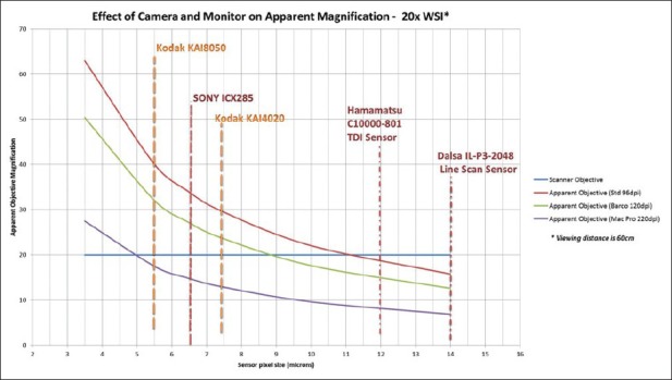
Graph depicting the effect of the camera sensor size and monitor display on apparent magnification. In this example, the images are acquired with a ×20 microscope objective lens, shown as a blue horizontal line. The X-input axis is the sensor pixel size and Y-output axis is the apparent magnification, as viewed on the screen at a distance of 60 cm. The apparent magnification on the screen increases as the sensor pixel size is decreased, as shown by the family of curves. The red curve is apparent magnification using a standard 96 dpi monitor, the purple curve using a 220 dpi Mac Retina display – these two representing the current extremes in monitor resolution
The magnification of an image on a computer screen is further influenced by one more element: How far the person viewing the image is sitting from the screen. Simply put, the further away you sit, the smaller the image seems. To illustrate, the graphs in Figure 3 bring together the complex relationship between sensor pixel size, objective lens magnification and the type of workstation monitor used to view the image. The bright blue, horizontal line represents the magnification due to the objective lens used in the scanner, which in this example is ×20. The family of curves represents the apparent magnification of the image as seen with different types of monitors, when seated 60 cm from the screen. The red curve is for a standard digital display with 96 dpi and the green curve shows the result for a higher density medical grade Barco™ 120 dpi computer monitor. Finally, the blue curve is the apparent magnification of an image when using the most advanced “retinal” display monitor with 220 dpi, as is now available with certain Apple Macintosh computers. With a standard 96 dpi display, a system with an 11 micron pixel will have the same apparent magnification as the standard light microscope. An advanced WSI scanner, such as vendor 1 with a 5.5 micron pixel sensor, provides an equivalent ×40 magnification.
CONCLUSION
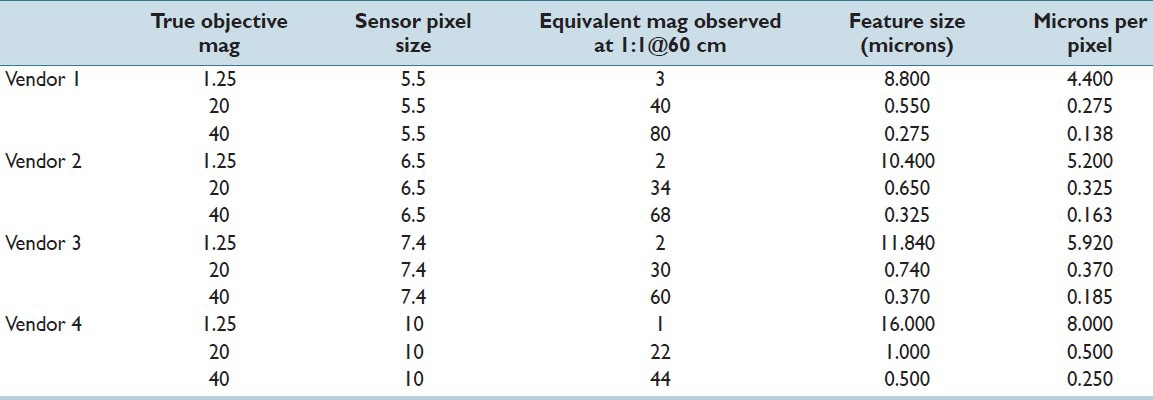
Magnification of images related to digital pathology imaging systems is complex. There are differences between digital pathology image viewing and traditional microscopy, which can lead to confusion by both vendors and pathologists. Therefore, it is important to remember that WSI differs from traditional microscopy because of the extra sensor and monitor aspects. Traditional glass microscope image quality monikers may no longer apply; rather than “×40” perhaps it should be preferable to refer to image quality by other labels that can be compared across different systems. As digital pathology systems continue to evolve, microns/pixel will likely be more widely and appropriately used to denote characteristic features within a digital image. Microns/pixel may be a better, vendor-neutral descriptor for “magnification” or image resolution quality for digital pathology systems and should be considered a standard for future digital pathology systems and should be considered by standards - creation efforts (e.g., Digital Imaging and Communications in Medicine). Microns/pixel, shown in the rightmost column of Table 1 and in Figure 4, relates directly to how the glass slide is digitized and will unambiguously represent increases in optical magnification or smaller pixels sizes. Microns/pixel is expected to become the standard of annotating pathology images and will most likely be used for diagnostic and prognostic considerations. The number of mitosis per high power field or mm2 is an example of a diagnostic consideration. Another area that the display of micro per pixel can impact is automated diagnostics, which is the use of computer aided algorithms (CAD) to detect and quantify cellular processes and disease. The resolution between the various digital pathology systems on the market can vary and the display of micron/pixel can help standardize the CAD results, since it indicates the resolution. Finally, WSI technologies that offer lower micron/pixel values allow pathologists to get more information from the image, which could lead to considerable time savings if pathologists are able to render a faster diagnosis since less image review time is needed.
Table 1.
The effect of monitor resolution and camera sensor on apparent magnification. Microns per pixel best capture increasing resolving power of scanner. A standard monitor resolution of 96 dpi was used in the calculations

Figure 4.

Image viewing software that includes a display of micron/pixel in upper right corner micron/pixel are expected to become the standard of annotating pathology images and will most likely be used for diagnostic and prognostic considerations
Footnotes
Available FREE in open access from: http://www.jpathinformatics.org/text.asp?2013/4/1/21/116866
REFERENCES
- 1.Park S, Pantanowitz L, Parwani AV. Digital imaging in pathology. Clin Lab Med. 2012;32:557–84. doi: 10.1016/j.cll.2012.07.006. [DOI] [PubMed] [Google Scholar]
- 2.Parwani AV, Feldman M, Balis U, Pantanowitz L. Digital imaging. In: Pantanowitz L, Balis UJ, Tuthill JM, editors. Pathology Informatics: Theory and Practice. Vol. 15. Canada: ASCP Press; 2012. pp. 231–56. [Google Scholar]
- 3.Agarwal G. ntroduction to biological light microscopy. In: Rittscher J, Machiraju R, Wong ST, editors. Microscopic Image Analysis for Life Science Applications. Boston: Artech House; 2008. pp. 1–18. [Google Scholar]
- 4.Piston DW. Choosing objective lenses: The importance of numerical aperture and magnification in digital optical microscopy. Biol Bull. 1998;195:1–4. doi: 10.2307/1542768. [DOI] [PubMed] [Google Scholar]
- 5.Kayser K, Molnar B, Weinstein RS. Berlin: VSV Interdisciplinary Medical Publishing; 2006. Virtual Microscopy-Fundamentals, Applications-Perspectives of Electronic Tissue-based Diagnosis. [Google Scholar]
- 6.Kayser K, Görtler J, Borkenfeld S, Kayser G. Interactive and automated application of virtual microscopy. Diagn Pathol. 2011;6(Suppl 1):S10. doi: 10.1186/1746-1596-6-S1-S10. [DOI] [PMC free article] [PubMed] [Google Scholar]
- 7.Leong FJ. Thesis: University of Oxford; 2002. An Automated Diagnostic System for Tubular Carcinoma of the Breast: A Model for Computer-based Cancer Diagnosis. [Google Scholar]
- 8.Balis UJ. Optical considerations in digital imaging. Clin Lab Med. 1997;17:189–200. [PubMed] [Google Scholar]
- 9.Yagi Y, Gilbertson JR. The importance of optical optimization in whole slide imaging (WSI) and digital pathology imaging. Diagn Pathol. 2008;3(Suppl 1):S1. doi: 10.1186/1746-1596-3-S1-S1. [DOI] [PMC free article] [PubMed] [Google Scholar]


