Abstract
When systematic movement errors occur, the brain responds with a systematic change in motor behavior. This type of adaptive motor learning can transfer intermanually; adaptation of movements of the right hand in response to training with a perturbed visual signal (visuomotor adaptation) may carry over to the left hand. While visuomotor adaptation has been studied extensively, it is unclear whether the cerebellum, a structure involved in adaptation, is important for intermanual transfer as well. We addressed this question with three experiments in which subjects reached with their right hands as a 30° visuomotor rotation was introduced. Subjects received anodal or sham transcranial direct current stimulation (tDCS) on the trained (Experiment 1) or untrained (Experiment 2) hemisphere of the cerebellum, or, for comparison, motor cortex (M1). After the training period, subjects reached with their left hand, without visual feedback, to assess intermanual transfer of learning aftereffects. Stimulation of the right cerebellum caused faster adaptation, but none of the stimulation sites affected transfer. To ascertain whether cerebellar stimulation would increase transfer if subjects learned faster as well as a larger amount, in Experiment 3 anodal and sham cerebellar groups experienced a shortened training block such that the anodal group learned more than sham. Despite the difference in adaptation magnitude, transfer was similar across these groups, although smaller than in Experiment 1. Our results suggest that intermanual transfer of visuomotor learning does not depend on cerebellar activity, and that the number of movements performed at plateau is an important predictor of transfer.
Keywords: Reaching, cerebellum, motor adaptation, brain stimulation
Introduction
Behavioral task, physical properties of the body, and the environment in which a person moves all undergo frequent changes, which must be compensated for if movements are to be accurate (1–3). When systematic movement errors occur, the brain may respond with a systematic change in motor behavior. This type of motor learning, called adaptation, leads to a reduction in movement errors in the presence of a perturbation. If the perturbation is suddenly removed, behavioral aftereffects can be observed. This form of learning involves the modification of an internal model (4,5). In a visuomotor rotation task, for example (6), subjects make reaching movements from a central position to a series of targets, viewing a cursor to indicate hand position. When the cursor is rotated counterclockwise, subjects will initially move toward the target but observe themselves making a counterclockwise error. Through repeated exposure to this perturbation, healthy subjects will adapt; i.e., shift their trajectory clockwise such that the cursor moves toward the target. When the rotation is suddenly removed, subjects will initially move clockwise and observe clockwise errors (negative aftereffects).
Adaptive motor learning can transfer intermanually. In other words, adaptation of movements of the right hand may carry over to some extent, causing savings (7) or aftereffects (8) in the left hand. Transfer of savings occurs if the untrained hand, on exposure to the same perturbation that the trained hand received, adapts at a faster rate than the trained hand did. Transfer of aftereffects, examined in the present study, occurs if the untrained hand exhibits negative aftereffects despite no exposure to a perturbation. This is an important process because it saves time and energy for either hand to have access to knowledge gained from the other hand. Such transfer has been found to occur for force perturbations (9,10) as well as visuomotor perturbations (7,11,12). The degree to which motor adaptation transfers from one hand to the other depends on several factors. For example, Sainburg and Wang (13) found that in a visuomotor rotation task, dominant arm training improved final position accuracy but not initial movement direction in the non-dominant arm, and non-dominant arm training had the opposite effect on the dominant arm (13). Transfer also depends on the workspace locations in which each hand trains (7) and the degree of handedness of the subject (14). Whether a visuomotor perturbation is introduced abruptly or gradually, intermanual transfer of aftereffects occurs throughout adaptation but is always incomplete, reaching 33–50% of the magnitude of adaptation (15).
Visuomotor adaptation is thought to involve the cerebellum (16,17). For instance, people with cerebellar damage have difficulty adapting to prism offsets, visuomotor rotation, force fields, and other tasks requiring adaptation to motor or sensory perturbations (18–22). In addition, enhancing cerebellar excitability leads to faster error reduction in visuomotor adaptation (23). The primary motor cortex (M1) is also thought to be involved in the process of adaptive learning (33–36). Interestingly, the role of M1 in human studies has been linked to retention mechanisms. Specifically, transcranial magnetic stimulation studies interfering with M1 activity showed impaired retention rather than effects on acquisition of the adaptation (28). Similarly, enhancing the excitability of M1 via transcranial direct current stimulation (tDCS) elicited an improvement of memory retention in a visual rotation paradigm (23).
While motor adaptation has been studied extensively, less is known about the neural basis of intermanual transfer of such adaptation. For example, when one hand trains, a new representation for the learned movement is formed in the corresponding hemisphere (i.e. ipsilateral for cerebellum). It has been proposed that a weaker version of the representation is independently and simultaneously stored in the untrained hemisphere, and it is this weaker representation that accounts for intermanual transfer (24).
Here we examined the involvement of right and left cerebellar and M1 hemispheres in intermanual transfer of the aftereffects of visuomotor adaptation. Subjects adapted to a 30-degree visuomotor rotation while receiving anodal or sham transcranial direct current stimulation (tDCS) to either the trained or untrained cerebellar or M1 hemisphere. Anodal tDCS is a noninvasive form of brain stimulation known to increase cerebral (25) and cerebellar (26) excitability, as well as speed up adaptation when applied over the cerebellum (23,26). After the adaptation period, the visuomotor rotation and visual feedback were removed and subjects continued the task with their left and right hands to assess intermanual transfer of aftereffects and retention. We first sought to confirm that in this paradigm, one hand’s training results in movement aftereffects for the other hand. If so, we would expect to see aftereffects in the untrained hand after adaptation of the trained hand during sham tDCS. To assess cerebellar and M1 involvement in intermanual transfer, we next asked whether stimulating the training cerebellum or M1 increases transfer to the untrained hand. We additionally asked if stimulating the untrained cerebellum or M1 increases transfer to the untrained hand, which would support the cross-activation model of intermanual transfer. Finally, we assessed the contribution of adaptation magnitude vs. the number of movements performed at plateau to intermanual transfer.
Methods
Experiment 1 examined the contribution of the trained hemisphere of M1 (right) and the cerebellum (left) on intermanual transfer of aftereffects. Experiment 2 tested the involvement of the untrained hemisphere of M1 (left) and the cerebellum (right) in intermanual transfer. In Experiment 3, the adaptation period was truncated to allow us to compare the importance of adaptation rate vs. adaptation magnitude to intermanual transfer when the training cerebellar hemisphere was stimulated.
Subjects
79 subjects (mean age 25.3 years; 32 men) participated in one of three experiments (45 in Experiment 1, 18 in Experiment 2, 16 in Experiment 3). All were right-handed according to the Edinburgh Handedness Inventory, with a mean score of 78.3 (28) and reported that they were neurologically healthy. All procedures were in accordance with the Helsinki Declaration of 1975, as revised in 2000, and subjects gave informed consent approved by the Johns Hopkins Institutional Review Board. After each session, subjects rated their quality of sleep the previous night, level of attentiveness during the experiment, fatigue from the experiment, and perceived pain from tDCS on a scale from 1 to 10, with 10 being the most sleep, attention, fatigue, or pain. All but four subjects completed these ratings.
Visuomotor Rotation Task
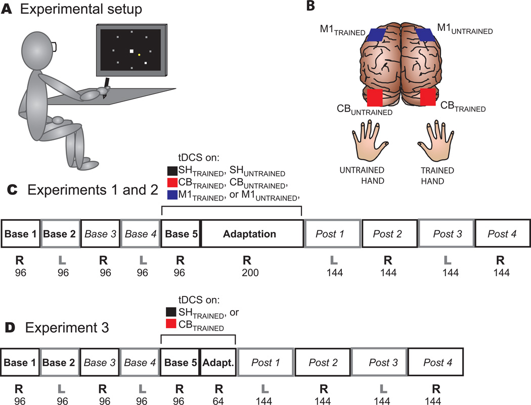
Subjects were seated with a horizontal digitizing tablet (62 × 46 cm, Inuoso4) at waist height and a computer monitor (1280 × 1024 pixel resolution) at eye level (Figure 1A), and wore goggles that obscured their view of the tablet. They held a pen that controlled a cursor on the monitor (2 mm green dot) and performed a center-out movement task (23,29,30) controlled by a custom Matlab program (Mathworks). Subjects were instructed to move the cursor from the home position in the center of the display (3 mm white square) through the target (2 mm white square) that appeared in one of 8 locations around the home position (Figure 1A). Targets were presented pseudorandomly, with each set of 8 targets including all 8 locations. Targets were 10 cm away from the home position, for the cursor as well as the hand. Subjects were told to move in a straight line with a rapid “shooting” movement and to go through the target rather than stopping on it, to reduce the likelihood of corrective movements.
Figure 1. Experimental protocol.
A. Experimental setup. Subjects were seated in front of a digitizing tablet and computer monitor, wearing goggles to prevent seeing their hands. Subjects held a digitizing pen and made rapid shooting movements on the tablet to make a cursor on the screen (small yellow dot) move from the center home position (white square) through a target (large white dot) that appeared in one of 8 positions (grey dots; not visible to subject). B. Representation of the back of the brain with locations of anodal tDCS stimulation on the trained and untrained cerebellum and M1 (red and blue electrodes respectively). C. Experiment 1 and 2 protocol. Subjects performed 10 blocks of trials. Bold type: continuous visual error feedback was present. Italics: no visual feedback. The final row indicates number of trials in each block. tDCS was on throughout Blocks 5 and 6, over the trained or untrained hemisphere. In Block 6 (Adaptation), a 30° clockwise rotation was introduced to the cursor, such that subjects experienced large clockwise errors and had to adjust their movements to compensate. D. Experiment 3 protocol. Identical to Experiment 1, except Block 6 (Adaptation) was only 64 trials.
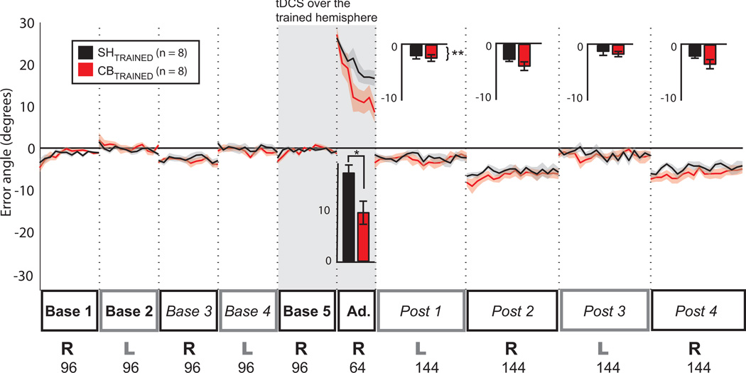
All three experiments consisted of 10 blocks with varying numbers of trials; Experiment 3 differed from Experiments 1 and 2 only in that Adaptation had 64 trials instead of 200 (Figure 1D). The first 5 blocks were baselines: right and left hand with visual feedback about hand position, right and left without visual feedback, and right with visual feedback and tDCS stimulation (Fig. 1C,D). In the sixth block (Adaptation) a 30° clockwise rotation of the cursor was introduced to assess visuomotor adaptation in the right hand (e.g.,(23)). Blocks 7–10 were post-adaptation blocks with no visual feedback, alternating left and right hands, to assess transfer of aftereffects to the left hand and retention in the right hand (Fig. 1C and D).
Subjects received visual feedback about the cursor position during the reach only during Base 1, 2, 5 and Adaptation. The cursor was visible during the movement toward the target (online visual feedback), but was frozen at 10 cm from the home position. In the no-feedback blocks (Base 3, 4, and all Post blocks), the cursor disappeared as soon as it was moved out of the home position so there was no visual feedback about performance. In all blocks, after a trial was finished and the subject moved back toward the home position, the cursor reappeared only within 2 cm of the home position.
To encourage consistency across subjects in movement speed, subjects heard a high-pitched tone if their movement was too fast (< 275 ms) and a low-pitched tone if their movement was too slow (> 375 ms). If subjects slowed their movements or began to make online corrections, they were reminded to move in a straight line and stay within the speed limits.
Transcranial Direct Current Stimulation (tDCS)
Current was delivered at 2 mA (23,26,31,32) with a Phoresor II Auto (Model No. PM850; IOMED) through two sponge electrodes (surface area 25 mm2, current density 0.08 mA/cm2; (31) that had been soaked in saline solution. Experiments 1 and 2 included three stimulation groups (Cerebellum, M1, and Sham) while Experiment 3 comprised only Cerebellum and Sham (Fig. 1B). For cerebellar stimulation, the anode was placed 3 cm to the right (CBTRAINED, Experiment 1 and 3) or left (CBUNTRAINED, Experiment 2) of the inion and the cathode on the corresponding buccinator muscle (23,26). For M1 groups, the anode was centered over the left (M1TRAINED) or right (M1UNTRAINED) motor hotspot for first dorsal interosseus, as determined by single pulses of transcranial magnetic stimulation (Magstim BiStim2; Whitland) at suprathreshold intensities. The cathode was placed on the supraorbital region contralateral to the anode (23,25).
In Experiments 1 and 2, half the Sham (SHTRAINED or SHUNTRAINED) subjects had electrodes placed in the M1 configuration and half in the cerebellar configuration. In Experiment 3, all Sham subjects had electrodes placed in the cerebellar configuration. For all subjects, current was increased gradually over 30s to full strength before the beginning of Base 5. For non-sham subjects, current was decreased to zero at the end of Adaptation. For Sham subjects, once current reached full intensity it was gradually decreased to zero before beginning Base 5. This method is effective at blinding subjects to whether they are receiving sham or real tDCS (23,37).
Data Collection and Analysis
Data collection was similar to Galea et al.(23). Hand position on the digitizing tablet was continuously recorded at 75 Hz by a custom Matlab program (Mathworks). Kinematic data were filtered at 10Hz with a low-pass Butterworth filter and differentiated to calculate movement velocity (23). The point at which movement velocity crossed 5% of peak velocity was defined as movement onset.
Subjects’ angular error at 80 ms (initial deviation of the cursor from the target angle) and at the cursor’s final position (angular deviation from the target when the cursor was 10 cm from home position) was recorded. Given that the patterns of results with the two types of error were the same, we focused on final angular error to be consistent with Galea et al. (2011). Positive values indicate angular errors that deviated clockwise from the target. Every set of 8 trials was averaged to create an epoch, and epochs were averaged across subjects in each group to create line plots. Trials that lasted more than 550 ms were excluded from analysis.
To quantify any differences in adaptation or transfer among groups, we averaged a number of epochs within each block to obtain a mean error value in that block for each subject. For baseline blocks (Base 1 through Base 5), epochs 2–6 were averaged for each subject (23,38). For each post-adaptation block (Post 1 through Post 4), epochs 2–11 were averaged. In Experiments 1 and 2, epochs 6–7 of Adaptation were averaged to capture the part of the adaptation curve in which learning occurs most rapidly. We chose these epochs instead of epochs 2–11, as done in in the past (23), because subject performance in the present study was more variable, even in the sham tDCS group. Indeed, variance of the first 16 trials of the block preceding Adaptation (block 5 in the present study vs. block 2 in Galea et al. (23)) was significantly greater in the present data (F(15,15) = 4.09, p < 0.01). This difference is likely due to the different study design. Here, participants were exposed to 5 baseline blocks including right and left arm performance as well as with and without visual feedback, rather than 2 baseline blocks with visual feedback using the right arm. Because this data set is more variable, we focused on the period of largest effect difference to maximize statistical power. In Experiment 3, the last epoch of Adaptation (epoch 8) was averaged across subjects to capture any difference in magnitude of adaptation. These averages were used in all group statistical analyses except where otherwise noted.
To eliminate any individual biases, for the bar plots only, the mean of baseline epochs 2–6 was subtracted from the appropriate epochs in Adaptation through Post 4 for each subject: the Base 5 error value (right hand with feedback and tDCS) was subtracted from the Adaptation value; Base 4 error (left hand without feedback) was subtracted from Posts 7 and 9; Base 3 error (right hand without feedback) was subtracted from Posts 8 and 10. Note that baselines were subtracted for bar graphs for clarity, but not for line plots or statistical analysis.
Statistical Analysis
Performance effects of tDCS
To assess any effects of tDCS on general motor performance, we conducted a separate mixed model repeated measures ANOVA (ANOVARM) in each experiment. Here, we compared the mean error in epoch 2–6 of Base 1 to the mean error in epoch 2–6 during Base 5 (within-subjects factor TIME) across CB, M1, and SH groups (between-subjects factor GROUP).
Adaptation
To determine whether the groups differed during adaptation, we performed separate ANOVARM in Experiments 1 and 2 comparing the mean error in epoch 6–7 of Adaptation to the mean error in epoch 2–6 during Base 5 (within-subjects factor TIME) across CB, M1, and SH groups (between-subjects factor GROUP). To assess whether each group had achieved a similar magnitude of learning by the end of the Adaptation block, in each of the three experiments we performed an ANOVARM with one within-subjects factor TIME: mean error of epoch 2–6 in Base 5 vs. the last epoch of Adaptation, and one between-subjects factor GROUP. Because CBTRAINED in Experiment 3 appeared to have a similar adaptation magnitude as SHTRAINED in Experiment 1, despite the different Adaptation block length (64 vs, 200 trials respectively), we also performed ANOVARM comparing adaptation magnitude in those two groups to find out if they were different.
Transfer and retention
We performed separate ANOVARM in each of the three experiments (within-subjects factor TIME, Base 4 vs. Post 1, and between-subjects factor GROUP) to assess whether significant intermanual transfer occurred and whether it differed across stimulation groups. To determine whether the amount of retention in the right hand differed across groups, we performed separate ANOVARM for each experiment comparing error in Base 3 (right hand baseline with no visual feedback) vs. Post 2.
In Experiment 1, 5 CB and 9 SH subjects performed an earlier version of the task that lacked baselines with no visual feedback (Base 3 and Base 4). However, among subjects who did the full Experiment 1 (10 CB, 6 SH, 16 M1), baseline error with no feedback was very similar to the baseline with visual feedback. Indeed, a 3-way ANOVA with group vs. visual feedback vs. hand in those who completed all baseline measurements showed no interactions (all p > 0.2), and there was no effect of visual feedback (p > 0.07). Therefore, in those subjects who did not have data for Base 3 and 4, we substituted Base 1 and 2, respectively, for within-subject comparisons (ANOVARM factor TIME).
Finally, to determine whether the amount of intermanual transfer is influenced by the duration of the Adaptation block, we performed an ANOVARM comparing Base 4 vs. Post 1 (within-subjects factor TIME) across the sham groups of Experiment 1 and 3 (long vs. short Adaptation block, between-subjects factor GROUPS). We also examined the effect of CB stimulation in a 2-way ANOVARM comparing Base 4 to Post 1 (TIME) across stimulation condition CB vs. SH (STIMULATION) in the two Adaptation block durations (EXPERIMENT).
Plateau Length Analysis
To estimate how much of the Adaptation block represented movements at a plateau level, we calculated the number of epochs that fell within 2SD of the final Adaptation epoch (39). In other words, for each individual we calculated 2SD of the last epoch in the Adaptation block and counted backwards until we reached an epoch that fell outside this range. This number of epochs represented the approximate duration that each individual performed at plateau. We then averaged the plateau length across subjects within each group. To determine whether plateau length differed between Experiment 1 and 3 we performed a 2-way factorial ANOVA with factors ADAPTATION LENGTH (long or short) and STIMULATION (CB, M1, or SH).
Sample size
We wanted to know if the lack of difference across groups in magnitude of transfer was due to small sample size. To determine how many subjects would be needed per group to find a significant difference in intermanual transfer for each experiment, we bootstrapped the existing Base 4 and Post 1 data to create larger sample sizes and performed an ANOVARM with within-subjects factor TIME and between-subjects factor GROUP. We repeated this 1000 times with each sample size and calculated the percentage of the 1000 repeats that yielded an ANOVARM group-time interaction p value < 0.05. We increased the bootstrap per-group sample size until we found a sample size that yielded a 90% chance of getting a group-time interaction p < 0.05. This is analogous to a sample size analysis on a statistical test that has already been performed (40).
Subject ratings
After each session, subjects rated their quality of sleep the previous night, level of attentiveness during the experiment, fatigue from the experiment, and perceived pain from tDCS on a scale from 1 to 10, with 10 being the most sleep, attention, fatigue, or pain. We used 1-way ANOVAs to compare each of these ratings, as well as subject age and handedness score, across groups.
Results
Anodal tDCS over the training cerebellar hemisphere facilitates adaptation
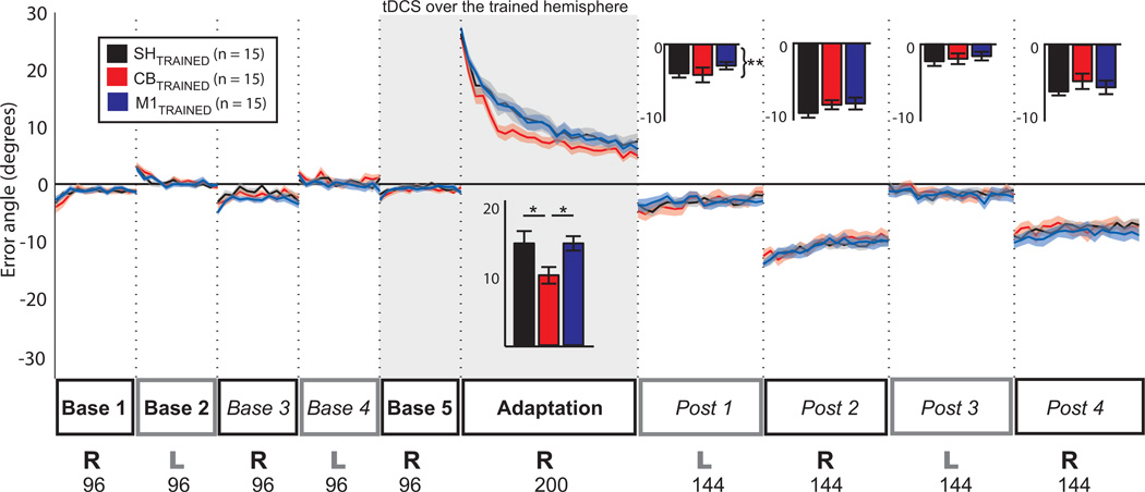
There was no significant interaction of GROUP x TIME for any of the three experiments (all p > 0.4) when comparing Base1 to Base5, indicating that baseline performance was not affected by tDCS. In Experiment 1, cerebellar tDCS on the trained (right) hemisphere (CBTRAINED) sped up learning in the Adaptation block (Fig. 2) relative to M1 and Sham groups (M1TRAINED and SHTRAINED). Indeed, an ANOVARM comparing the mean errors with factors TIME (Base 5 vs. epochs 6–7 of the Adaptation block) and GROUP (CBTRAINED, M1TRAINED, SHTRAINED) showed significant effects for both time and group (F(89,1) = 271.9, p < 0.001; F(89,2) = 4.5, p = 0.017) as well as a time by group interaction (F(89,2) = 3.7, p = 0.034; Figure 2). Post-hoc tests showed that learning in the CBTRAINED group at this point in the Adaptation block was significantly greater than both the SHTRAINED group (p < 0.024) and the M1TRAINED group (p = 0.023).
Figure 2. Experiment 1 results.
Anodal tDCS on the trained hemisphere of cerebellum or M1 does not increase intermanual transfer. SHTRAINED (black) and M1TRAINED (blue) tDCS had similar effects on error angle throughout the entire session. CBTRAINED (red) tDCS, however, increased the speed of learning in the Adaptation block. Inset bars represent averages and standard error of epochs 6–7 for Adaptation and 2–11 for the four Post-adaptation blocks, with baselines subtracted. *Post-hoc tests showed Adaptation in the CBTRAINED group was significantly different from SHTRAINED and M1TRAINED. **Please note that all groups showed significant transfer of aftereffects in Post 1 (ANOVARM on un-subtracted data, effect of time, p < 0.01).
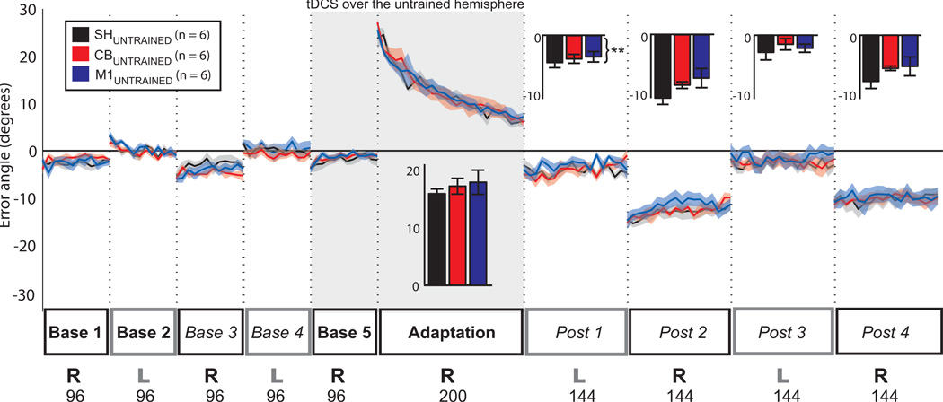
In Experiment 2, tDCS on CBUNTRAINED and M1UNTRAINED did not affect adaptation speed relative to SHUNTRAINED. Here, ANOVARM showed a significant effect of time (F(1,35) = 371, p < 0.001), but no significant group effect (F(2,35) = 0.91, p = 0.42) or group by time interaction (F(2,35) = 0.44, p = 0.66), indicating that the three groups were perturbed, but did not adapt differently (Fig. 3).
Figure 3. Experiment 2 results.
Anodal tDCS on the untrained hemisphere of cerebellum or M1 does not increase intermanual transfer. SHUNTRAINED (black), CBUNTRAINED (red), and M1UNTRAINED (blue) tDCS had similar effects on error angle in Adaptation. Inset bars represent averages and standard error of epochs 6–7 for Adaptation and 2–11 for the four Post-adaptation blocks, with baselines subtracted. **All groups showed significant transfer of aftereffects in Post 1 (ANOVARM on un-subtracted data, effect of time, p < 0.001).
We also compared the final epoch in Adaptation (epoch 25) with Base 5 to determine whether there was any difference in the magnitude of adaptation between groups. In Experiment 1, an ANOVARM showed a strong effect of time (F(1,89) = 105, p < 0.001), indicating that subjects did not reach baseline error levels by the end of adaptation. However, there was no significant group effect or interaction of time by group (F(2,89) =1.47, p = 0.24; F(2,89) = 0.81, p = 0.45), indicating that the three groups had learned a similar amount at the end of the Adaptation block in Experiment 1. In Experiment 2, an ANOVARM comparing Adaptation epoch 25 to Base 5 also showed an effect of time (F(1,35) = 190, p < 0.001), but no group effect and no interaction (F(2,35) = 0.48, p = 0.63; F(2,35) = 0.27, p = 0.77). These results suggest that stimulation of the trained or untrained cerebellum or M1 did not impact the magnitude of adaptation, even in the CBTRAINED group where adaptation was faster. In other words, tDCS over the training cerebellum sped up the adaptation process to compensate for the visuomotor rotation, but the total amount of compensation was similar at the end of the Adaptation block.
Anodal tDCS over the training or non-training cerebellar or M1 hemispheres does not affect transfer to the untrained hand
Stimulation over either the training or non-training hemisphere of the cerebellum or M1 did not affect intermanual transfer when assessing aftereffects in the left hand. In Experiment 1 (tDCS over the training hemisphere), an ANOVARM comparing mean errors in Post 1 relative to Base 4 showed no effect of group and no significant interaction of group by time (F(2,89) = 0.01, p = 0.99; F(2,89) = 0.83, p = 0.44), suggesting that there was no difference among the groups in terms of intermanual transfer of aftereffects. However, there was a strong effect of time (F(1,89) = 75, p < 0.001), indicating that all groups experienced significant intermanual transfer. The negative sign of errors in Post 1 suggests that transfer of aftereffects took place in an extrinsic (rather than intrinsic) reference frame, which is consistent with other studies (13,41,42).
To determine whether the lack of difference in magnitude of transfer in Experiment 1 could be due to small sample size, we performed a bootstrap analysis for ANOVARM comparing Post 1 to Base 4 across groups. We found that we would need 91 subjects per group (273 total) to have a 90% chance of getting a significant group-time interaction. The fact that so many subjects would be needed to detect a difference supports the idea that any behavioral difference across groups is small. Together, these results suggest that tDCS over the trained cerebellum or M1 does not change the aftereffects of adaptive learning in the untrained hand. It also suggests that the beneficial effect of tDCS on learning with the trained hand (CBTRAINED) does not influence the magnitude of aftereffects transfer.
In Experiment 2 (tDCS over the non-training hemisphere), we found no significant differences in transfer magnitude across ANUNTRAINED and SHUNTRAINED when comparing Post 1 to Base 4. An ANOVARM revealed that mean error was not statistically different for group (F(2,35) = 0.89, p = 0.43) or the interaction group by time (F(2,35) = 0.35, p = 0.71). However, again, we found a significant effect of time (F(2,35) = 69.6, p < 0.001), indicating that both groups experienced significant intermanual transfer of aftereffects. To determine whether the lack of difference in magnitude of transfer in Experiment 2 could be due to small sample size, we performed a bootstrap analysis for ANOVARM comparing Post 1 to Base 4 across groups. We found that we would need 101 subjects per group (303 total) to have a 90% chance of getting a significant group-time interaction. Again, the fact that so many subjects would be needed to detect a difference supports the idea that any difference between groups in magnitude of transfer is small. This suggests that modulating the excitability of the untrained cerebellum and M1 hemispheres does not affect intermanual transfer.
Increasing adaptation speed and magnitude with tDCS over the trained cerebellum does not increase transfer to the untrained hand
To determine whether the observed lack of transfer despite faster adaptation in Experiment 1 was due to similar magnitude of adaptation when transfer was assessed (i.e. at the end of the Adaptation block), we performed Experiment 3. Here, subjects performed a shorter Adaptation block to allow testing of transfer when the CBTRAINED group has corrected for more errors than the SHTRAINED group.
In Experiment 3 we found, similarly to Experiment 1, that CBTRAINED adapted faster relative to SHTRAINED. With the shortened Adaptation block in Experiment 3, CBTRAINED resulted in subjects learning significantly more than SHTRAINED by the end of the block (final error angle in Adaptation was 16.4° for SHTRAINED and 9.0° for CBTRAINED). An ANOVARM comparing mean errors at Base 5 to the last epoch of Adaptation showed significant differences for time, group, and their interaction (F(1,31) = 88.9, p < 0.001; F(1,31) = 7.48, p = 0.016; F(1,31) = 7.48, p = 0.016), indicating that CBTRAINED and SHTRAINED changed differently from Base 5 to the end of Adaptation.
Similar to Experiments 1 and 2, both groups in Experiment 3 had significant intermanual transfer. ANOVARM comparing error in Post 1 to Base 4 showed a strong effect of time (F(1,31) = 26.6, p < 0.001). However, there was no effect of group or time by group interaction (F(1,31) < 0.21, p = 0.66; F(1,31) = 0.24, p = 0.64). This shows that intermanual transfer was not affected even with the facilitation of speed and magnitude of adaptation resulting from tDCS over the training cerebellum. Indeed, a bootstrap analysis for ANOVARM comparing Post 1 to Base 4 showed that we would need 315 subjects per group (630 total) to have a 90% chance of getting a significant group-time interaction. Again, given that so many subjects would be needed to detect a difference in magnitude of transfer, cerebellar modulation with tDCS does not seem to influence intermanual transfer of adaptation.
To determine the effect of magnitude of learning on intermanual transfer we compared the errors in Post 1 vs. Base 4 in the SHTRAINED groups from Experiment 3 (short Adaptation block) vs. Experiment 1 (long Adaptation block). This revealed a significant effect of time (F(1,45) = 54.9, p < 0.001), indicating that both sham groups had significant intermanual transfer of aftereffects. We found no group effect, but a robust group by time interaction (F(1,45) = 0.02, p = 0.89; F(1,45) = 4.6, p = 0.045). This interaction suggests that the length of the Adaptation block or difference in adaptation magnitude significantly affected how much transfer occurred in the SHTRAINED groups. Magnitude of adaptation was significantly different for the two sham groups (ANOVARM time, group, and interaction effects: F(1,45) = 101, p < 0.001; F(1,45) = 13.2, p = 0.0016; F(1,45) = 13.8, p = 0.0013).
Magnitude of transfer may depend on duration of training at plateau rather than simply adaptation magnitude
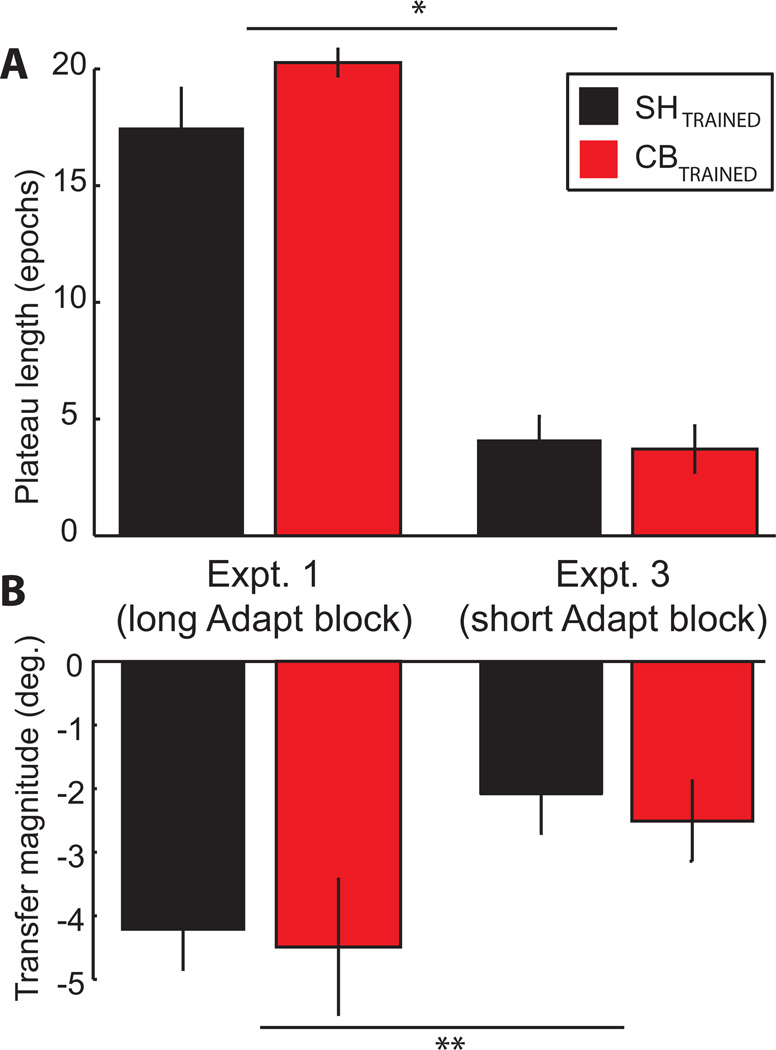
A comparison between Experiments 1 and 3 yielded puzzling results. The sham groups in Experiment 1 vs. Experiment 3 appeared to show an effect of adaptation magnitude on intermanual transfer (in other words, adapting more is associated with transferring more). However, the CBTRAINED group in Experiment 3 and the SHTRAINED group in Experiment 1 had similar magnitudes of adaptation (ANOVARM group by time interaction F(1,45) = 0.018, p > 0.65), yet subjects in Experiment 1 transferred significantly more than subjects in Experiment 3 across stimulation conditions (2-way ANOVARM experiment by time interaction F(1,91) = 4.75, p = 0.035; all other effects N.S.). We therefore wondered if it was the number of reaches performed at plateau, rather than the magnitude of adaptation, that was related to transfer. Thus, we estimated an individual’s plateau length as the number of epochs in the Adaptation block within 2SD of the final Adaptation epoch. For SHTRAINED and CBTRAINED in Experiment 1 this averaged 17.7 and 20.5 epochs, respectively (Fig. 5A). In Experiment 3, plateau length was only 4.1 epochs for SHTRAINED and 3.7 epochs for CBTRAINED (Fig. 5A). A 2-way ANOVA indicated no effect on plateau length of stimulation (real or sham) and no stimulation x Adaptation block duration interaction (F(1,44) = 0.70, p = 0.41; F(1,44) = 1.27, p = 0.27). However, we found a robust effect of Adaptation block duration (F(1,44) = 112.8, p < 0.001). This indicates that the difference in transfer between CBTRAINED in Experiment 3 and SHTRAINED in Experiment 1 (Fig. 5B) could be due to the number of reaches performed at plateau (plateau length) rather than adaptation magnitude.
Figure 5. Comparison of plateau length and transfer in Experiments 1 and 3.
AIn the Adaptation block, subjects performed more trials at plateau in Experiment 1 than in Experiment 3. *Significant effect of Adaptation block duration on plateau length (2-way ANOVA effect of Adaptation block duration F(1,44) = 112.8, p < 0.001). B. Subjects also transferred more in Experiment 1 than in Experiment 3. **Significant interaction of Adaption block duration and time (2-way ANOVARM on un-subtracted data in Post 1 vs. Base 4 showed an experiment by time interaction F(1,91) = 4.75, p = 0.035).
Adaptation aftereffects in the training hand are similar across groups
We also examined retention of aftereffects in the right hand (comparing Post 2 to Base 3) in all three experiments, but found no differences between groups. In Experiment 1, an ANOVARM showed a strong effect of time (F(1,89) = 460.6, p < 0.001), indicating significant aftereffects in the right hand for CBTRAINED, M1TRAINED, and SHTRAINED, but no group effect or interaction (F(2,89) = 0.39, p = 0.68; F(2,89) = 0.97, p = 0.39). In Experiment 2, an ANOVARM also showed a strong effect of time (F(1,35) = 171.2, p < 0.001) with no group effect or interaction (F(2,35) = 0.31, p = 0.74; F(2,35) = 2.15, p = 0.15). Similarly, an ANOVARM in Experiment 3 demonstrated a strong time effect (F(1,31) = 59, p < 0.001), but no group effect or interaction (F(1,31) = 0.54, p = 0.48; F(1,31) = 2.09, p = 0.17). These results indicate that in each experiment, all groups expressed significant aftereffects in their right hand, but cerebellar and M1 groups (both trained and untrained hemisphere) were similar to sham in this regard.
We compared subjects’ Edinburgh handedness inventory scores (Oldfield et al. 1971) across all groups and found no significant differences in handedness (1-way ANOVA F(5,56) = 1.9, p = 0.11). In addition, subjects were similar across groups in age (1-way ANOVA F(5,57) = 0.38, p = 0.86). After each session, subjects rated their quality of sleep the previous night, level of attentiveness during the experiment, fatigue from the experiment, and perceived pain from tDCS on a scale from 1 to 10, with 10 being the most sleep, attention, fatigue, or pain. Across all subjects, these scores were 7.3, 7.2, 4.4, and 2.2, respectively. There was no difference across groups in rating of sleep quality (F(5,54) = 0.57, p = 0.73), attentiveness (F(5,54) = 1.01, p = 0.42), fatigue (F(5,54) = 0.65, p = 0.66), or pain (F(5,53) = 1.58, p = 0.19).
Discussion
In this study we examined the role of the cerebellum and primary motor cortex in transfer of the aftereffects of visuomotor adaptation to the untrained hand. We found significant intermanual transfer in all groups, confirming previous findings (10) that the untrained hand has access to an internal model modified by the other hand’s training. However, transfer was not affected by excitatory anodal tDCS over the trained or untrained cerebellar or M1 hemisphere, suggesting that intermanual transfer of aftereffects might depend on different neural substrates. We did find that anodal tDCS over the trained cerebellar hemisphere facilitated visuomotor adaptation, consistent with previous results (23), but neither the increased speed nor magnitude of adaptation led to greater intermanual transfer, further supporting the idea that the cerebellum is not directly responsible for this process.
Visuomotor adaptive learning is thought to involve the cerebellum; for instance, people with cerebellar damage have difficulty adapting to prism offsets, visuomotor rotation, force fields, and other tasks requiring adaptation to motor or sensory perturbations (18–22,30,43). In addition, enhancing cerebellar excitability leads to faster error reduction in visuomotor adaptation (23). Primary motor cortex (M1) is also thought to play a role in the process of adaptive learning, although in humans this area may be more involved in retention than acquisition (33–35). For example, enhancing the excitability of M1 via tDCS elicited an improvement of memory retention in a visual rotation paradigm (23).
While the involvement of the cerebellum and M1 in visuomotor adaptation is well established (18,19,23), it is less clear whether these structures are important for the transfer of adaptive learning from one hand to the other. Understanding transfer processes is important because it allows for efficiency when learning new behaviors with different effectors and can potentially be exploited to optimize rehabilitation of motor impairment in one limb following brain lesions.
Intermanual transfer has been investigated and described when subjects are exposed to force perturbations (9,10) and in visuomotor perturbations tasks (7,11,12). These investigations demonstrated intermanual transfer by assessing changes in motor learning savings or aftereffects. Transfer of savings occurs if the untrained hand, on exposure to the same perturbation as the trained hand received, adapts at a faster rate than the trained hand did. Transfer of aftereffects, examined in the present study, occurs if the untrained hand exhibits aftereffects despite no exposure to a perturbation.
Here we took advantage of the effects of anodal tDCS when applied over the cerebellum, a noninvasive form of stimulation known to modulate the excitability of the cerebellum (26) and to speed up visuomotor adaptation (23). By delivering anodal tDCS over the training or non-training hemisphere of the cerebellum or M1 during adaptation we investigated the role of these structures in transfer.
Intermanual transfer of aftereffects was not affected by stimulation of either the training or non-training hemisphere of cerebellum or M1
We asked in Experiment 1 whether stimulating the trained hemisphere of the cerebellum or M1 with tDCS increases transfer of adaptation aftereffects to the untrained hand. Consistent with a previous study (23), we found that stimulation of the training hemisphere of the cerebellum caused subjects to adapt to the perturbation faster. While we did observe significant transfer in all groups, there was no difference between cerebellar, M1, and sham stimulation.
We additionally asked in Experiment 2 if stimulating the untrained hemisphere of the cerebellum or M1 increases transfer to the untrained hand. However, although significant intermanual transfer of aftereffects was again observed for all groups, there was no difference in transfer. Importantly, in this experiment we did not see other behavioral changes across stimulation groups because subjects were not exposed to a perturbation in the untrained hand and they had no visual feedback of errors when reaching with their untrained hand. In other words, there was no direct adaptive learning taking place in the stimulated hemisphere.
Finally, in Experiment 3 we asked whether stimulating the training cerebellum had an effect on transfer if the adaptation block was stopped when the cerebellar group had learned about twice as much as the sham group (64 adaptation trials instead of 200). Even with this difference in adaptation magnitude, there was no difference in transfer between cerebellar and sham groups.
Interestingly, we found that the sham groups of Experiment 1 and 3 (long vs. short adaptation blocks), which had significantly different magnitudes of adaptation, did display different amounts of transfer. In other words, transfer of aftereffects had not reached a ceiling by the end of 64 adaptation trials (short group), and appeared proportional to the magnitude of adaptation. However, Experiment 1 sham and Experiment 3 anodal cerebellar groups had similar magnitude of adaptation but different transfer. Therefore, the different transfer of aftereffects may be explained by the number of reaches performed at plateau (significantly larger in Experiment 1 groups vs. Experiment 3 groups) rather than magnitude of adaptation.
Based on our results, it appears that the cerebellum, which plays a major role in visuomotor adaptation, is not directly involved in the process of intermanual transfer of adaptation aftereffects. Consistent with this finding, Werner et al. (44) determined that cerebellar patients adapted less than controls, while maintaining similar amount of transfer relative to healthy controls (44). Thus, it is possible that the neural network that carries out transfer of aftereffects may be distributed among multiple areas such that changes in excitability of cerebellum are compensated for. Importantly, even if the cerebellum is not directly involved in transfer of aftereffect, it is possible that this structure is important for the intermanual transfer of savings. Of note, because we were unable to measure an effect of M1 stimulation on performance at any phase of the task, we can’t determine whether M1 is involved in intermanual transfer or not.
Models of intermanual transfer should take into account time practicing at plateau
The cross-activation model of intermanual transfer (24) suggests that when one hand trains, a representation of what is being learned forms in the corresponding hemisphere, while a weaker version of the representation is simultaneously and independently stored in the non-training hemisphere. Thus, when the untrained hand begins to perform, it has access to the weaker representation. We predicted that if transfer occurs by this mechanism, anodal tDCS over the untrained hemisphere should increase the magnitude of intermanual transfer. The lack of difference in transfer among the untrained hemisphere stimulation groups indicates that our results do not support a cross-activation mechanism mediated by the cerebellum. The mechanism best supported by our data is that practicing at plateau leads to strengthening of a remapping between movement direction and the external visual environment. Latash (65) concluded from mirror-writing that, depending on the task, motor adaptation may take place in intrinsic joint space or extrinsic Cartesian space, which affects the kind and degree of transfer. Given that all of our subjects exhibited transfer in extrinsic space, it seems reasonable to suggest that more repetition of trials at plateau acted to better reinforce the new extrinsic mapping of visual space to movement direction, leading to greater intermanual transfer in extrinsic space. This mechanism goes beyond the cross-activation model, in that modification of the mapping between extrinsic visual space and movement direction likely involves more brain areas than simple storage of the movement representation in the untrained hemisphere. A complex remapping involving visual space would likely be reflected in a neural network that includes bilateral parietal as well as motor areas.
A more recent model of motor learning, involving a fast-learning and fast-forgetting process along with a slow-learning and slow-forgetting process (66; reviewed in 67), is likely relevant to our results as well. Intermanual transfer presumably involves the slow-forgetting component of adaptation, which is most active later in the adaptive processes, where subjects in the present study were experiencing plateau.
Orban de Xivry et al. (68) showed that more repetition of motor commands (i.e., at plateau) is associated with a greater contribution of M1 to the later stages of adaptation. One might wonder why, then, if repetition of trials at plateau is important for transfer, M1 anodal tDCS did not increase transfer in the present study. One possibility is that this effect does not play a role in our task because we used 8 targets instead of 2 (68); thus, the actual motor commands, in terms of both joint angles and kinematics, were not very repetitive even at plateau. Second, Orban de Xivry et al. also suggest that M1 is more involved in feed-forward force production than in feedback-dependent kinematic changes such as we studied here (68). A better test of M1 involvement in transfer might be a task with only two target directions so that motor commands are more repetitive at plateau.
Other factors that may have impacted intermanual transfer
tDCS excitability effects can outlast the stimulation period (26,45) as well as affect excitability in other connected areas (i.e. the homologous motor cortex (46)). The combination of these phenomena, such as long lasting effects on connected areas rather than the stimulated one, has not been described with anodal tDCS. However, if both interhemispheric effects and persistence of excitability changes occurred in the present study, it could have masked only symmetric bilateral cerebellar or M1 involvement in transfer. For example, if we enhanced transfer with anodal tDCS over the trained cerebellum, we might not know it because when we test transfer in the untrained hand; a cross-hemispheric effect of the tDCS could linger, suppressing untrained cerebellum during the post-adaptation blocks. However, we found similar magnitude of transfer when we tested tDCS on both trained and untrained hemispheres and the two hemispheres are unlikely to be involved in transfer in exactly equal proportions. In any case, it is not likely that stimulating one cerebellar hemisphere will result in inhibition of the contralateral cerebellum given the lack of fibers connecting directly both cerebellar hemispheres, although this physiological effect has not been assessed to our knowledge.
One aspect of our results that is somewhat puzzling is the lack of difference across groups in post-adaptation right hand blocks. These blocks were included to assess the decay of aftereffects in the trained right hand. Previous literature suggests that M1 anodal stimulation improves the retention of aftereffects (23). A potential explanation for this discrepancy is that our experiment differs from Galea et al. (23) in that a left-hand block takes place between the end of adaptation and the first right-hand post-adaptation block. Perhaps the left-hand post-adaptation block, or the passage of time, washed out the retention benefit induced by M1 tDCS.
We do not think the effect of cerebellar anodal tDCS on adaptation rate is due to extra-cerebellar effects (i.e. via stimulation of the vestibular nuclei). Jayaram et al. (52) found that cerebellar tDCS had no effect on walking trajectory or center of pressure when standing, two parameters that are known to change with vestibular stimulation (47,48). Furthermore, Galea et al. showed that tDCS over the right cerebellum did not affect brainstem or V1 excitability (23,26). We also do not think the lack of difference in either transfer or retention across groups is due to insufficient intensity of stimulation, since we used the same stimulation parameters as Galea et al. (23), and found a clear effect during the adaptation block with cerebellar tDCS.
Although many studies of transfer have focused on savings, in some cases it is difficult to exclude cognitive components of transfer in a savings task. Indeed, Malfait & Ostry (49) found robust transfer of savings when a force perturbation was abrupt, but no transfer when it was gradual, suggesting that interlimb transfer of force field adaptation savings may have a cognitive component when the perturbation is suddenly introduced. On the other hand, similar amounts of transfer of aftereffects have been found for abrupt vs. gradual visuomotor perturbations, suggesting that awareness of the perturbation did not affect this type of transfer (15,50). Here, we focused on aftereffects after an abrupt perturbation to reproduce results found in Galea et al. (23) while minimizing any potential influence of cognitive factors on transfer.
While the cerebellum and M1 have been shown to play an important role in visuomotor adaptation, there are other areas also relevant in adaptation that could influence intermanual transfer, including dorsal premotor cortex (PMd), posterior parietal cortex (PPC) and dorsolateral prefrontal cortex (DLPFC). For example, Lee and van Donkelaar (51) found that PMd generates on-line error corrections during sensorimotor adaptation, and Anguera et al. (52) found that early adaptation was associated with frontal and parietal activation, including bilateral PMd. However, in a TMS study, Hadipour-Niktarash et al. (29) showed that PMd disruption did not affect retention of adaptation effects. The basal ganglia are another potential substrate of intermanual transfer; they have been implicated in motor learning (53) as well as intermanual coordination (54). Posterior parietal cortex has also been implicated in visuomotor adaptive learning, with evidence from both imaging (17,55,56) and lesion (57) studies. Finally, DLPFC is thought to be important in the early phases of adaptive motor learning, when arbitrary sensorimotor relationships are being learned (58). Of note, bilateral activity in this region is associated with transfer of tool-use learning in monkeys (59). Further research is needed to assess whether other areas tied to adaptation are important for intermanual transfer.
Conclusion
Our results show that the untrained hand has access to an internal model that has been altered by visuomotor adaptation of the other hand, and that the amount of learning that transfers intermanually is dependent on the number of movements performed at plateau rather than the magnitude of adaptation. However, the neural network responsible for the intermanual transfer, as evidenced here by aftereffects in the untrained hand, does not appear to include the cerebellum or M1.
Figure 4. Experiment 3 results.
Faster and greater extent of adaptation caused by anodal tDCS on trained cerebellum does not influence intermanual transfer. Inset bars represent averages and standard error of epoch 8 for Adaptation and 2–11 for the four Post-adaptation blocks, with baselines subtracted. *By the end of an Adaptation block of 64 trials, subjects with ANTRAINED (red) tDCS had learned significantly more than subjects with SHTRAINED (black) tDCS. However, these effects did not alter transfer to the left hand. **Both groups showed significant transfer of aftereffects in Post 1 (ANOVARM on un-subtracted data, effect of time, p < 0.001).
Acknowledgments
Grants
This work was supported by NIH grants R21 HD 060169 and T32 HD007414-16
Footnotes
Conflict of Interest Statement
The authors have no conflicts of interest to disclose.
References
- 1.Lackner JR, DiZio P. Motor control and learning in altered dynamic environments. Curr. Opin. Neurobiol. 2005 Dec;15(6):653–659. doi: 10.1016/j.conb.2005.10.012. [DOI] [PubMed] [Google Scholar]
- 2.Duff SV, Sainburg RL. Lateralization of motor adaptation reveals independence in control of trajectory and steady-state position. Exp Brain Res. 2007 Jun;179(4):551–561. doi: 10.1007/s00221-006-0811-1. [DOI] [PMC free article] [PubMed] [Google Scholar]
- 3.Vasudevan EVL, Torres-Oviedo G, Morton SM, Yang JF, Bastian AJ. Younger is not always better: development of locomotor adaptation from childhood to adulthood. J. Neurosci. 2011 Feb 23;31(8):3055–3065. doi: 10.1523/JNEUROSCI.5781-10.2011. [DOI] [PMC free article] [PubMed] [Google Scholar]
- 4.Bizzi E, Mussa-Ivaldi FA. Neural basis of motor control and its cognitive implications. Trends Cogn. Sci. (Regul. Ed.) 1998 Mar 1;2(3):97–102. doi: 10.1016/s1364-6613(98)01146-2. [DOI] [PubMed] [Google Scholar]
- 5.Heuer H, Hegele M. Constraints on visuo-motor adaptation depend on the type of visual feedback during practice. Exp Brain Res. 2008 Feb;185(1):101–110. doi: 10.1007/s00221-007-1135-5. [DOI] [PubMed] [Google Scholar]
- 6.Mistry S, Contreras-Vidal JL. Learning multiple visuomotor transformations: adaptation and context-dependent recall. Motor Control. 2004 Oct;8(4):534–546. doi: 10.1123/mcj.8.4.534. [DOI] [PubMed] [Google Scholar]
- 7.Wang J, Sainburg RL. The symmetry of interlimb transfer depends on workspace locations. Exp. Brain Res. 2006 Apr;170(4):464–471. doi: 10.1007/s00221-005-0230-8. [DOI] [PMC free article] [PubMed] [Google Scholar]
- 8.Choe CS, Welch RB. Variables affecting the intermanual transfer and decay of prism adaptation. J Exp. Psychol. 1974 Jun;102(6):1076–1084. doi: 10.1037/h0036325. [DOI] [PubMed] [Google Scholar]
- 9.Criscimagna-Hemminger SE, Donchin O, Gazzaniga MS, Shadmehr R. Learned dynamics of reaching movements generalize from dominant to nondominant arm. J Neurophysiol. 2003 Jan;89(1):168–176. doi: 10.1152/jn.00622.2002. [DOI] [PubMed] [Google Scholar]
- 10.Wang J, Sainburg RL. Interlimb transfer of novel inertial dynamics is asymmetrical. J. Neurophysiol. 2004 Jul;92(1):349–360. doi: 10.1152/jn.00960.2003. [DOI] [PMC free article] [PubMed] [Google Scholar]
- 11.Wang J. A dissociation between visual and motor workspace inhibits generalization of visuomotor adaptation across the limbs. Exp. Brain Res. 2008 May;187(3):483–490. doi: 10.1007/s00221-008-1393-x. [DOI] [PMC free article] [PubMed] [Google Scholar]
- 12.Balitsky Thompson AK, Henriques DY. Visuomotor adaptation and intermanual transfer under different viewing conditions. Exp. Brain Res. 2010 May;202(3):543–552. doi: 10.1007/s00221-010-2155-0. [DOI] [PubMed] [Google Scholar]
- 13.Sainburg RL, Wang J. Interlimb transfer of visuomotor rotations: independence of direction and final position information. Exp. Brain Res. 2002;145(4):437–447. doi: 10.1007/s00221-002-1140-7. [DOI] [PMC free article] [PubMed] [Google Scholar]
- 14.Chase C, Seidler R. Degree of handedness affects intermanual transfer of skill learning. Exp. Brain Res. 2008;190(3):317–328. doi: 10.1007/s00221-008-1472-z. [DOI] [PMC free article] [PubMed] [Google Scholar]
- 15.Taylor JA, Wojaczynski GJ, Ivry RB. Trial-by-trial analysis of intermanual transfer during visuomotor adaptation. J. Neurophysiol. 2011 doi: 10.1152/jn.01008.2010. [Internet] (0022–3077 (Linking)). Available from: PM:21917998. [DOI] [PMC free article] [PubMed] [Google Scholar]
- 16.Ghilardi M, Ghez C, Dhawan V, Moeller J, Mentis M, Nakamura T, et al. Patterns of regional brain activation associated with different forms of motor learning. Brain Res. 2000 Jul 14;871(1):127–145. doi: 10.1016/s0006-8993(00)02365-9. [DOI] [PubMed] [Google Scholar]
- 17.Krakauer JW, Ghilardi MF, Mentis M, Barnes A, Veytsman M, Eidelberg D, et al. Differential cortical and subcortical activations in learning rotations and gains for reaching: a PET study. J Neurophysiol. 2004 Feb;91(2):924–933. doi: 10.1152/jn.00675.2003. [DOI] [PubMed] [Google Scholar]
- 18.Weiner MJ, Hallett M, Funkenstein HH. Adaptation to lateral displacement of vision in patients with lesions of the central nervous system. Neurology. 1983 Jun;33(6):766–772. doi: 10.1212/wnl.33.6.766. [DOI] [PubMed] [Google Scholar]
- 19.Martin TA, Keating JG, Goodkin HP, Bastian AJ, Thach WT. Throwing while looking through prisms. I. Focal olivocerebellar lesions impair adaptation. Brain. 1996;119(Pt 4):1183–1198. doi: 10.1093/brain/119.4.1183. (0006–8950 (Print)) [DOI] [PubMed] [Google Scholar]
- 20.Diedrichsen J, Verstynen T, Lehman SL, Ivry RB. Cerebellar involvement in anticipating the consequences of self-produced actions during bimanual movements. J. Neurophysiol. 2005 Feb;93(2):801–812. doi: 10.1152/jn.00662.2004. [DOI] [PubMed] [Google Scholar]
- 21.Rabe K, Livne O, Gizewski ER, Aurich V, Beck A, Timmann D, et al. Adaptation to visuomotor rotation and force field perturbation is correlated to different brain areas in patients with cerebellar degeneration. J. Neurophysiol. 2009 Apr;101(4):1961–1971. doi: 10.1152/jn.91069.2008. [DOI] [PubMed] [Google Scholar]
- 22.Chapman HL, Eramudugolla R, Gavrilescu M, Strudwick MW, Loftus A, Cunnington R, et al. Neural mechanisms underlying spatial realignment during adaptation to optical wedge prisms. Neuropsychologia. 2010 Jul;48(9):2595–2601. doi: 10.1016/j.neuropsychologia.2010.05.006. [DOI] [PubMed] [Google Scholar]
- 23.Galea JM, Vazquez A, Pasricha N, de Xivry J-JO, Celnik P. Dissociating the roles of the cerebellum and motor cortex during adaptive learning: the motor cortex retains what the cerebellum learns. Cereb. Cortex. 2011 Aug;21(8):1761–1770. doi: 10.1093/cercor/bhq246. [DOI] [PMC free article] [PubMed] [Google Scholar]
- 24.Parlow SE, Kinsbourne M. Asymmetrical transfer of training between hands: implications for interhemispheric communication in normal brain. Brain Cogn. 1989;11(1):98–113. doi: 10.1016/0278-2626(89)90008-0. [DOI] [PubMed] [Google Scholar]
- 25.Nitsche MA, Paulus W. Excitability changes induced in the human motor cortex by weak transcranial direct current stimulation. J. Physiol. 2000;527(Pt 3):633–639. doi: 10.1111/j.1469-7793.2000.t01-1-00633.x. 0022–3751 (Linking) [DOI] [PMC free article] [PubMed] [Google Scholar]
- 26.Galea JM, Jayaram G, Ajagbe L, Celnik P. Modulation of cerebellar excitability by polarity-specific noninvasive direct current stimulation. J. Neurosci. 2009 Jul 15;29(28):9115–9122. doi: 10.1523/JNEUROSCI.2184-09.2009. [DOI] [PMC free article] [PubMed] [Google Scholar]
- 27.Oldfield RC. The assessment and analysis of handedness: the Edinburgh inventory. Neuropsychologia. 1971 Mar;9(1):97–113. doi: 10.1016/0028-3932(71)90067-4. [DOI] [PubMed] [Google Scholar]
- 28.Hadipour-Niktarash A, Lee CK, Desmond JE, Shadmehr R. Impairment of retention but not acquisition of a visuomotor skill through time-dependent disruption of primary motor cortex. J. Neurosci. 2007 Dec 5;27(49):13413–13419. doi: 10.1523/JNEUROSCI.2570-07.2007. [DOI] [PMC free article] [PubMed] [Google Scholar]
- 29.Tseng YW, Diedrichsen J, Krakauer JW, Shadmehr R, Bastian AJ. Sensory prediction errors drive cerebellum-dependent adaptation of reaching. J Neurophysiol. 2007 Jul;98(1):54–62. doi: 10.1152/jn.00266.2007. [DOI] [PubMed] [Google Scholar]
- 30.Iyer MB, Mattu U, Grafman J, Lomarev M, Sato S, Wassermann EM. Safety and cognitive effect of frontal DC brain polarization in healthy individuals. Neurology. 2005 Mar 8;64(5):872–875. doi: 10.1212/01.WNL.0000152986.07469.E9. [DOI] [PubMed] [Google Scholar]
- 31.Ferrucci R, Marceglia S, Vergari M, Cogiamanian F, Mrakic-Sposta S, Mameli F, et al. Cerebellar transcranial direct current stimulation impairs the practice-dependent proficiency increase in working memory. J Cogn Neurosci. 2008 Sep;20(9):1687–1697. doi: 10.1162/jocn.2008.20112. [DOI] [PubMed] [Google Scholar]
- 32.Wise SP, Moody SL, Blomstrom KJ, Mitz AR. Changes in motor cortical activity during visuomotor adaptation. Exp Brain Res. 1998 Aug;121(3):285–299. doi: 10.1007/s002210050462. [DOI] [PubMed] [Google Scholar]
- 33.Li CS, Padoa-Schioppa C, Bizzi E. Neuronal correlates of motor performance and motor learning in the primary motor cortex of monkeys adapting to an external force field. Neuron. 2001 May;30(2):593–607. doi: 10.1016/s0896-6273(01)00301-4. [DOI] [PubMed] [Google Scholar]
- 34.Paz R, Boraud T, Natan C, Bergman H, Vaadia E. Preparatory activity in motor cortex reflects learning of local visuomotor skills. Nat. Neurosci. 2003;6(8):882–890. doi: 10.1038/nn1097. [DOI] [PubMed] [Google Scholar]
- 35.Mandelblat-Cerf Y, Novick I, Vaadia E. Expressions of Multiple Neuronal Dynamics during Sensorimotor Learning in the Motor Cortex of Behaving Monkeys. PLoS ONE. 2011 Jul 6;6(7):e21626. doi: 10.1371/journal.pone.0021626. [DOI] [PMC free article] [PubMed] [Google Scholar]
- 36.Gandiga PC, Hummel FC, Cohen LG. Transcranial DC stimulation (tDCS): a tool for double-blind sham-controlled clinical studies in brain stimulation. Clin. Neurophysiol. 2006 Apr;117(4):845–850. doi: 10.1016/j.clinph.2005.12.003. [DOI] [PubMed] [Google Scholar]
- 37.Krakauer JW, Ghez C, Ghilardi MF. Adaptation to Visuomotor Transformations: Consolidation, Interference, and Forgetting. J. Neurosci. 2005 Jan 12;25(2):473–48. doi: 10.1523/JNEUROSCI.4218-04.2005. [DOI] [PMC free article] [PubMed] [Google Scholar]
- 38.Malone LA, Bastian AJ. Thinking about walking: effects of conscious correction versus distraction on locomotor adaptation. J. Neurophysiol. 2010 Apr;103(4):1954–1962. doi: 10.1152/jn.00832.2009. [DOI] [PMC free article] [PubMed] [Google Scholar]
- 39.Zar JH. Biostatistical Analysis. 4th ed. Upper Saddle River, NJ: Prentice Hall; 1999. Power and Sample Size in Analysis of Variance; pp. 189–195. [Google Scholar]
- 40.Cunningham HA, Welch RB. Multiple concurrent visual-motor mappings: implications for models of adaptation. J Exp Psychol Hum Percept Perform. 1994 Oct;20(5):987–999. doi: 10.1037//0096-1523.20.5.987. [DOI] [PubMed] [Google Scholar]
- 41.Imamizu H, Shimojo S. The locus of visual-motor learning at the task or manipulator level: implications from intermanual transfer. J Exp Psychol Hum Percept Perform. 1995 Aug;21(4):719–733. doi: 10.1037//0096-1523.21.4.719. [DOI] [PubMed] [Google Scholar]
- 42.Smith MA, Shadmehr R. Intact ability to learn internal models of arm dynamics in Huntington’s disease but not cerebellar degeneration. J Neurophysiol. 2005 May;93(5):2809–2821. doi: 10.1152/jn.00943.2004. [DOI] [PubMed] [Google Scholar]
- 43.Werner S, Bock O, Timmann D. The effect of cerebellar cortical degeneration on adaptive plasticity and movement control. Exp. Brain Res. 2009 Feb;193(2):189–196. doi: 10.1007/s00221-008-1607-2. [DOI] [PubMed] [Google Scholar]
- 44.Taylor HG, Heilman KM. Left-hemisphere motor dominance in righthanders. Cortex. 1980 Dec;16(4):587–603. doi: 10.1016/s0010-9452(80)80006-2. [DOI] [PubMed] [Google Scholar]
- 45.Anguera JA, Russell CA, Noll DC, Seidler RD. Neural correlates associated with intermanual transfer of sensorimotor adaptation. Brain Res. 2007 Dec;14(1185):136–151. doi: 10.1016/j.brainres.2007.09.088. 0006–8993 (Linking) [DOI] [PubMed] [Google Scholar]
- 46.Camus M, Ragert P, Vandermeeren Y, Cohen LG. Mechanisms controlling motor output to a transfer hand after learning a sequential pinch force skill with the opposite hand. Clin. Neurophysiol. 2009 Oct;120(10):1859–1865. doi: 10.1016/j.clinph.2009.08.013. [DOI] [PMC free article] [PubMed] [Google Scholar]
- 47.Morton SM, Lang CE, Bastian AJ. Inter- and intra-limb generalization of adaptation during catching. Exp. Brain Res. 2001 Dec;141(4):438–445. doi: 10.1007/s002210100889. [DOI] [PubMed] [Google Scholar]
- 48.Laszlo JI, Baguley RA, Bairstow PJ. Bilateral transfer in tapping skill in the absence of peripheral information. J Mot. Behav. 1970;2(4):261–271. doi: 10.1080/00222895.1970.10734884. [DOI] [PubMed] [Google Scholar]
- 49.Grafton ST, Hazeltine E, Ivry RB. Abstract and effector-specific representations of motor sequences identified with PET. J. Neurosci. 1998 Nov 15;18(22):9420–9428. doi: 10.1523/JNEUROSCI.18-22-09420.1998. [DOI] [PMC free article] [PubMed] [Google Scholar]
- 50.Nitsche MA, Paulus W. Sustained excitability elevations induced by transcranial DC motor cortex stimulation in humans. Neurology. 2001 Nov 27;57(10):1899–1901. doi: 10.1212/wnl.57.10.1899. [DOI] [PubMed] [Google Scholar]
- 51.Fregni F, Boggio PS, Mansur CG, Wagner T, Ferreira MJ, Lima MC, et al. Transcranial direct current stimulation of the unaffected hemisphere in stroke patients. Neuroreport. 2005;16(14):1551–1555. doi: 10.1097/01.wnr.0000177010.44602.5e. [DOI] [PubMed] [Google Scholar]
- 52.Jayaram G, Tang B, Pallegadda R, Vasudevan EVL, Celnik P, Bastian A. Modulating locomotor adaptation with cerebellar stimulation. J. Neurophysiol. 2012 Jun;107(11):2950–2957. doi: 10.1152/jn.00645.2011. [DOI] [PMC free article] [PubMed] [Google Scholar]
- 53.Séverac Cauquil A, Martinez P, Ouaknine M, Tardy-Gervet MF. Orientation of the body response to galvanic stimulation as a function of the inter-vestibular imbalance. Exp Brain Res. 2000 Aug;133(4):501–505. doi: 10.1007/s002210000434. [DOI] [PubMed] [Google Scholar]
- 54.Deshpande N, Patla AE. Postural responses and spatial orientation to neck proprioceptive and vestibular inputs during locomotion in young and older adults. Exp Brain Res. 2005 Dec;167(3):468–474. doi: 10.1007/s00221-005-0182-z. [DOI] [PubMed] [Google Scholar]
- 55.Malfait N, Ostry DJ. Is interlimb transfer of force-field adaptation a cognitive response to the sudden introduction of load? J Neurosci. 2004;24(37):8084–8089. doi: 10.1523/JNEUROSCI.1742-04.2004. [DOI] [PMC free article] [PubMed] [Google Scholar]
- 56.Wang J, Joshi M, Lei Y. The extent of interlimb transfer following adaptation to a novel visuomotor condition does not depend on awareness of the condition. J. Neurophysiol. 2011 Jul;106(1):259–264. doi: 10.1152/jn.00254.2011. [DOI] [PMC free article] [PubMed] [Google Scholar]
- 57.Lee JH, van Donkelaar P. The human dorsal premotor cortex generates on-line error corrections during sensorimotor adaptation. J Neurosci. 2006 Mar 22;26(12):3330–3334. doi: 10.1523/JNEUROSCI.3898-05.2006. [DOI] [PMC free article] [PubMed] [Google Scholar]
- 58.Toni I, Passingham RE. Prefrontal-basal ganglia pathways are involved in the learning of arbitrary visuomotor associations: a PET study. Exp Brain Res. 1999 Jul;127(1):19–32. doi: 10.1007/s002210050770. [DOI] [PubMed] [Google Scholar]
- 59.Verschueren SM, Swinnen SP, Dom R, De Weerdt W. Interlimb coordination in patients with Parkinson’s disease: motor learning deficits and the importance of augmented information feedback. Exp Brain Res. 1997 Mar;113(3):497–508. doi: 10.1007/pl00005602. [DOI] [PubMed] [Google Scholar]
- 60.Clower DM, Hoffman JM, Votaw JR, Faber TL, Woods RP, Alexander GE. Role of posterior parietal cortex in the recalibration of visually guided reaching. Nature. 1996 Oct 17;383(6601):618–621. doi: 10.1038/383618a0. [DOI] [PubMed] [Google Scholar]
- 61.Tanaka H, Sejnowski TJ, Krakauer JW. Adaptation to visuomotor rotation through interaction between posterior parietal and motor cortical areas. J. Neurophysiol. 2009 Nov;102(5):2921–2932. doi: 10.1152/jn.90834.2008. [DOI] [PMC free article] [PubMed] [Google Scholar]
- 62.Mutha PK, Sainburg RL, Haaland KY. Left parietal regions are critical for adaptive visuomotor control. J. Neurosci. 2011 May 11;31(19):6972–6981. doi: 10.1523/JNEUROSCI.6432-10.2011. [DOI] [PMC free article] [PubMed] [Google Scholar]
- 63.Shadmehr R, Holcomb HH. Neural correlates of motor memory consolidation. Science. 1997;277(5327):821–825. doi: 10.1126/science.277.5327.821. [DOI] [PubMed] [Google Scholar]
- 64.Obayashi S, Suhara T, Kawabe K, Okauchi T, Maeda J, Nagai Y, et al. Fronto-parieto-cerebellar interaction associated with intermanual transfer of monkey tool-use learning. Neurosci. Lett. 2003 Mar 20;339(2):123–126. doi: 10.1016/s0304-3940(02)01477-5. [DOI] [PubMed] [Google Scholar]
- 65.Latash ML. Mirror writing: Learning, transfer, and implications for internal inverse models. J. Mot. Behav. 1999;31(2):107–111. doi: 10.1080/00222899909600981. [DOI] [PubMed] [Google Scholar]
- 66.Smith MA, Ghazizadeh A, Shadmehr R. Interacting adaptive processes with different timescales underlie short-term motor learning. PLoS Biol. 2006;4(6):1035–1043. doi: 10.1371/journal.pbio.0040179. [DOI] [PMC free article] [PubMed] [Google Scholar]
- 67.Shadmehr R, Smith MA, Krakauer JW. Error correction, sensory prediction, and adaptation in motor control. Annu. Rev. Neurosci. 2010;33:89–108. doi: 10.1146/annurev-neuro-060909-153135. [DOI] [PubMed] [Google Scholar]
- 68.Orban de Xivry JJ, Criscimagna-Hemminger SE, Shadmehr R. Contributions of the motor cortex to adaptive control of reaching depend on the perturbation schedule. Cerebral Cortex. 2011;21:1475–1484. doi: 10.1093/cercor/bhq192. [DOI] [PMC free article] [PubMed] [Google Scholar]


