
Figure 2.
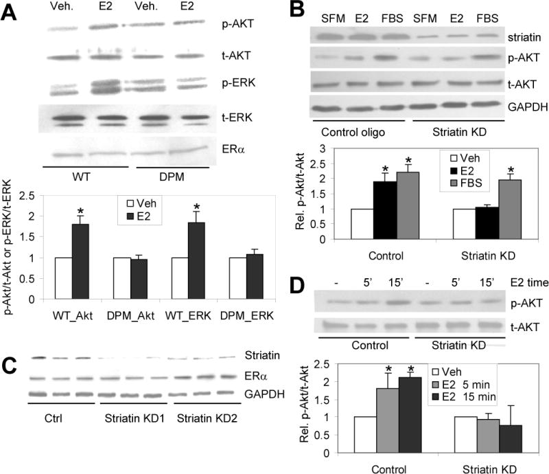
Rapid signaling is lost in cells from DPM mice, and in primary and immortalized cells lacking striatin. (A) Primary lung endothelial cells from WT and DPM mice were progressively starved for serum over ~12 hours, before treatment with 10 nM E2 or vehicle for 20 minutes. The total cell lysates were immunoblotted with the indicated antibodies. (B) Primary bovine aortic endothelial cells (BAECs) were cultured in DMEM supplemented with 10% FBS and striatin expression was knocked down (KD) via transient shRNA transfection. Cells were grown for 24 hrs in serum-free media then treated with vehicle (SFM), 10nM E2 or 10%FBS for 20 min, and immunoblotted with the indicated antibodies. (C) Western blot showing the reduction of striatin protein in stably transfected EAhy926 knock down cell lines (StriatinKD 1 & 2) but not in an empty-vector control cell line (Ctrl). Shown are results from 3 separate cultures derived from 2 independent clones for each cell line. Lower panels: immunoblots for ERα (showing ERα protein levels are unaffected by striatin shRNAs) and for GAPDH (loading control). (D) Stably-transfected striatin knock down or empty vector control EAhy926 cells were cultured in DMEM supplemented with 10% FBS, followed by treating with SFM for 24 h, followed by treatment with 10 nM E2 for 5 or 15 minutes. Cell lysates were immunoblotted for the indicated antibodies. Experiments were done in triplicate (for B and D) or quadruplicate (for A). Bars: standard deviation. “*”: p. <.05. Veh = Vehicle treated; E2- 17β-estradiol treated.