
Figure 5.
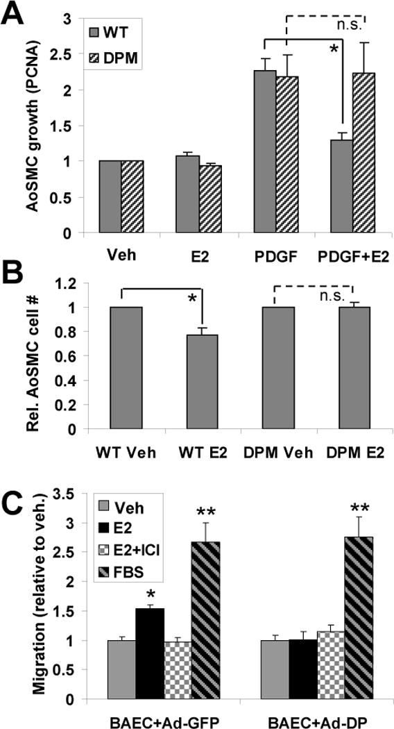
The ERα176-253 striatin-blocking peptide eliminates the effect of E2 on SMC growth and endothelial cell migration. (A) Cultured aortic smooth muscle cells (AoSMCs) from WT and DPM mice were treated with E2 and/or PDGF as indicated. Cell growth relative to untreated controls was measured by quantitative RT-PCR for the DNA-replication sliding clamp protein, PCNA, whose expression is tightly coupled to cell growth 7, with normalization to GAPDH. Each experiment was repeated four times, with two technical replicates per experiment. (B) As in (A), but with cell growth after 2 days measured by counting of trypsin-released cells using a hemocytometer. The experiment was repeated twice with three technical replicates, and data were normalized to the vehicle control value for WT or for DPM. (C) BAECs were infected with control adenovirus containing GFP (Ad-GFP) or ERα176-253-FLAG peptide (Ad-DP) expression vectors, and assayed for migration under the indicated conditions in a scratch-wound assay. Each experiment was repeated three times, with four technical replicates. Bars: standard error of the mean. “**”: p<.001 (relative to EtOH control), “*”: p<.02, “n.s.”: no significant difference.