Abstract
This study aimed to determine the frequency of cervical lung lobe herniation (CLLH) in dogs evaluated fluoroscopically and to identify associated characteristics. Reports of diagnostic procedures and patient summaries from 2008 to 2010 were reviewed retrospectively. Signalment, body weight, duration of cough, presence of heart murmur and airway collapse, and radiographic findings were compared between dogs with and without CLLH. Of the 121 dogs that were examined, CLLH occurred in 85 (70%). The extra-thoracic trachea kinked during herniation in 33 (39%) dogs with CLLH. Collapse of the intra-thoracic trachea (assessed fluoroscopically or bronchoscopically) and collapse of major bronchi (assessed fluoroscopically) were strongly associated with CLLH. Although redundant dorsal tracheal membrane on radiographs was associated with CLLH, extra-thoracic tracheal collapse, assessed fluoroscopically or bronchoscopically, was not. No other associations were found. Cervical lung lobe herniation was present in most dogs evaluated during cough and was associated with intra-thoracic large airway collapse, but not duration of cough.
Résumé
Herniation du lobe pulmonaire cervical chez les chiens identifié par fluoroscopie. Cette étude a visé à déterminer la fréquence de l’herniation du lobe pulmonaire cervical (HLPC) chez les chiens évalués par fluoroscopie et à identifier les caractéristiques connexes. Des rapports des procédures diagnostiques et des sommaires des patients de 2008 à 2010 ont été examinés rétrospectivement. Le signalement, le poids corporel, la durée de la toux, la présence d’un souffle cardiaque et de l’affaissement des voies aériennes ainsi que les constatations radiographiques ont été comparés entre les chiens avec et sans HLPC. Parmi les 121 chiens qui ont été examinés, HLPC s’est produite dans 85 cas (70 %). La trachée extra-thoracique s’est tordue durant l’herniation chez 33 (39 %) des chiens atteints de HLPC. L’affaissement de la trachée intra-thoracique (évalué par fluoroscopie ou bronchoscopie) et l’affaissement des bronches majeures (évalué par fluoroscopie) étaient fortement associés à HLPC. Même si la membrane trachéale dorsale redondante sur les radiographies était associée à HLPC, l’affaissement trachéal extra-thoracique, évalué par fluoroscopie ou bronchoscopie, ne l’était pas. Aucune autre association n’a été trouvée. L’herniation du lobe pulmonaire cervical était présente chez la plupart des chiens évalués durant la toux et était associée à l’affaissement des grandes voies aériennes intra-thoraciques, mais non à la durée de la toux.
(Traduit par Isabelle Vallières)
Introduction
Lung lobe herniation is the protrusion of lung parenchyma beyond the musculoskeletal thorax (1–4). It has rarely been reported in dogs (5–7). In humans, lung lobe hernias are classified by location as cervical, thoracic, or diaphragmatic (1–4). They are further classified as congenital or acquired, with acquired etiologies including iatrogenic, traumatic, pathologic (e.g., resulting from neoplasia or inflammation), or spontaneous disease (1–4,8). Approximately one-third of lung lobe hernias in humans are cervical (1). Cervical lung lobe herniation (CLLH) in humans is most often congenital, due to a defect in Sibson’s fascia, which extends from the first rib to the last cervical vertebra, resulting in an area of weakness between the sternocleidomastoid and anterior scalene muscles (1–4,8). Congenital CLLH is observed in infants during crying and usually resolves spontaneously by 1 y of age (2,4). Spontaneously resolving cases in young children are considered by some to be protrusions rather than true hernias, and either a normal finding or anatomic variant (9).
Cervical lung lobe hernias in humans can be acquired as a result of increased intra-thoracic pressures in combination with a localized weakness in the thorax (2,3). Cough, particularly in association with chronic obstructive pulmonary disease (COPD), is one such cause (1–4). Presumably chronic coughing, and in some cases associated emphysema or hyperinflation, results in weakening and stretching of the cervical tissues (4). The lung rarely becomes entrapped in CLLH in humans, returning to its normal location when intra-thoracic pressures are no longer elevated. Although surgical correction is possible, treatment is generally aimed at minimizing increases in intra-thoracic pressures, such as through control of underlying airway disease and avoidance of Valsalva maneuvers.
The authors are aware of reports of CLLH in 3 dogs (5,6). All cases were associated with chronic cough and increased expiratory efforts. The dogs were 13–14 years of age, and 2 were brachycephalic breeds (pug and Pekingese). Intermittent swelling in the neck was reported by the client in 2 cases, and observed during physical examination in all cases. The diagnosis was confirmed by thoracic radiography, with images obtained during expiration, and by fluoroscopy.
It was the authors’ experience that CLLH is a commonly identified abnormality of dogs evaluated fluoroscopically. Therefore, this retrospective study was performed in order to determine the frequency of CLLH in these dogs, and to identify associated factors. We hypothesized that CLLH would be a frequent finding, and would be associated with duration of cough, brachycephalic conformation, and airway collapse.
Materials and methods
A computerized search of the radiology database of the North Carolina State University — Veterinary Health Complex (NCSU-VHC) was performed to identify all dogs that underwent tracheal fluoroscopy between January 1, 2008 and December 31, 2010. Radiology reports of tracheal fluoroscopy and thoracic radiography, and discharge summaries from the time of fluoroscopy were reviewed for each dog and the results recorded in a spreadsheet. Results were also recorded from bronchoscopy reports for dogs that had undergone this procedure.
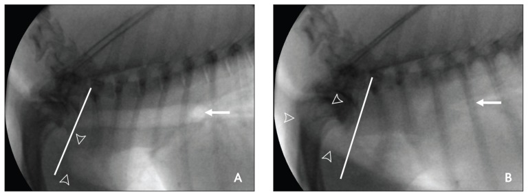
Reports of tracheal fluoroscopy were reviewed to determine whether the evaluation included only tidal breathing, or if cough or forceful expiration was observed. Dogs that were not imaged during cough or forceful expiration were eliminated from the study. The findings of CLLH and kinking of the trachea at the thoracic inlet, and the location of any airway collapse were recorded. The definition used for the diagnosis of CLLH by the radiology service at the NCSU-VHC is obvious protrusion of lung lobes cranial to a line drawn between the cranial aspect of the first thoracic vertebra and the cranial aspect of the manubrium. The definition for airway collapse is a decrease in airway diameter by 50% or more.
From the discharge summaries, signalment, body weight, body condition score, duration of cough, mention in the history or physical examination of a swelling at the thoracic inlet, and heart murmur and grade were recorded. From thoracic radiography reports, tracheal redundancy, abnormal pulmonary pattern, cardiomegaly, left atrial enlargement, and hepatomegaly were noted. From the bronchoscopy report, the presence or absence of tracheal collapse and its location (intra-thoracic and/or extra-thoracic) and bronchial collapse were noted.
Tabulated findings were compared between dogs with CLLH and dogs without CLLH. Chi-square, using Yates correction for continuity, or Fisher’s exact test was used for categorical data. Continuous data were not normally distributed and were compared using the Mann-Whitney rank sum test, and presented as median (25-/75- percentiles). Statistics were calculated using SigmaPlot (Systat Software, San Jose, California, USA). A value of P ≤ 0.05 was considered to be significant.
Results
During the 3-year study period tracheal fluoroscopy was performed on 167 dogs, 121 of which had documentation of evaluation during cough or forced expiration. Of the 121 dogs, 85 (70%) had CLLH (Figure 1). The median (25-/75- percentiles) age of dogs in both groups was approximately 10 y [dogs with CLLH, 10.5 (7.1/12.6) y; dogs without CLLH, 10.3 (6.4/12.3) y; P = 0.477]. Pomeranians (n = 18), Yorkshire terriers (n = 17), Chihuahuas (n = 13), and miniature poodles (n = 11) were the breeds most commonly evaluated. No association was found between any specific breed, or between being brachycephalic, and CLLH. However, all 8 of the Maltese dogs evaluated had CLLH (P = 0.103). The median weight of dogs in both groups was approximately 6 kg [dogs with CLLH, 6.0 (4.2/8.6) kg; dogs without CLLH, 5.5 (3.2/7.3) kg; P = 0.199]. The median body condition score in both groups, as noted for 75 dogs, was 6 on a scale of 1 to 9 (P = 0.627). A swelling in the neck or thoracic inlet was not noted in the summary of history and physical examination findings in any dog.
Figure 1.
Static images captured from recorded fluoroscopy of a dog with cervical lung lobe herniation. During inspiration (A), no abnormalities are seen. The line approximates the cranial extent of the thoracic cavity, extending from the cranial aspect of the first thoracic vertebra to the cranial aspect of the manubrium. The open arrowheads point to the cranial aspect of the cranial lung lobes. The intra-thoracic trachea and carina (solid arrow) are of normal size. At peak expiration during cough (B) the cervical lung is extruded past the sternum, well into the neck (open arrowheads). The intra-thoracic trachea and carina (solid arrow) are collapsed and displaced cranially.
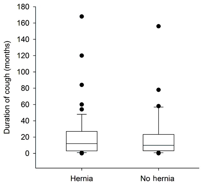
Nearly all dogs evaluated had a history of cough (116 of 121; 96%). The 5 dogs without cough were evaluated because of other respiratory signs, such as tachypnea, panting, gagging, stertorous breathing, and wheezing. Duration of cough was reported in 109 of these 116 dogs (94%). There was no association between duration of cough and CLLH. Dogs with CLLH had a median duration of cough of 12 mo (3/27), and dogs without CLLH had a median duration of cough of 10 mo (3/23; P = 0.792; Figure 2) Duration of cough was ≤ 1 mo in 8 (7%) dogs with CLLH for which these data were reported. The shortest duration of cough in a dog with CLLH was 2 d.
Figure 2.
There was no association between duration of cough and presence of cervical lung lobe herniation. The box and whiskers plot demonstrates the median (line), 25th and 75th percentiles (box), 10th and 90th percentiles (whiskers), and outlying values.
The presence or absence of a heart murmur was recorded in 84 of the 85 dogs with CLLH and in all dogs without CLLH. Heart murmurs were present in a nearly equal proportion of dogs with CLLH (52 dogs, 62%) and in dogs without CLLH (22 dogs, 63%; P = 0.913). Heart murmurs in dogs with CLLH were louder (median grade, 4; 25-/75- percentiles, 2/4) compared with dogs without CLLH (median grade 2; 1/4), but statistical significance was not reached (P = 0.068).
Reports from thoracic radiography were available for all but 1 dog, a dog without CLLH. No dogs had CLLH or kinking of the trachea visible on thoracic radiographs. An abnormal pulmonary pattern was mentioned in 47 (39%) of all dogs, and was predominantly bronchial in 34 (72%) of these dogs. There was no difference between groups with regard to presence of any abnormal pattern (P = 0.619) or of a bronchial pattern (P = 0.853). The presence of a redundant dorsal tracheal membrane was noted more often in dogs with CLLH (40 dogs, 47%) compared with dogs without CLLH (9 dogs, 26%; P = 0.050). More dogs with CLLH had cardiomegaly (27 dogs, 32%) and left atrial enlargement (37 dogs, 44%), compared to dogs without CLLH [cardiomegaly, 6 dogs (17%); left atrial enlargement, 9 dogs (26%)], but the differences were not statistically significant (P = 0.160 and 0.106, respectively). There was also no association between hepatomegaly and CLLH (P = 0.281).
Based on fluoroscopy, 82 dogs (96%) with CLLH had airway collapse at 1 or more locations, compared with 32 dogs (89%) without CLLH (P = 0.227). Collapse of the intra-thoracic trachea was strongly associated with CLLH, as was collapse of the mainstem or caudal lobar bronchi. Collapse of the intra-thoracic trachea was seen in 59 (69%) dogs with CLLH and 13 (36%) dogs without CLLH (P = 0.001). Collapse of the mainstem or caudal lobar bronchi was seen in 78 (92%) dogs with CLLH and 25 (70%) dogs without CLLH (P = 0.004). Associations were not seen for extra-thoracic tracheal collapse, identified in 4 (5%) dogs with CLLH and 1 (3%) dog without CLLH (P = 0.990), or collapse at the thoracic inlet, noted in 9 (11%) dogs with CLLH and 2 (6%) dogs without CLLH (P = 0.593).
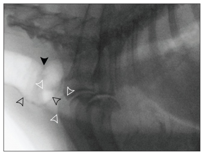
Kinking of the extra-thoracic trachea, which occurred during cough or forced expiration, was noted in 33 (39%) dogs with CLLH, and in no dogs without CLLH (Figure 3). Kinking was not associated with age (P = 0.534), weight (P = 0.509), duration of cough (P = 0.621), breed (including being brachycephalic; P = 0.276), left atrial enlargement (P = 0.610), murmur (P = 0.622), or hepatomegaly (P = 0.993).
Figure 3.
Kinking of the extra-thoracic trachea, in association with cervical lung lobe herniation, is apparent in this static image captured from recorded fluoroscopy at peak expiration during cough. The white arrowheads indicate the cranial extent of the cranial lung lobes. The black arrowheads indicate a dramatic kink in the trachea, with a visible fold dorsally (solid black arrowhead).
Bronchoscopy was performed in 22 dogs with CLLH and 9 dogs without. Of the 22 dogs with CLLH that had bronchoscopy, 8 had kinking of the extra-thoracic trachea based on fluoroscopy. Mucosal abnormalities of the trachea were present in 4 (50%) dogs with tracheal kinking (nodular irregularity, moderate hyperemia, moderate mucus, and mild hyperemia and mucus), but this was not statistically different than in dogs without tracheal kinking in which 9 of 23 (39%) had mucosal abnormalities (P = 0.689). Airway collapse was identified by bronchoscopy in more dogs than were identified by fluoroscopy (14 dogs with extra-thoracic tracheal collapse, 5 dogs with intra-thoracic tracheal collapse, and 3 dogs with bronchial collapse). Airway collapse was identified by fluoroscopy, but not bronchoscopy, in 4 dogs (1 with intra-thoracic tracheal collapse and 3 with bronchial collapse). Collapse of the intra-thoracic trachea as identified bronchoscopically was strongly associated with CLLH, and was present in 19 (86%) dogs with CLLH and 3 (33%) dogs without CLLH (P = 0.007). Collapse of the bronchi was seen in 20 (91%) dogs with CLLH and 6 (67%) dogs without CLLH (P = 0.131). No association was seen between extra-thoracic tracheal collapse, which was identified bronchoscopically in 14 (64%) dogs with CLLH and 5 (56%) dogs without CLLH (P = 0.704).
Discussion
This study confirms the frequent occurrence of CLLH in dogs evaluated fluoroscopically, the majority of which had a history of cough. While the study population is not representative of the general population of coughing dogs, the high rate of diagnosis proves that CLLH is not a rare complication. With increased awareness of this complication, clinicians can begin to make clinical correlations between the presence of herniation and progression of disease in their own practices, and future investigations can prospectively consider CLLH as a factor or co-factor in therapeutic studies. In addition, kinking of the trachea during herniation was seen in approximately one-third of dogs. In some dogs, the kinking was severe and tissue trauma was probable. Although this conjecture was not proven by our study, we recommend aggressive cough control in such dogs, including centrally acting cough suppressants, to stop this potential component of the cough-cough cycle.
Fluoroscopy is not available in most clinical settings, but it should be possible to identify CLLH on routine physical examination in most dogs. Observation or palpation of the neck during a cough would be required, but cough can often be induced with tracheal manipulation in dogs with airway disease. It is remarkable that such a finding was never noted in this study, particularly since swelling in the neck was reported in the 3 previously reported cases with CLLH (5,6). Possible explanations for the failure to note cervical swelling include data collection from discharge summaries rather than primary history or physical examination forms, and interference with visualization due to hair coat or fat deposits. In addition, as a rarely reported event, it is possible that clinicians are not actively evaluating for this abnormality. The typical position of the veterinarian standing above and lateral to the patient during much of a routine examination is not optimal for visualizing the ventral cervical region. In severely affected dogs, herniation may be diagnosed by thoracic radiography. Routine inspiratory views will not show the defect. Rather, radiographs should be taken during maximal expiration with the forelimbs pulled caudally, if evaluation for CLLH and kinking is warranted (5,6).
Nearly all study dogs had a history of cough. This finding is consistent with the association in humans between CLLH and cough (1–4). It is also a result of the study design. Fluoroscopic evaluations at the NCSU-VHC are usually requested for patients with: chronic cough, particularly when lower airway disease or collapse is suspected; suspected intra- or extra-thoracic large airway obstruction; or possible tracheal or pulmonary masses. The requirement that the fluoroscopic evaluation included cough further biased the patient population to those with historic cough. In such patients, cough is more easily induced with tracheal palpation, and more effort to elicit a cough is made. Nevertheless, this patient population was appropriate for the goals of this study. In order to identify characteristics associated with CLLH, dogs needed to be assigned to groups as accurately as possible. Including dogs that were evaluated only during tidal breathing would have resulted in dogs being erroneously classified in the group of dogs without CLLH.
In humans, CLLH is associated with COPD (1–4). Based on our results, lung lobe herniation in dogs is strongly associated with intra-thoracic large airway collapse. Both intra-thoracic large airway collapse and small airway disease result in increased resistance to air flow that is most pronounced during expiration. Obstruction resulting from large airway collapse is more severe during forceful expiratory efforts, including cough. One hypothesis for the frequent occurrence of CLLH in these dogs is that their obstructive disease prevents the usual relief of pressure from cough through the expiration of air through the glottis, allowing greater than usual pressures to build up within the thorax. Although an association was also found between redundant dorsal membrane as identified on thoracic radiography and CLLH, an association between CLLH and extra-thoracic tracheal collapse was not found based on fluoroscopic or bronchoscopic findings. A previous study found that thoracic radiography had a specificity of only 0.48 for collapse of the extra-thoracic trachea using fluoroscopic findings as the standard (10). Another study from the same institution also found that bronchoscopy and fluoroscopy did not always yield identical results with respect to identifying airway collapse, with bronchoscopy identifying collapse more often than fluoroscopy in a small number of cases in which both were performed (11). This is not surprising since the resolution of fluoroscopic images is low, all lobar bronchi are not usually visible, patient motion can interfere with interpretation, and narrowing of the airway lumen by at least 50% has been traditionally recommended for the fluoroscopic diagnosis of collapse. The advantage of fluoroscopy over bronchoscopy in assessing for airway collapse is lower cost and risk. Despite some differences in results between fluoroscopy and bronchoscopy in individual dogs, a strong association between CLLH and intra-thoracic airway collapse was found by either method of assessment in the current study.
Most dogs in this study were older and had a prolonged history of cough, but no distinction could be made between dogs with and without CLLH. Acquired CLLH in humans is typically seen in association with chronic elevations in intra-thoracic pressure. In addition to humans with chronic respiratory disease, an association is seen with professions such as glass-blowing, playing a wind instrument, or lifting heavy weights (1–4,8). There are reports of CLLH occurring acutely in humans (12). One of the dogs in this study had a cough of only 2 days duration.
In addition to high intra-thoracic pressures, another possible contributing factor to the development of CLLH is congenitally weak fascia or musculature. In a report of 6 humans with CLLH, 2 had a history of hernia repairs, suggesting the possibility of congenital weakness (8). The breeds of dogs that most often had CLLH were similar to breeds known to have relatively high rates of chronic cough and tracheobronchomalacia (10,11,13). However no specific breed associations could be made with CLLH, and there were no obvious associations with chest conformation. Associations with individual specific breeds could have been missed as a result of the small number of dogs of any 1 breed represented in this study. Another possibility is that dogs without cough, particularly of specific breeds such as the Maltese, all have CLLH with enough increase in intra-thoracic pressures. No normal dogs were evaluated, though a third of all study dogs with cough did not have CLLH.
The potential contribution of CLLH to clinical signs in humans with COPD has not been addressed in detail, perhaps because CLLH is an unusual diagnosis. Lateral deviation of the trachea was seen radiographically in 5 of 6 humans; 6 of whom had cough during herniation and another had dysphagia, presumably from compression of trachea or esophagus, respectively (8). Cough and hoarseness have been reported as potential signs resulting from airway compression in human reviews (2,3) and CLLH resulted in upper airway obstruction and stridor in a 6-year-old boy (14). It is concerning that over one-third of dogs with CLLH had kinking of the trachea noted during herniation. Trauma to the trachea as a result of kinking was not identified bronchoscopically. However, trauma could be occurring that is not visible either due to relative insensitivity of gross examination or due to damage to deeper tissues. As demonstrated in Figure 3, the distortion of normal anatomy can be severe.
Potential limitations of this study include its retrospective nature, its reliance on discharge summaries for history and physical examination findings, and its reliance on radiology reports for fluoroscopic and radiographic findings. This study design was chosen to maximize the study population and give strength to our findings. The discharge summaries at the NCSU-VHC are part of the electronic medical record, while the history and physical examination sheets are not. It is the authors’ experience that the records of the history and physical examinations within these summaries are usually complete. Radiographic reports are also part of the electronic medical record. Further, archived fluoroscopic images would not always accurately reflect the findings of the full study. Fluoroscopic studies are not recorded in their entirety. Earlier studies, in particular, were performed using equipment without the capability of prolonged image capture. Short-duration abnormalities (such as collapse or herniation) are sometimes observed during cough before the recording feature can be activated. While the reliance on written reports could introduce variability in interpretation of findings among radiologists, several factors should minimize the likelihood of such variability significantly affecting the results.
Our hospital runs a Respiratory Focus Clinic, and fluoroscopic studies are regularly performed by board-certified radiologists and radiology residents using a consistent protocol. Radiology residents are closely supervised by 1 of several board-certified radiologists. Assignment of personnel to cases is random, based solely on clinic scheduling, so the relative over- or under-interpretation of findings by 1 or more individuals would not be expected to bias findings between groups. Although available for only a quarter of the study population, bronchoscopic findings supported the association between intra-thoracic tracheal collapse and CLLH that was identified by fluoroscopy.
In summary, CCLH and kinking of the trachea during herniation are complications of cough which could possibly impact progression of disease, and the diagnostic evaluation and treatment of dogs with cough. Physical examination for CLLH should become routine practice for dogs with cough due to its frequent diagnosis fluoroscopically, and its association with intra-thoracic large airway collapse and kinking of the trachea. Prospective studies are needed to further characterize the development of CLLH, and determine its role in the progression and prognosis of cough in dogs. CVJ
Footnotes
An abstract of this study was presented at the Veterinary Comparative Respiratory Society Meeting in Vienna, Austria, September 2011 and at the ACVIM Forum in New Orleans, Louisiana, May 2012.
Use of this article is limited to a single copy for personal study. Anyone interested in obtaining reprints should contact the CVMA office (hbroughton@cvma-acmv.org) for additional copies or permission to use this material elsewhere.
This work was not supported by a specific grant.
References
- 1.Minai OA, Hammond G, Curtis A. Hernia of the lung: A case report and review of literature. Conn Med. 1997;61:77–81. [PubMed] [Google Scholar]
- 2.Moncada R, Vade A, Gimenez C, et al. Congenital and acquired lung hernias. J Thorac Imaging. 1996;11:75–82. doi: 10.1097/00005382-199601110-00008. [DOI] [PubMed] [Google Scholar]
- 3.Scullion DA, Negus R, al-Kutoubi A. Case report: Extrathoracic herniation of the lung with a review of the literature. Br J Radiol. 1994;67:94–96. doi: 10.1259/0007-1285-67-793-94. [DOI] [PubMed] [Google Scholar]
- 4.Bhalla M, Leitman BS, Forcade C, Stern E, Naidich DP, McCauley DI. Lung hernia: Radiographic features. Am J Roentgenol. 1990;154:51–53. doi: 10.2214/ajr.154.1.2104725. [DOI] [PubMed] [Google Scholar]
- 5.Guglielmini C, De Simone A, Valbonetti L, Diana A. Intermittent cranial lung herniation in two dogs. Vet Radiol Ultrasound. 2007;48:227–229. doi: 10.1111/j.1740-8261.2007.00233.x. [DOI] [PubMed] [Google Scholar]
- 6.Coleman MG, Warman CG, Robson MC. Dynamic cervical lung hernia in a dog with chronic airway disease. J Vet Intern Med. 2005;19:103–105. doi: 10.1892/0891-6640(2005)19<103:dclhia>2.0.co;2. [DOI] [PubMed] [Google Scholar]
- 7.Weaver AD. Severe traumatic pneumothorax and lung prolapse in a Jack Russell bitch. Vet Rec. 1982;111:505. doi: 10.1136/vr.111.22.505. [DOI] [PubMed] [Google Scholar]
- 8.McAdams HP, Gordon DS, White CS. Apical lung hernia: Radiologic findings in six cases. Am J Roentgenol. 1996;167:927–930. doi: 10.2214/ajr.167.4.8819385. [DOI] [PubMed] [Google Scholar]
- 9.Currarino G. Cervical lung protrusions in children. Pediatr Radiol. 1998;28:533–538. doi: 10.1007/s002470050405. [DOI] [PubMed] [Google Scholar]
- 10.Macready DM, Johnson LR, Pollard RE. Fluoroscopic and radiographic evaluation of tracheal collapse in dogs: 62 cases (2001–2006) J Am Vet Med Assoc. 2007;230:1870–1876. doi: 10.2460/javma.230.12.1870. [DOI] [PubMed] [Google Scholar]
- 11.Johnson LR, Pollard RE. Tracheal collapse and bronchomalacia in dogs: 58 cases (7/2001–1/2008) J Vet Intern Med. 2010;24:298–305. doi: 10.1111/j.1939-1676.2009.0451.x. [DOI] [PubMed] [Google Scholar]
- 12.Mao A, Kammen BF. Dynamic cervical lung herniation in a 10-year-old girl with cough. Pediatr Radiol. 2007;37:1058. doi: 10.1007/s00247-007-0504-3. [DOI] [PubMed] [Google Scholar]
- 13.Hawkins EC, Clay LD, Bradley JM, Davidian M. Demographic and historical findings, including exposure to environmental tobacco smoke, in dogs with chronic cough. J Vet Intern Med. 2010;24:825–831. doi: 10.1111/j.1939-1676.2010.0530.x. [DOI] [PMC free article] [PubMed] [Google Scholar]
- 14.Gonzalez del Rey J, Cunha C. Cervical lung herniation associated with upper airway obstruction. Ann Emerg Med. 1990;19:935–937. doi: 10.1016/s0196-0644(05)81574-4. [DOI] [PubMed] [Google Scholar]


