Abstract
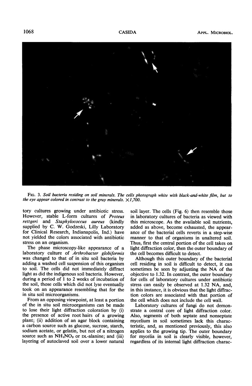
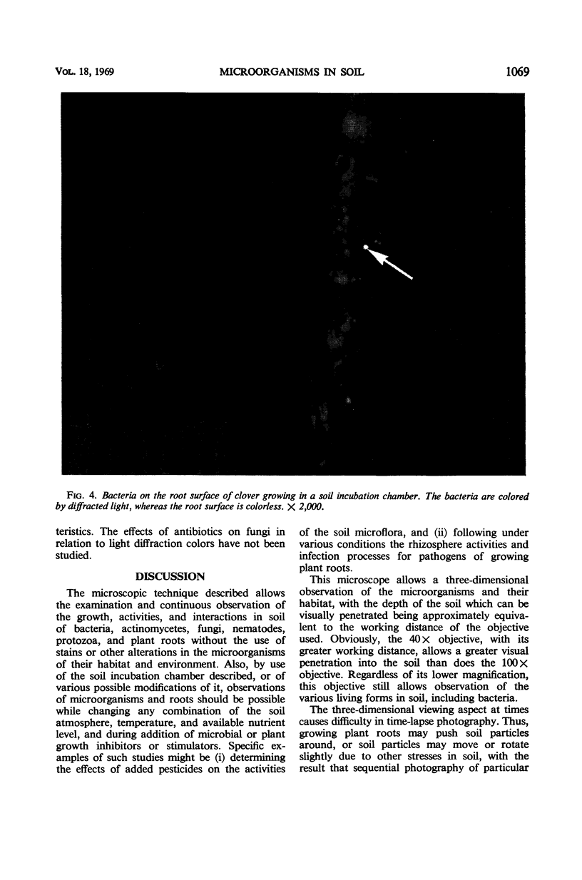
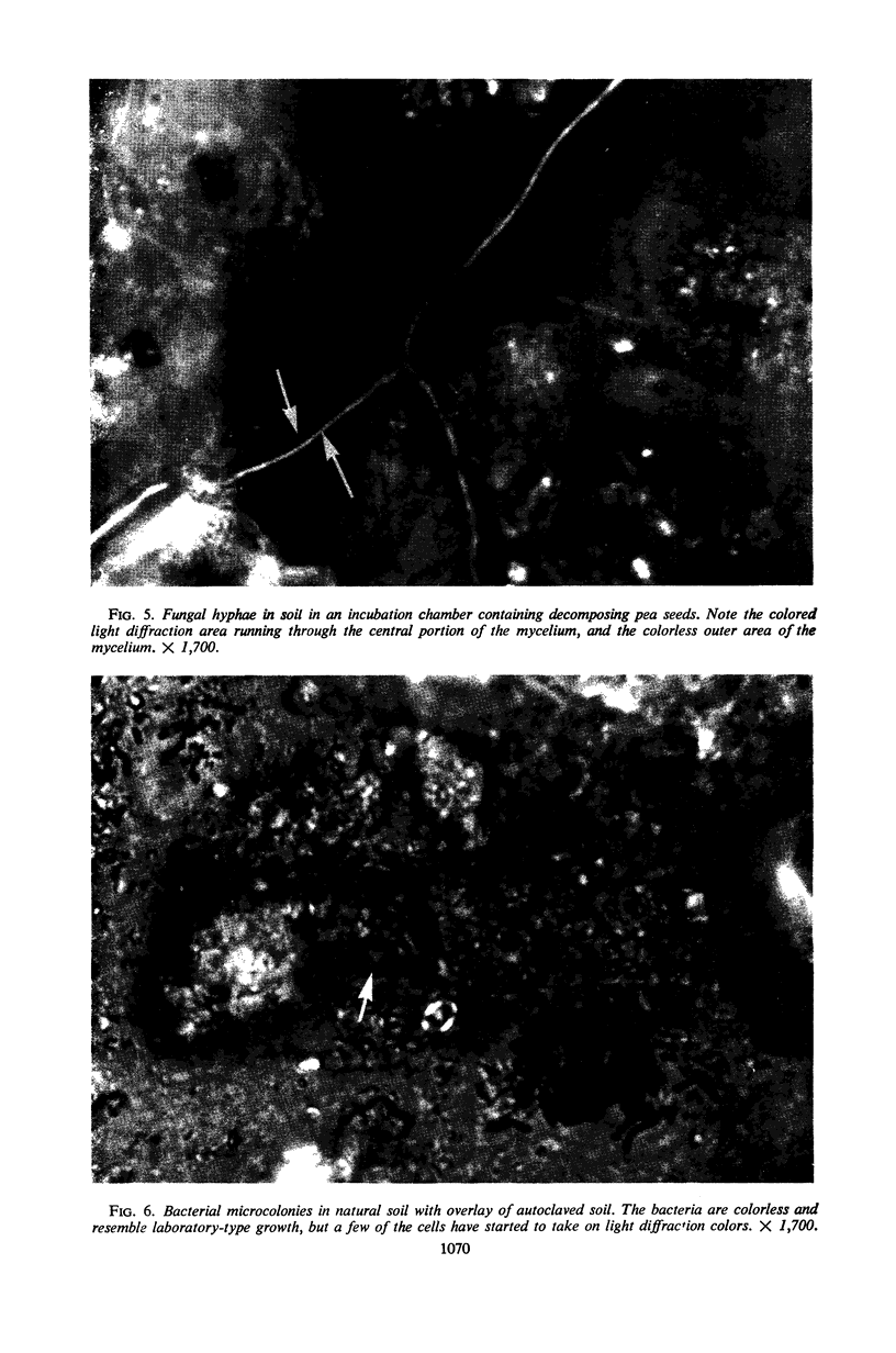
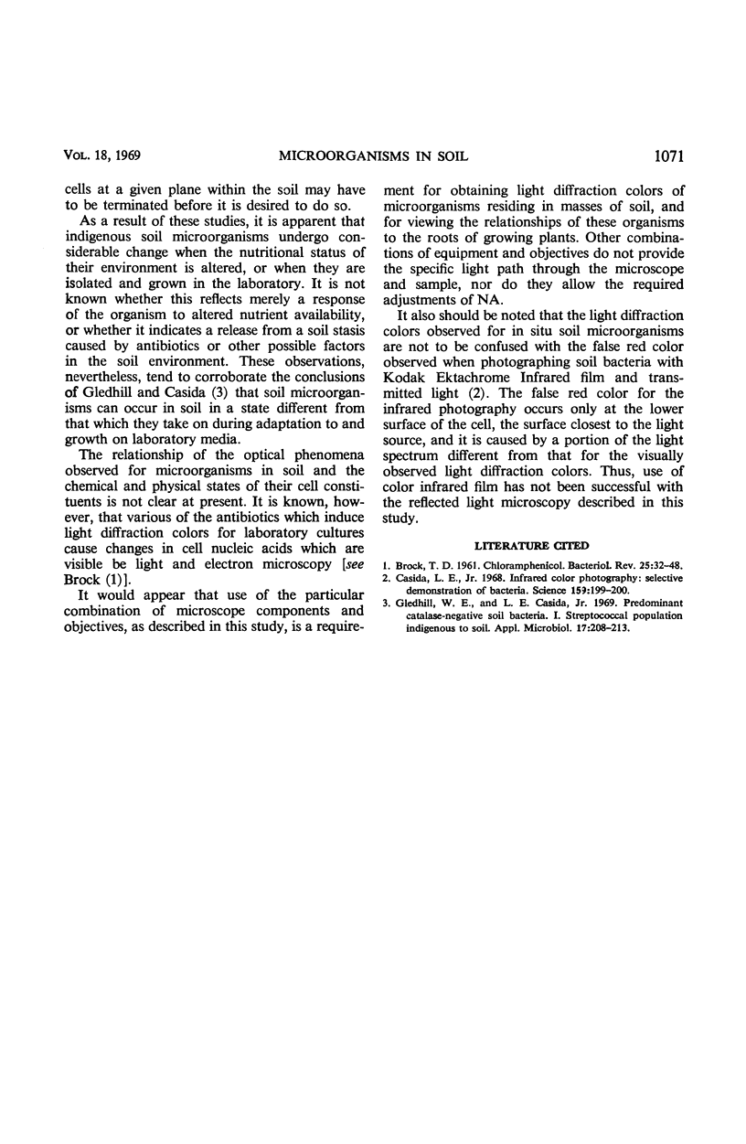
A procedure is described for visually observing and following the activities and interactions of bacteria, actinomyctes, fungi, protozoa, nematodes, and plant roots in masses of soil. Specific microscope components and objectives are used, and the numerical apertures are adjusted such that light diffraction colors are produced to allow differentiation of the various biological entities and their habitat materials. Strains or other alterations in the organisms and their habitat are not employed, and time-lapse photography can be used to follow the activities of soil microorganisms and plant roots. As a result of the use of this technique, it is apparent that in situ indigenous soil microorganisms differ from similar organisms grown in the laboratory, but that, under the proper conditions, the state of the organism in either habitat can be altered to match that which occurs in the contrasting habitat.
Full text
PDF
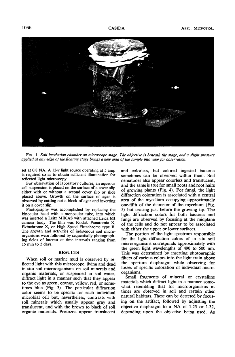
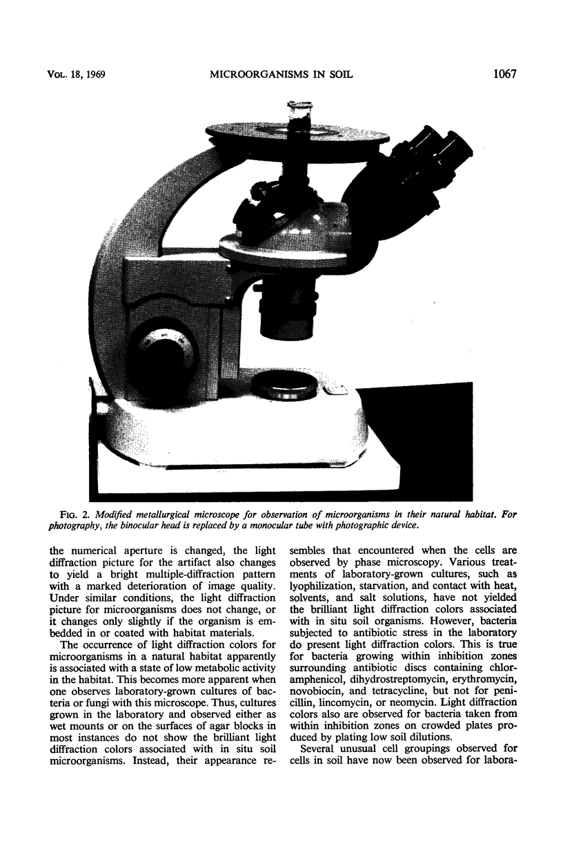
Images in this article
Selected References
These references are in PubMed. This may not be the complete list of references from this article.
- Brock T. D. CHLORAMPHENICOL. Bacteriol Rev. 1961 Mar;25(1):32–48. doi: 10.1128/br.25.1.32-48.1961. [DOI] [PMC free article] [PubMed] [Google Scholar]
- Casida L. E., Jr Infrared color photography: selective demonstration of bacteria. Science. 1968 Jan 12;159(3811):199–200. doi: 10.1126/science.159.3811.199. [DOI] [PubMed] [Google Scholar]
- Gledhill W. E., Casida L. E., Jr Predominant catalase-negative soil bacteria. I. Streptococcal population indigenous to soil. Appl Microbiol. 1969 Feb;17(2):208–213. doi: 10.1128/am.17.2.208-213.1969. [DOI] [PMC free article] [PubMed] [Google Scholar]


