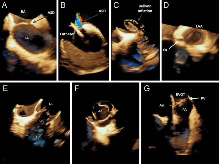
Echocardiography plays an essential role in guiding complex cardiovascular interventions: atrial septal defect (ASD) closure, left atrial appendage (LAA) occlusion, and transcatheter aortic valve implantation (TAVI). Most procedures are guided by three-dimensional (3D) transoesophageal echocardiography.
Intracardiac echocardiography (ICE) has the advantage of not requiring general sedation, does not interfere with fluoroscopy, and provides simultaneous guidance of the entire procedure. However, until now, ICE could only provide 2D images.
We report the first worldwide clinical use of 3D intracardiac echocardiography. Images were acquired using an Acuson SC 2000™ ultrasound unit (Siemens Medical Solutions, Erlangen, Germany) and a 10 F AcuNav V™, able to acquire a 60° × 15° volume, at a volume rate of 20 vps.
Images in Panels A–C and E–F were acquired with the catheter in the right atrium. Panel A shows the right and left atrium (RA, LA) and an ASD (see Supplementary data online, video S1). This view was used in this patient to guide the closure of the ASD: Panels B and C show 3D ICE images during the procedure. Panels E and F show a long- and short-axis view of the aortic valve (Ao), which can be used to guide TAVI.
The image in Panel D was acquired with the catheter advanced into the left atrium, showing the LAA opening (see Supplementary data online, video S2), which can be used to guide LAA occlusion procedures. The circumflex artery (Cx) can also be viewed.
Finally, Panel G shows the pulmonary valve (PV), right ventricle outflow tract (RVOT) and pulmonary artery (PA).
Supplementary data are available at European Heart Journal – Cardiovascular Imaging online.

Supplementary Material
Associated Data
This section collects any data citations, data availability statements, or supplementary materials included in this article.


