Abstract
We report a successful endovascular technique using a snare with a suture for retrieving a migrated broken peripherally inserted central catheter (PICC) in a chemotherapy patient. A 62-year-old male received monthly chemotherapy through a central venous port implanted into his right subclavian area. The patient completed chemotherapy without complications 1 mo ago; however, he experienced pain in the right subclavian area during his last chemotherapy session. Computed tomography on that day showed migration of a broken PICC in his left pulmonary artery, for which the patient was admitted to our hospital. We attempted to retrieve the ectopic PICC through the right jugular vein using a gooseneck snare, but were unsuccessful because the catheter was lodged in the pulmonary artery wall. Therefore, a second attempt was made through the right femoral vein using a snare with triple loops, but we could not grasp the migrated PICC. Finally, a string was tied to the top of the snare, allowing us to curve the snare toward the pulmonary artery by pulling the string. Finally, the catheter body was grasped and retrieved. The endovascular suture technique is occasionally extremely useful and should be considered by interventional cardiologists for retrieving migrated catheters.
Keywords: Port catheter, Catheter migration, Endovascular suture technique
Core tip: Catheter migration has been reported as a delayed complication of peripherally inserted central catheter (PICC). Retrieval by the endovascular technique using a snare is usually attempted in cases of PICC migration, but there have been some difficulties in retrieving the broken catheter. We encountered a patient with an ectopic PICC in the left pulmonary artery; the ectopic PICC could not be retrieved by the usual method using a snare, but was successful retrieved using a snare and suture technique. The endovascular suture technique is a useful method to retrieve a dislocated or broken catheter and should be considered by interventional cardiologists.
INTRODUCTION
Chemotherapy drug administration through a peripherally inserted central catheter (PICC) has been widely used because of several advantages such as easier PICC insertion and improved patient satisfaction[1]. The main complications of PICC are bloodstream infection and venous thrombosis[1,2], although catheter migration has been reported as a delayed complication[1,3,4]. In cases of PICC migration to the heart or pulmonary artery, retrieval by the endovascular technique using a snare is usually attempted[4,5], but some difficulties have been reported and the procedure requires several devices and advanced techniques[4-7]. We describe a patient with an ectopic PICC in the left pulmonary artery, which was successfully retrieved using a snare with a suture technique on the second attempt.
CASE REPORT
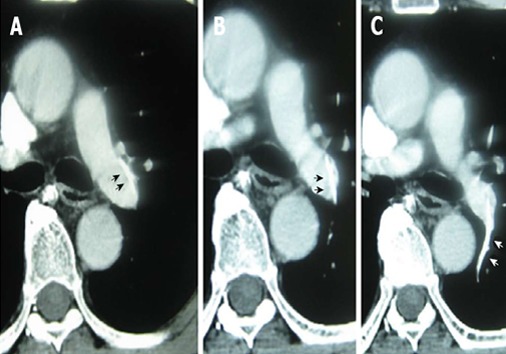
A 62-year-old male, who underwent surgeries for advanced colon cancer in November 2008 and for lung metastasis in February 2010, was receiving monthly chemotherapy through a central venous port to his right subclavian vein since March 2010. The last chemotherapy session was performed on November 22, 2010 and no problems occurred during this time. On December 20, 2010, he experienced pain in the right subclavian area during chemotherapy. Subsequent contrast-enhanced computed tomography (CT) showed an ectopic PICC in his left pulmonary artery (Figure 1); therefore, the patient was presented and admitted to our hospital for retrieval of the ectopic PICC on the same day. On admission, his vital signs were stable. Blood examination showed increase in D-dimer (3.6 mg/dL) and C-reactive protein (0.96 mg/dL) levels. In addition, he had poorly controlled diabetes mellitus, with hemoglobin A1C level of 8.4%.
Figure 1.

Contrast-enhanced computed tomography showing the proximal end of the catheter lodged in the wall of the left pulmonary artery trunk (A and B); the distal catheter end was in the small branch of left pulmonary artery (C). The broken catheter is indicated by arrows.
After informed consent was obtained, we attempted retrieval of the ectopic PICC through the right internal jugular vein using a 12-Fr sheath and 8-Fr guiding catheter. CT and pulmonary arteriography revealed that the distal end of the broken catheter was present in the left branch of the pulmonary artery and the proximal end was present in the left pulmonary artery trunk. Therefore, we decided to grasp the ectopic PICC from the proximal end of the catheter. Two goose neck snares (Amplatz GooseNeck, COVIDIEN) measuring 15 and 25 mm were used in our first attempt, but the PICC body could not be grasped because it was lodged in the pulmonary artery wall; we therefore discontinued the attempt because of the extended procedure time. The patient and his family were explained about the possible consequences of the presence of an ectopic PICC in the left pulmonary artery and they requested a second attempt at retrieval.
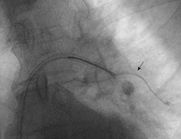
Three days later, the second retrieval attempt was made through the right femoral vein using 12 and 6-Fr long sheaths (Parent Plus, Medikit). At first, a balloon-occluded pulmonary arteriography (BOPA) was performed using a wedge balloon catheter (Selecon MP Catheter II, Terumo Clinical Supply), which showed thrombosis of the arterial branch housing the distal end of the ectopic PICC. The proximal end of the ectopic PICC could not be grasped using a snare equipped with triple loops (En Snare, SHEEN MAN). Other catheters such as a 4-Fr Judkins right catheter and pig-tail catheter were inserted through the left femoral vein to lift the proximal end of PICC lodged in the pulmonary artery wall. However, these attempts failed to grasp the ectopic PICC. Finally, an endovascular suture technique was attempted. Several 2.0 sutures were tightly combined and tied to the bottom of the snare loops (Figure 2A) to guide the snare downward. The 6-Fr guiding catheter was switched for an 8-Fr guiding catheter because the 2.0 sutures could not be inserted into a 6-Fr sheath. After the snare was inserted into the left pulmonary artery, it was curved downward toward the pulmonary artery by pulling the string, thus allowing us to grasp the proximal end of the ectopic PICC (Figure 3) and retrieve it (Figure 2B). Two days later, the patient was discharged without any complications.
Figure 2.

Snare with a suture technique and the retrieved catheter. A: A snare with a suture was curved downward by pulling the string; B: The retrieved catheter.
Figure 3.

Image of the procedure. The second attempt using the snare with a suture technique was successful to grasp of the body of broken catheter. The broken catheter is indicated by an arrow.
DISCUSSION
We encountered a patient with an ectopic PICC in the left pulmonary artery; the ectopic PICC could not be retrieved by the usual method using a snare, but was successfully retrieved using a snare/suture technique.
Although the main complications of PICC are bloodstream infection and venous thrombosis[1,2,8], migration of a broken catheter has been reported as a delayed complication[1,3]. Catheters can migrate at an estimated rate of 0%-3.1%[9,10] within 1.5 years[4]. The most common sign of catheter migration is irrigation resistance to infusion[4], which indicates that a few weeks have elapsed since the onset of an ectopic PICC. Even in the present case, the exact time of the ectopic PICC migration was unknown, and a few weeks may have elapsed before it was detected. The delayed discovery of the ectopic PICC in our case and the use of a thicker catheter, may have caused the catheter to lodge in the pulmonary artery wall, thereby complicating the retrieval procedure.
In general, ectopic PICC management includes percutaneous transcatheter retrieval, open thoracotomy, and long-term anticoagulation therapy[6]. However, cases of fatal cardiac tamponade following migration of a broken catheter have been reported[2]; therefore, percutaneous transcatheter retrieval is usually performed as the first treatment. In the present case, an ectopic PICC extended from the left pulmonary artery trunk to the branch of the left pulmonary artery, in which a thrombosis occurred. Although, cardiac tamponade and pulmonary artery perforation seldom occur, an ectopic PICC may cause an increased incidence of thrombosis in the left pulmonary artery, which may cause clinical symptoms or may act as a source of potential infection. Therefore, after we considered the patient’s requests and the possibility of complications, we attempted percutaneous transcatheter retrieval twice.
There have been several reported percutaneous transcatheter retrieval techniques using a snare, basket catheter, pigtail catheter, or ablation catheter[4-7]. However, it may be important to grasp the center of the catheter body to contain it within a guiding catheter or a long sheath. Contrast-enhanced CT and/or pulmonary arteriography, including BOPA, are useful to assist the surgeon in deciding the catheter end to be grasped. Furthermore, it may be more important to remove the end of catheter from the vessel wall or myocardium to enable the surgeon to grasp the catheter body using a snare. However, in the present case, other devices such as a pigtail catheter did not help to retrieve the broken catheter end because it was lodged in the vessel wall. Therefore, we had to guide the snare downward toward the vessel wall and subsequently use the snare with an endovascular suture technique[7]. The guidewire or catheter can be easily controlled by pulling the attached string. Although this technique is interesting and a useful method to control catheter movement, it may be associated with the risk of vascular injury and other unresolved problems such as the thickness and type of suture used.
In conclusion, the endovascular suture technique is occasionally an extremely useful method to retrieve a dislocated or broken catheter and should be considered by interventional cardiologists.
Footnotes
P- Reviewer Olsha O S- Editor Wen LL L- Editor A E- Editor Lu YJ
References
- 1.Chopra V, Anand S, Krein SL, Chenoweth C, Saint S. Bloodstream infection, venous thrombosis, and peripherally inserted central catheters: reappraising the evidence. Am J Med. 2012;125:733–741. doi: 10.1016/j.amjmed.2012.04.010. [DOI] [PubMed] [Google Scholar]
- 2.Amerasekera SS, Jones CM, Patel R, Cleasby MJ. Imaging of the complications of peripherally inserted central venous catheters. Clin Radiol. 2009;64:832–840. doi: 10.1016/j.crad.2009.02.021. [DOI] [PubMed] [Google Scholar]
- 3.Cheng CC, Tsai TN, Yang CC, Han CL. Percutaneous retrieval of dislodged totally implantable central venous access system in 92 cases: experience in a single hospital. Eur J Radiol. 2009;69:346–350. doi: 10.1016/j.ejrad.2007.09.034. [DOI] [PubMed] [Google Scholar]
- 4.Motta Leal Filho JM, Carnevale FC, Nasser F, Santos AC, Sousa Junior Wde O, Zurstrassen CE, Affonso BB, Moreira AM. Endovascular techniques and procedures, methods for removal of intravascular foreign bodies. Rev Bras Cir Cardiovasc. 2010;25:202–208. doi: 10.1590/s0102-76382010000200012. [DOI] [PubMed] [Google Scholar]
- 5.Kawata M, Ozawa K, Matsuura T, Kuroda M, Hirayama Y, Adachi K, Matsuura A, Sakamoto S. Percutaneous interventional techniques to remove embolized silicone port catheters from heart and great vessels. Cardiovasc Interv Ther. 2012;27:196–200. doi: 10.1007/s12928-012-0100-9. [DOI] [PubMed] [Google Scholar]
- 6.Cope C. Novel Endovascular Suture Techniques for Aortic and Femoral Branch Arteries. J Invasive Cardiol. 1998;10:443–446. [PubMed] [Google Scholar]
- 7.Liem TK, Yanit KE, Moseley SE, Landry GJ, Deloughery TG, Rumwell CA, Mitchell EL, Moneta GL. Peripherally inserted central catheter usage patterns and associated symptomatic upper extremity venous thrombosis. J Vasc Surg. 2012;55:761–767. doi: 10.1016/j.jvs.2011.10.005. [DOI] [PubMed] [Google Scholar]
- 8.Kock HJ, Pietsch M, Krause U, Wilke H, Eigler FW. Implantable vascular access systems: experience in 1500 patients with totally implanted central venous port systems. World J Surg. 1998;22:12–16. doi: 10.1007/s002689900342. [DOI] [PubMed] [Google Scholar]
- 9.Charvát J, Linke Z, Horáèková M, Prausová J. Implantation of central venous ports with catheter insertion via the right internal jugular vein in oncology patients: single center experience. Support Care Cancer. 2006;14:1162–1165. doi: 10.1007/s00520-006-0073-2. [DOI] [PubMed] [Google Scholar]
- 10.Orme RM, McSwiney MM, Chamberlain-Webber RF. Fatal cardiac tamponade as a result of a peripherally inserted central venous catheter: a case report and review of the literature. Br J Anaesth. 2007;99:384–388. doi: 10.1093/bja/aem181. [DOI] [PubMed] [Google Scholar]


