Abstract
Context:
Skin thickness of type-2 diabetic insulin naïve adult patients.
Background:
We have limited data on skin and subcutaneous tissue thickness of Indian type-2 diabetic population. Objective of this study was to assess skin and subcutaneous tissue thickness in insulin naïve type-2 diabetic patients as this information may be useful for insulin injection technique.
Aims:
To assess the skin and subcutaneous tissue thickness at insulin injection sites in insulin naïve, type-2 diabetic adult population across different body mass index (BMI).
Settings and Design:
Observational study carried out at our institute.
Materials and Methods:
One hundred and one insulin naïve type-2 diabetic subjects underwent skin thickness measurement using ultrasound at insulin administration sites. Skin and subcutaneous tissue thickness were measured and prints taken. Though, the sample size to be taken for the study was not calculated, the results obtained clearly show that the power of the study was 80%.
Results:
At arm and thigh, the mean skin thickness was more in males as compared to females in the BMI range <23 kg/m2 (P < 0.05). At abdomen, skin thickness was more in males in the BMI range 19-23 kg/m2 (P < 0.05). Across all the BMIs, mean skin plus subcutaneous thickness at arm was more in females (P < 0.05) except for BMI >25 kg/m2 where thickness in males was comparable. At thigh, the skin plus subcutaneous tissue thickness was more in females (P < 0.05), across all BMI ranges. At abdomen, thickness was more in females for the BMI ranges 17-19 kg/m2 and 23-25 kg/m2, while it was comparable across all other BMI ranges (P > 0.05).
Conclusions:
Skin and subcutaneous tissue thickness can be estimated by BMI. In general it is higher in females.
Keywords: Insulin naïve diabetic adult population, skin thickness, ultrasonographic evaluation of skin thickness
INTRODUCTION
The human skin comprises of the epidermis and dermis. The subcutaneous tissue is found below the layer of the dermis. Information about skin and subcutaneous thickness may be used to make recommendations on insulin injection technique in type-2 diabetes. In type-2 diabetes, there is insulin resistance which may itself affect subcutaneous tissue thickness. In addition, insulin therapy can cause lipohypertrophy and thus can affect subcutaneous tissue thickness. Keeping these facts in consideration, it was planned do a study involving insulin naïve type-2 diabetic population only and measure skin and subcutaneous tissue thickness at popular insulin injection areas.
Since data on effect of body mass index (BMI) and gender on epidermis and dermis thickness in Indian type-2 diabetic population are very scarce and no data is available in the Indian diabetic population, specifically insulin naïve population and thus need to undertake such a study arose.
This observational study was conducted to definitively assess the skin and subcutaneous tissue thickness in insulin naïve type-2 diabetic adult population across different BMI and to find out difference in skin and subcutaneous tissue thickness of type-2 diabetic population and to generate reliable data in relation to BMI and gender.
MATERIALS AND METHODS
The present study to evaluate the skin and subcutaneous tissue thickness of Indian, insulin naïve, type-2 diabetic adult population was conducted at our institute from August 2010 to December 2011. This was a prospective, non-invasive, observational study.
Subjects attending the out-patient department, with known case of diabetes mellitus and require the use of insulin for the 1st time were enrolled for this study. In all, 101 insulin naïve subjects of either gender were selected for analysis who fulfilled all the inclusion and exclusion criteria. Of these 68 were males and 33 were females.
Subjects’ volunteering to participate in this observational study were given consent form in the language (Hindi/English language) preferred by the subject. And only after getting their consent, their demographic information including name, initials, gender, date of birth, height, weight, diabetes status, duration of diabetes, etc., were collected and then were assigned a subject identifier (ID) for identification purpose.
Subject's skin thickness was measured using ultrasound machine (11 MHz probe)[1,2,3] at specific locations (used for insulin administration) [Figure 1] for measurements based on bony landmarks where possible to reduce inter-subject measurement variability:
Figure 1.
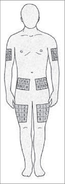
Sites for insulin delivery
Rear upper arm (mid-section between the acromion and olecranon processes)
Anterior upper thigh (mid-distance between the iliac crest and the top edge of the patella)
Anterior abdomen (midway between the umbilicus and the iliac crest).
The sonographic image prints of skin and subcutaneous tissue thickness of arm, thigh and abdomen of each subject were taken. Images clearly show the measurement marks on the prints. The sonological measurements of skin and subcutaneous tissue thickness were transcribed onto the proforma designed specifically for the purpose [Figures 2-4]. The complete filled proforma was reviewed by the co-ordinator, co-investigator and then finally by the investigator before the data was transferred on to the computer for analysis.
Figure 2.
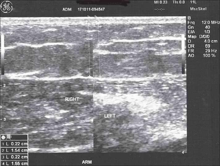
Sonographic image of the arm with clear markings for measurement of skin + subcutaneous tissue thickness
Figure 4.
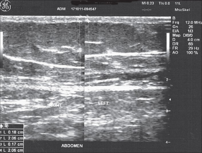
Sonographic image of the abdomen with clear markings for measurement of skin + subcutaneous tissue thickness
Figure 3.
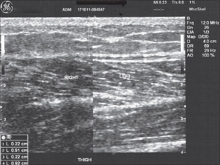
Sonographic image of the thigh with clear markings for measurement of skin + subcutaneous tissue thickness
Statistical analysis
Excel spreadsheet and SPSS Statistical Software Version 10.0 were used for analyzing the data. Chi-square test was applied when comparing the BMI among both the genders. For comparing the skin thickness between the arm, thigh and the abdomen in relation to BMI, Student t test and Pearson correlation were used. A P < 0.05 was taken as significant and P > 0.05 was taken as non-significant. Wherever useful, tabular representation has been done to provide information.
An external agency was hired to perform the statistical analysis. All the data that could identify the subject's personal information were hidden and only a generated number to identify the patient (subject ID) was provided to the agency along with other collected data.
All subjects who fulfilled the inclusion criteria were considered for this study. We did not calculate the sample size, but with these results power of study is more than 80%.
Patient selection criteria
Known type-2 diabetics belonging to the age group of 18-65 years, who have not yet initiated insulin treatment for the management of type-2 diabetes and are willing to provide voluntary consent for participation in the study, were included in the present study. Subjects considered as ineligible by investigator/are unwilling or unable to provide information were not taken into the survey.
Ethical and regulatory requirements
The present study including the protocol and informed consent form was submitted to the Institutional Ethics Committee before the initiation of the study for their approval. The study was initiated in the institution, only after getting approval.
The institution bore the cost of the ultrasound that was conducted on the subjects. No additional costs were taken from the subjects for the present study. This study was not sponsored by any external agency/pharmaceutical company.
RESULTS
Baseline characteristics
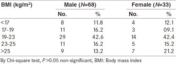
In the present study, there were 68 males and 33 females who voluntarily participated in this study. The male: female ratio was 1:0.49. There were 11.8% male and 12.1% females in the BMI range <17 kg/m2. In the BMI range 17-19 kg/m2 there were 16.2% males and 9.1% females, in 19-23 kg/m2 there were 42.6% males and 42.4% females, in 23-25 kg/m2 there were 16.2% males and 15.2% females and in the BMI range >25 kg/m2 there were 13.2% males and 21.2% females. The distribution of males and females in our series are not comparable across various BMIs (P > 0.05) [Table 1].
Table 1.
Gender-wise distribution of patients according to body mass index
Skin thickness
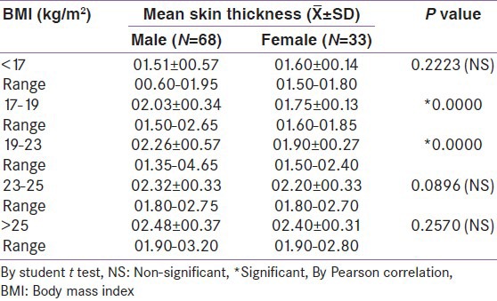
At arm, the mean skin thickness was more in males as compared to females in the BMI range 17-19 kg/m2 and 19-23 kg/m2 (P < 0.05), whereas in other BMI ranges, skin thickness was comparable (P > 0.05). The mean skin thickness of males ranged from 0.60 mm to 3.20 mm and in females it ranged from 1.50 mm to 2.80 mm [Table 2].
Table 2.
Gender-wise distribution of mean skin thickness of arm according to body mass index
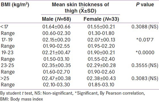
At thigh, the mean skin thickness was more in males as compared to females in the BMI range 17-19 kg/m2 and 19-23 kg/m2 (P < 0.05), whereas in other BMI ranges, the skin thickness was comparable (P > 0.05). The mean skin thickness of males ranged from 0.6 mm to 3.30 mm and in females it ranged from 1.30 mm to 3.10 mm [Table 3].
Table 3.
Gender-wise distribution of mean skin thickness of thigh according to body mass index
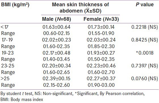
At abdomen, the mean skin thickness was more in males as compared to females in the BMI range 19-23 kg/m2 (P < 0.05), whereas in other BMI ranges, the skin thickness was comparable (P > 0.05). The mean skin thickness of males ranged from 0.6 mm to 2.60 mm and in females it ranged from 1.55 mm to 3.00 mm [Table 4].
Table 4.
Gender-wise distribution of mean skin thickness of abdomen according to body mass index
Subcutaneous tissue thickness
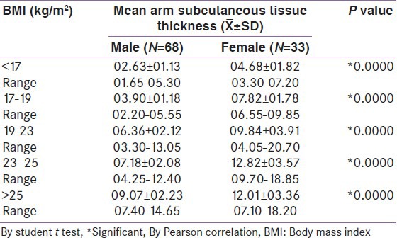
At arm, the subcutaneous tissue thickness is more in females as compared to that of males across all BMI ranges (P < 0.05). The subcutaneous tissue thickness range in males is from 1.65 mm to 14.65 mm, whereas it is from 3.30 mm to 18.20 mm in females. The subcutaneous tissue thickness increases as the BMI increases [Table 5].
Table 5.
Gender-wise distribution of mean arm subcutaneous tissue thickness according to body mass index
At thigh, the subcutaneous tissue thickness is more in females as compared to that of males across all BMI ranges (P < 0.05). The subcutaneous tissue thickness range in males is from 1.65 mm to 18.35 mm, whereas it is from 2.70 mm to 25.20 mm in females. The subcutaneous tissue thickness increases as the BMI increases [Table 6].
Table 6.
Gender-wise distribution of mean thigh subcutaneous tissue thickness according to body mass index

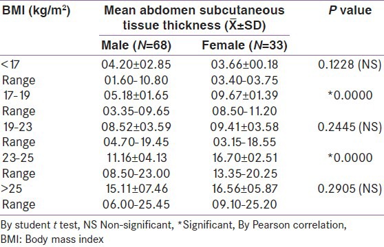
At abdomen, the subcutaneous tissue thickness is more in females as compared to that of males in the BMI ranges 17-19 kg/m2 and 23-25 kg/m2 (P < 0.05), whereas is it comparable in other BMI ranges (P > 0.05). The subcutaneous tissue thickness range in males is from 1.60 mm to 25.45 mm, whereas it is from 3.40 mm to 25.20 mm in females. The subcutaneous tissue thickness increases as the BMI increases [Table 7].
Table 7.
Gender-wise distribution of mean abdomen subcutaneous tissue thickness according to body mass index
Skin + subcutaneous tissue thickness
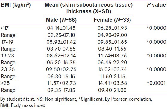
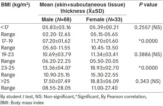
Gender wise distribution of mean (skin + subcutaneous tissue thickness) of arm according to BMI. Mean arm total (skin + subcutaneous) thickness was more in female cases than in male among all the ranges of BMI and difference between them was statistically significant (P < 0.05). The mean (skin + subcutaneous) thickness increases with BMI in both the genders. Thus, the subcutaneous tissue thickness of females is more than that of males across all BMI ranges. The range of skin + subcutaneous tissue thickness at arm is 2.25-17.85 mm in males and 4.90-21.00 mm in females [Table 8].
Table 8.
Gender-wise distribution of mean (skin+subcutaneous tissue thickness) of arm according to body mass index
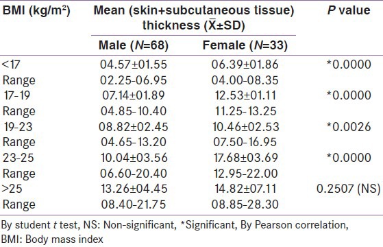
Gender wise distribution of mean (skin + subcutaneous tissue thickness) of anterior upper thigh according to BMI. The mean thickness (skin + subcutaneous tissue thickness) was less in males as compared to females, across all the BMIs (P < 0.05), which is statistically significant; except for BMI >25 kg/m2 where the thickness among males and females was comparable and statistically not significant (P > 0.05). The skin + subcutaneous thickness increases with BMI in both the genders. Thus, the subcutaneous tissue thickness of females is more than that of males across all BMI ranges. The range of skin + subcutaneous tissue thickness at thigh is 2.25-21.75 mm in males and 4.00-28.30 mm in females [Table 9].
Table 9.
Gender-wise distribution of mean (skin+subcutaneous tissue thickness) of anterior upper thigh according to body mass index
Gender wise distribution of mean (skin + subcutaneous tissue thickness) of abdomen according to BMI. The mean thickness (skin + subcutaneous tissue thickness) was comparable in the BMI range <17; 19-23 and >25 in both the genders, which is statistically not significant (P > 0.05). While in the BMI range 17-19 and 23-25, there is a significant difference between the mean thickness (skin + subcutaneous tissue thickness) between males and females, which is statistically significant (P < 0.05). The skin + subcutaneous thickness increases with BMI in both the genders. Thus, the subcutaneous tissue thickness of females is more than males in the BMI range 17-19 kg/m2 and 23-25 kg/m2 (P < 0.05), and comparable across all other BMI ranges (P > 0.05). The range of skin + subcutaneous tissue thickness at abdomen is 2.20-28.05 mm in males and 5.15-27.40 mm in females [Table 10].
Table 10.
Gender-wise distribution of mean (skin+subcutaneous tissue thickness) of abdomen according to body mass index
Overall distribution
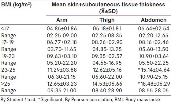
Table 11 depicts the overall distribution of mean skin + subcutaneous tissue thickness at arm, thigh and abdomen across all the BMI ranges. It clearly depicts that as the BMI increases the skin + subcutaneous tissue thickness also increases at all the experimental sites, i.e. arm, thigh and abdomen. There is no significant difference in the skin + subcutaneous tissue thickness at arm, thigh and abdomen upto the BMI range of 23 kg/m2 (P > 0.05). But in the BMI range >23 kg/m2, there is a significant skin + subcutaneous tissue thickness difference when comparing thickness of arm and abdomen (P < 0.05) [Table 11].
Table 11.
Overall distribution of mean skin+subcutaneous tissue thickness according to body mass index
DISCUSSION
This study was intended to understand skin and subcutaneous tissue thickness at insulin injection sites so insulin naïve type-2 diabetics were studied and for the same reason no control arm was included. In the present study, there was a male preponderance over female participants. There was unequal distribution of males and females over various BMI ranges. The mean arm skin thickness was more in males than females across all BMI ranges except for BMI range <17 kg/m2. While at thigh, skin thickness was more in males as compared to females across all BMI ranges. Thus males were having more skin thickness as compared to females. But at abdomen, the skin thickness in males is higher to females in the 19-23 kg/m2 BMI range, while at all other BMIs, it was comparable.
But at abdomen, the subcutaneous tissue thickness was more in males for the BMI range <17 kg/m2, while in all other BMI ranges, the subcutaneous tissue thickness was more in females than compared to males.
The total skin + subcutaneous tissue thickness at arm was more in females compared to males across all BMI ranges. At thigh the thickness was more in females in comparison to males, except for BMI >25 kg/m2, where it was comparable. At abdomen, the skin + subcutaneous tissue was comparable in the BMI ranges <17 kg/m2; 19-23 kg/m2 and >25 kg/m2.
Thus, our study clearly depicts that the skin thickness of males is more as compared to the female counterparts independent of BMI at arm, thigh and abdomen, while the subcutaneous tissue thickness is more in females as compared to their male counterparts in all the three areas. The combined thickness of skin + subcutaneous tissue also shows a higher thickness in females as compared to their male counterparts at arm and at thigh, while this thickness is higher in females at BMI 17-19 kg/m2 and 23-25 kg/m2 ranges. Tables 12-14 shows analysis of important observations. Thus, skin thickness may be higher in males in normal BMI range of 19-23 kg/m2 but total skin and subcutaneous tissue thickness is higher in females at all three insulin injection sites.
Table 12.
Overall distribution of mean skin thickness according to body mass index at three injection sites

Table 14.
Overall distribution of mean skin+subcutaneous thickness according to body mass index at three injection sites

Table 13.
Overall distribution of mean subcutaneous thickness according to body mass index at three injection sites

CONCLUSION
From the above we can conclude that the mean skin thickness at arm, thigh and abdomen is higher in males when BMI <23 kg/m2. The skin thickness increases with increase in BMI for arm, thigh and abdomen regions in both the genders.
The subcutaneous tissue thickness of females is higher than that of their male counterparts. Due to this, the skin + subcutaneous tissue thickness of females is higher as compared to males across all the BMIs at arm, thigh and abdomen regions.
In all type-2 diabetes patients, both genders, with BMI more than 23 kg/m2 had a combined skin plus subcutaneous tissue thickness of more than 6 mm at all three insulin injection sites. While all patients with BMI 19-23 kg/m2 had a combined skin plus subcutaneous tissue thickness of more than 4 mm at all three insulin injection sites. When BMI <19 kg/m2, all females had a combined skin plus subcutaneous tissue thickness of more than 4 mm at all three insulin injection sites while few males had less than 4 mm thickness.
ACKNOWLEDGMENT
We thankfully acknowledge the support provided by the staff of USG department of our institution during the study and other staff who helped in data collection in the questionnaires.
Footnotes
Source of Support: Nil
Conflict of Interest: No
REFERENCES
- 1.Alexander H, Miller DL. Determining skin thickness with pulsed ultra sound. J Invest Dermatol. 1979;72:17–9. doi: 10.1111/1523-1747.ep12530104. [DOI] [PubMed] [Google Scholar]
- 2.Black MM. A modified radiographic method for measuring skin thickness. Br J Dermatol. 1969;81:661–6. doi: 10.1111/j.1365-2133.1969.tb16204.x. [DOI] [PubMed] [Google Scholar]
- 3.Laurent A, Mistretta F, Bottigioli D, Dahel K, Goujon C, Nicolas JF, et al. Echographic measurement of skin thickness in adults by high frequency ultrasound to assess the appropriate microneedle length for intradermal delivery of vaccines. Vaccine. 2007;25:6423–30. doi: 10.1016/j.vaccine.2007.05.046. [DOI] [PubMed] [Google Scholar]


