

Abstract:
Mumps is an acute and self-limiting disease characterized by parotitis, however in some cases it leads to aseptic meningitis, deafness, encephalitis and orchitis, which is a serious health concern. MMR vaccination was successful in eradicating the disease however, recent reports question the efficacy of MMR vaccine and countless outbreaks are observed in vaccinated populations throughout the world. Lack of specific treatment methods for mumps infection and inefficiency of MMR vaccine in vaccinated populations accentuates the need for the development of novel drugs to control mumps virus mediated serious infections. It was with this backdrop of information that the anti-mumps virus activity of Mimosa pudica was evaluated. Suspected mumps cases were collected to isolate a standard mumps virus by systematic laboratory testing which included IgM antibody assays, virus isolation, RT-PCR and phylogenetic analysis. The virus was quantified by TCID50 assay and anti-mumps virus property was evaluated by CPE reduction assay and cytotoxicity of the extract was measured by MTT assay and phytochemical analysis was done by gas chromatography-mass spectroscopy. The RT-PCR and phylogenetic tree analysis of the SH gene sequence of the clinical isolate showed it to be mumps virus genotype C. 150 μg/ml concentration of M. pudica completely inhibited mumps virus and the drug was found to be non-toxic up to 2 mg/ml. M. pudica was thus found to be a potent inhibitor of MuV.
Keywords: Mumps virus, MMR vaccine, Genotype C, M. pudica, Vero cells, CPE
Introduction
Mumps is an acute, highly contagious, systemic, communicable viral infection found throughout the world, characterized by parotitis of one or both salivary glands (90 %), aseptic meningitis (~15 %), transient deafness (~4 %) and encephalitis (~0.1 %). Other clinical features include orchitis (20–38 % in postpubertal males), oophoritis (0.5–7 %) and respiratory symptoms (40–50 %). Mumps is a vaccine preventable childhood disease that tends to be mild; about 30 % of infections are asymptomatic. Transmission occurs through inhalation of respiratory droplets or by direct person-to-person contact, reinfection may occur either after natural infection or vaccination [13, 28].
The mumps virus (MuV) belongs to genus Rubulavirus, subfamily Paramyxovirinae, family Paramyxoviridae and order Mononegavirales. It is an enveloped RNA virus with a non-segmented single-stranded negative-sense genome of 15,384 nucleotides (nt). The genome contains seven transcription units that encode open reading frames for the nucleocapsid protein, phospho protein, matrix protein, fusion protein, small hydrophobic protein (SH), hemagglutinin, neuraminidase protein, and large proteins [3, 7]. The SH gene, which contains 316 nt and encodes 57 amino acids, is non-essential for viral replication, and its function is obscure. Since the SH gene is most genetically divergent, its sequence data have been mainly used for phylogenetic analysis of MuVs [11, 28].
Mumps, though historically a disease of childhood, is self limiting and MMR vaccination was successful in eradicating the disease. A present outbreak of mumps predominantly involved young adults, nearly all of whom had been vaccinated, most with the two-dose schedule [23]. There is no specific treatment for mumps. Currently, mumps treatment consists of managing the symptoms while the body fights off the infection. Such treatment called supportive care includes warm compresses and medication to help control fever or pain. Symptoms may be relieved by the application of intermittent ice or heat to the affected neck/testicular area and by acetaminophen/paracetamol (tylenol) for pain relief. However, in some cases it leads to aseptic meningitis, deafness, encephalitis and orchitis, which is a serious health concern [14, 25, 28]. Since there is a lack of specific treatment for mumps infection and MMR inefficiency in vaccinated populations this study was conducted to study anti-mumps activity of Mimosa pudica (M. pudica), which is popular for its unique seismonastic response movements and circadian rhythms [22].
M. pudica, Linn. is an annual or perennial herb belonging to family Mimosaceae, common plant in moist waste ground, lawns, open plantation and weedy thickets, in which the leaves close and the petiole hangs down in response to certain stressors such as a wound, wind, vibration, touch, hot or cold stimulus, drought or change in illumination (Fig. 1). Seismonastic movements such as response to touch, appear to be regulated by electrical and chemical signal transduction, spreading the stimulus throughout the plant [2]. M. pudica, is commonly known as ‘lajjalu’ in Hindi, ‘touch-me-not’ in English and ‘thottalsenungi’ in Tamil. M. pudica has been reported to contain mimosin (an alkaloid), free amino acids, beta-sitosterol, linoleic acid and oleic acid [12]. The known medicinal properties of M. pudica are antivenom properties [26], arresting bleeding, skin diseases, wound healing activity [12], alleviating headache, insomnia, diarrhea, dysentery, fever, piles, fistula, anticonvulsant activity [16], anti-hyperglycemic and hepato-protective activity [22] and antiviral activity [20]. Only few pharmacological studies have been reported on this plant, claimed in traditional medicine to treat many diseases related to the nervous system [20]. This study investigates the anti-mumps activity of M. pudica.
Fig. 1.
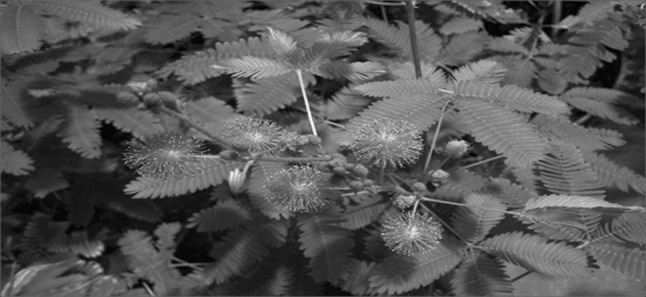
M. pudica: Compound leaves which gets sensitive on touching, spinous stipules and globose purple flower heads, grows as weed in almost all parts of the country
Materials and methods
Preparation of standard MuV
Patient recruitment and sample collection
WHO guidelines were adopted for selection of clinical cases of mumps. An informed consent, proforma and human ethical clearance for sample collection were obtained (Human Ethical No: IEC-NI/10/OCT/19/34). Blood (for antibody analysis) and buccal swab (for virus isolation and RT-PCR) were collected from patients at the outpatient department of V.K. Nursing Home. The sample was collected 1–7 days after onset (average = 3 days) and stored at −86 °C until use.
Measurement of mumps specific IgM antibody
An IgM EIA kit (Labor Diagnostika Nord GmbH & Co. KG, Germany) was used to determine mumps specific IgM levels in patient’s sera. The assay was performed and interpreted according to the manufacturer’s instructions. The IgM antibody titer was designated as an antibody index by calculating the ratio between the average optical density value of the sample and that of the cut-off, provided with the kit. An antibody index exceeding 1.1 was determined as positive.
Virus isolation and hemadsorption assay
MuV was isolated from the throat swab soaked in viral transport medium (Himedia, Cat. No: MS1760D) containing 2 ml HBSS with productive protein and antibiotics and was centrifuged at 1,500 rpm for 10 min. After sterilization through a 0.2 μm syringe filter, the supernatant was used for virus infection into Vero, MCF-7, MDCK and Hep-2 cells maintained in minimum essentials medium (MEM) (Sigma-Aldrich, Cat. No. M4655) with 10 % FBS, 2 mM l-glutamine, 100 U/ml penicillin and 100 μg/ml streptomycin (Hi-media, Cat. No. A001A) [1]. The cells were cultured at 37 °C at 5 % CO2, observed for 14 days and harvested when the cytopathic effects (CPE) became prominent. The culture harvests were aliquoted and stored at −86 °C until processed further. Two blind passages were performed on all CPE-negative tissue cultures. For the hemadsorption assay, Hep-2 cells were grown in 6 well culture plates for 48 h after which the medium was removed from the young monolayer, infected with a clinical isolate of the MuV, incubated at 37 °C, at 5 % CO2 for 90 min. The cells were washed with PBS (pH 7.2) and a suspension of 4 % guinea pig red cells was added to the cells and incubated for 1 h at 4 °C. The cells were thoroughly washed with PBS and observed under an inverted microscope (Micros, Austria).
RNA extraction and RT-PCR synthesis
The total RNA was extracted as per Jelena et al. [10]. Briefly, viral genomic RNA was extracted from 140 μl of throat swab samples, culture supernatant and MMR vaccine strain by using a QIAamp viral RNA minikit (Qiagen, Cat. No: 52904) according to the manufacturer’s protocol. The extracted total RNA was quantified by using Nanodrop 2000 (Thermo scientific Inc.), to amplify the SH gene region followed by reverse transcription of viral RNA into cDNA done by using an Omni script RT kit (Qiagen, Cat. No: 205111) according to the manufacturer’s instructions. To obtain the complete SH gene, a primer of 506 bp was designed from L-Zagreb vaccine strain (AY685920) (SHLZ-F: 5′-CAAGTAGTGTCGATGATCTCATCAGG-3′/SHLZ-R: 5′-GTGAAGAGTTTCGAGGGC TCATC-3′). Each PCR mixture included 1 U of Taq DNA polymerase, 5× PCR buffer (50 mM KCl, 1.5 mM MgCl2, 10 mM Tris–HCl, pH 9.0), 20 pmol of each primer and 10 mM dNTP mix in a total volume of 10 μl. All the PCR chemicals were purchased from Fermentas, USA. PCR was performed by 35 cycles of denaturation at 94 °C for 45 s, annealing at 65 °C for 45 s, extension at 72 °C for 45 s, and a final extension step of 3 min at 72 °C.
Automated DNA sequencing and phylogenetic analysis
The cycle sequencing reaction was performed using BigDye terminator V3.1 cycle sequencing Kit containing Ampli Taq polymerase (from Applied Biosystems, P/N: 4337457). The sequencing reaction-mix was prepared by adding 1 μl of BigDye v3.1, 2 μl of 5× sequencing buffer and 1 μl of 50 % DMSO. To 4 μl of sequencing reaction-mix, 4 pmol of primer (2 μl) and sufficient amount of plasmid was added. The constituted reaction was denatured at 95 °C for 5 min. Cycling began with denaturation at 90 °C for 30 s, annealing at 52 °C for 30 s and extension for 4 min at 60 °C and repetition for a total 30 cycles in a MWG thermocycler. The reaction was then purified on sephadex plate (Edge Biosystems) by centrifugation to remove unbound labelled and unlabelled nucleotides and salts. The purified reaction was loaded on to the 96 capillary ABI 3700 DNA analyzer and electrophoresis was carried out for 4 h. Generated sequence data were deposited in NCBI database. For phylogenetic analysis the SH genes sequences of virus were compared with WHO mumps update 2012 [28] and previously published MuV sequences by MEGA5 program [24] that uses the neighbour-joining method. The resulting dendrograms were used to verify previously proposed genotype assignments and identify areas for clarification. The phylogenetic tree is displayed.
Quantification of MuV
MuV quantification was done by tissue culture infective dose (TCID50) assay by end point dilution assay [4]. The plates were incubated at 37 °C in CO2 incubator and observed on day 5 and day 7. CPE was recorded and TCID50 was calculated using Karber’s formula = L − d (S − 0.5). L—least dilution, d—log difference in dilution, S—sum of proportion of wells shown CPE, 0.5—standard deduction.
M. pudica extract preparation
M. pudica was collected from Puducherry union territory, India and authenticated at the Department of Botany, University of Madras. The whole plant was rinsed in sterile distilled water, shade dried, powdered and stored at room temperature until use. A methanolic extract of M. pudica was prepared by adding 50 g of whole plant powder in 500 ml of methanol and filtering using Whatman No. 1 filter paper. The filtrate was allowed to evaporate for about 2–3 days. The dried filtrate was collected, weighed and stored at 4 °C until use.
Anti-mumps activity by CPE reduction assay
Vero cells were incubated at 37 °C on 24-well plates at a density of 2 × 10⁶ cells per well, with 5 % CO2 in a humidified atmosphere with 10 % MEM. After 24 h, 50 μl of MuV (5000 TCID50) were infected on an 80 % confluent monolayer and incubated for 90 min in 37 °C at 5 % CO2. Varying concentrations of the filtered methanolic extracts (25–200 μg/ml) were added to infected wells and incubated for 10 days at 37 °C in 5 % CO2 environment. Plates were microscopically examined everyday for reduction of CPE. Cell control and virus control were maintained [18].
MTT assays to measure the toxicity of extract
3-(4,5-Dimethylthiazol-2-yl)-2,5-diphenyltetrazolium bromide (MTT) assay was used for the screening the toxicity of extracts using standard protocol [21]. Briefly, Vero cells were incubated at 37 °C on 12-well plates at a density of 2 × 10⁶ cells per well, with 5 % CO2 in a humidified atmosphere with 10 % MEM. After 24 h, extracts were added on monolayer (70–80 % confluency) so that the final concentrations ranged from 50 μg to 10 mg/ml. The plates were incubated for further 5 days under the same conditions mentioned above. 200 μl MTT (Sigma-Aldrich, Catalogue No. M2003) solutions (5 mg/ml in phosphate buffer) was added to each well and incubated at 37 °C for 4 h. The MTT solution was decanted and formazan was extracted from the cells with 250 μl of DMSO in each well. Color was measured with a 12-well ELISA plate reader at 550 nm. The toxicity control used was 1 % Triton X -100 (Qualigens, Cat. No. 10655). All assays were done in triplicates.
Photochemical analysis of M. pudica extract by gas chromatography–mass spectroscopy (GC–MS)
Phytochemical analysis of M. pudica extract was performed at the Department of Sophisticated Analytical Instrument Facility (SAIF) IIT, Chennai. Preliminary screening was carried out to decipher the presence or absence of various phytocompounds, and GC–MS was performed to identify the major compounds [9, 19]. In this experiment methanolic extracts were evaluated for the active principle compounds which are associated for the observed bioactivities. M. pudica extracts were subjected for compounds identification by injecting 1 μl of extracts into GC–MS (JEOL GC mate) instrument. After running for 40 min, major compounds were identified by comparing with standard references.
Results
Characterization of MuV from the clinical specimens
Virus was isolated from five patients who had acute mumps infection with clinical manifestations of parotitis and swelling of the parotid glands. These patients also manifested other symptoms such as high fever, head-ache and loss of appetite. Mumps specific IgM antibody was found to be positive. A number of continuous cell lines were used for identification, isolation and growth of the MuV. Among the cell lines which were used Vero cells were found to be the most suitable for isolation of MuV. Vero cells were infected with buccal swab samples and incubated for 7 days in 5 % CO2 at 37 °C. At the end of the incubation period, the cells were examined for morphological changes. CPE characteristic to MuV was observed (Fig. 2a, b). MCF-7, MDCK and Hep-2 cell did not show any morphological changes. Infection of the Hep-2 cells, was confirmed by hemadsorption test suggesting that though the virus infected the cells, it failed to produce CPE on Hep-2 cells, (which is often observed in myxoviruses that mature by budding from the plasma membrane) (Fig. 2c).
Fig. 2.

Result of virus isolation and RT-PCR: a CPE of mumps virus rounding with vacuolation, b control, c hemadsorption test (RBC s adsorb onto the virus infected cells), d the polymerase chain reaction products of the SH gene (506 bp), lane 1 100 bp DNA marker. Lanes 2, 3 were cluneal isolates, lane 4 was L-Zagreb vaccine strain (control)
Genotypic analysis of the MuV strains isolated from the clinical specimens
The cDNA was amplified by RT-PCR and the PCR products (Fig. 2d) were subjected for purification and sequencing. DNA sequencing showed that the virus isolates were MuV genotype C and was named as MuVi/Chennai.IND [C] (GenBank accession nos. JX392385, JX392386, JX894238, JX894239, JX894240) and our isolated virus sequences were matched with WHO reference strains. Two isolates (JX392385, JX392386) and other three isolates (JX894238, JX894239, JX894240) were 99 % identical to each other based on their SH gene (316 bp) sequences. The sequences of our isolates exhibited 97 % homology with WHO genotype C (GenBank accession no. EU370206, JQ945268) and 45 % homology with C type reported from UK which was found to be originally isolated from a patient from India (GenBank accession no. AF142765). A phylogenetic tree comparison of the SH gene sequences of MuV isolates around the world is shown in Fig. 3.
Fig. 3.

Phylogenetic trees of MuV genotypes based on the 316 nucleotides of the entire SH gene: Neighbour-joining method of MEGA5 program was used. The parameter employed was Kimura 2-parameter model and the robustness of the internal branches was determined by 500 bootstrap replications. The horizontal length of the bar denotes percentage difference between sequences (see scale at bottom) and the bootstrap numbers (%) are given at each node
Antiviral examination by inhibition of viral replication
Vero cells which were infected with 5000 TCID50 of MuV and treated with 25–200 μg/ml methanolic extracts of M. pudica, was found to be inhibited at a concentration of 150 μg/ml of M. pudica (Fig. 4). Continuous treatment for 7 days prevented the development of CPE. The continuous presence of the extracts in the cell culture medium is apparently essential for continuous protection of Vero cells against viral CPE development. This observation was based on absence of CPE, as well as the inhibition of virus replication.
Fig. 4.

Antiviral examination by CPE reduction assay: Vero cells were infected with 5,000  of mumps virus and treated with 25 to 200 μg/ml extract of M. pudica immediately after the post infection. 150 μg/ml of M. pudica extracts potentially possess virus inhibition, such as affecting viral replication after the cell is infected
Cytotoxic effect of M. pudica on Vero cell viability
The cytotoxic effect of the extract was tested by measuring the viability of Vero cells after treatment. Vero cells were exposed to varying concentrations of plant extracts and incubated for 72 h. Cells were tested for percentage viability by MTT assay every 24 h. As shown in Fig. 5 there was 100 % viability seen from concentrations 50 μg/ml to 2 mg/ml at 72 h. From 3 to 10 mg/ml minor cytotoxicity was observed. However, none of these concentrations showed 100 % toxicity.
Fig. 5.

Drug toxicity assay for Mimosa pudica
Active principle compound of M. pudica extract
Initial screening of the compound showed the presence of alkaloids, flavonoids, saponins, carbohydrates, phenols, steroids, tannins, diterpenes and glycosides. GC-MS study of the extract of M. pudica showed several compounds of which one of the compounds was found to be predominant. One of the compounds known as 2-[2-methyl-5-nitro-imidazol-1-yl]-N-phenethyl-acetamide (chemical formula C14H16N4O3; molecular weight 288.30184 kDa) showed a peak area with RT of 13.85 (Fig. 6; Table 1). This compound was most abundant in M. pudica extract and could be responsible for the observed antiviral activity.
Fig. 6.

Phytochemical constituents of M. pudica extract (GC-MS): a spectrum of methanolic extract of M. pudica with retention time (RT). b Structure of the five major compounds with retention time
Table 1.
Phytochemical constituents of M. pudica extract
| S. No. | RT | Name of the compound | Molecular formula | Molecular weight |
|---|---|---|---|---|
| 1 | 13.65 | 2-[2-Methyl-5-nitro-imidazol-1-yl]-N-phenethyl-acetamide | C14H16N4O3 | 288.30184 |
| 2 | 15.07 | Cholan-24-oic acid, 3, 12-dihydroxy-, methyl ester, [3a, 5a, 12a]- | C27H44O5 | 448.64 |
| 3 | 11.43 | Propanoic acid, 2-(3-acetoxy-4,4,14-trimethylandrost-8-en-17-yl)- | C27H42O4 | 430.61998 |
| 4 | 13.85 | 4-(5-Pentyl-3a,4,5,7a-tetrahydro-4-indanyl)butanoicacid, methyl ester (stereoisomer 1) | C19H32O2 | 292.45618 |
| 5 | 14.55 | 2,7-Diphenyl-1,6-dioxopyridazino [4,5:2′,3′] pyrrolo [4,5′-d] pyridazine | C20H13N5O2 | 355.34952 |
Discussion
Typically known to be a childhood disease, mumps infection is usually self-limited, although rarely complications such as meningitis, transient deafness, facial palsy, orchitis or oophoritis might arise [8, 15, 17, 28]. Mumps was not considered a major problem because prophylaxis was brought about by the MMR vaccine and hence there was no requirement for development of specific drugs against MuV [27]. There are no reports regarding development of drugs against MuV till date. But recently, reports of resurgence of mumps in MMR vaccinated populations and its complications in children and adults have been established globally [5, 6, 23]. This current situation has warranted the need for effective drugs against mumps infection. It was with this notion that we studied an Indian plant M. pudica, known to possess varied medicinal benefits, including anti-hepatitis B activity, for its effectiveness against MuV.
As standard strains of MuV were not available for testing, we used strains which were isolated from patients with acute mumps infection, who had also tested positive for anti-mumps IgM antibody. Upon genotypic characterization, it was found that these strains belonged to genotype C (first report in India). Sequences were submitted to GenBank and phylogenetic analyses were done (GenBank accession no. JX392385, JX392386, JX894238, JX894239, JX894240). Assays done with varying concentrations of M. pudica methanolic extracts showed that 150 μg/ml of M. pudica extracts inhibited viral replication and prevented about 95 % of CPE development, when exposed to extract for 7 days. Toxicity of the extract was studied using the MTT assay. It was found that the extracts were non-toxic up to 2 mg/ml concentration and cells had 100 % viability. It has also been widely observed and accepted that the medicinal value of plants lies in the bioactive phytocomponents present in the plants. In the present investigation, the active principle of the phytocomponents of M. pudica were studied and the anti viral activity of the plant extract was tested against MuV in different concentrations of the extract to assess the most effective concentrations.
Mumps infection was not regarded to be a serious problem as the MMR vaccine was thought to be effective, but reports suggest otherwise. Currently, patients are given only symptomatic treatment for acute mumps infection and there have been no reports of development of specific anti-mumps medication. This is the first report which has portrayed the activity of M. pudica against mumps virus and it is non-toxic.
Acknowledgments
We sincerely thank Dr. V. Srinivasan and Dr. S. Chitra from V.K. Nursing Home, Valasaravakkam, Chennai-600 087 for providing mumps samples and Dr. B. Ananthan and Mr. Vishnu Prabu, Department of Genetics, University of Madras, Chennai-600 113 for his help in sequencing and phylogenetic analysis.
References
- 1.Afzal MA, Dussupt V, Minor PD, Pipkin PA, Fleck R, Hockley DJ, Stacey GN. Assessment of mumps virus growth on various continuous cell lines by virological, immunological, molecular and morphological investigations. J Virol Methods. 2005;126:149–156. doi: 10.1016/j.jviromet.2005.01.032. [DOI] [PubMed] [Google Scholar]
- 2.Alexander G, Volkov JC, Foster, Vladislav S, Markin Signal transduction in Mimosa pudica: biologically closed electrical circuits. Plant Cell Environ. 2010;33:816–827. doi: 10.1111/j.1365-3040.2009.02108.x. [DOI] [PubMed] [Google Scholar]
- 3.Carbone KM, Wolinsky JS. Mumps Virus. In: Knipe DM, Howley PM, editors. Fields Virology, 4th edn, vol 1. Philadelphia: Lippincott Williams & Wilkins; 2001. p. 1381–41.
- 4.Changqing Q, Jizhang Z, Xiaoan C, Guozhen L, Fuying Z, Xiaowei G. Immunization trials with an avian chlamydial MOMP gene recombinant adenovirus. Bioeng Bugs. 2010;1(4):267–273. doi: 10.4161/bbug.1.4.12115. [DOI] [PMC free article] [PubMed] [Google Scholar]
- 5.Dayan GH, Quinlisk MP, Parker AA, Barskey AE, Harris ML, Schwartz JM. Recent resurgence of mumps in the United States. N Engl J Med. 2008;358(15):1580–1589. doi: 10.1056/NEJMoa0706589. [DOI] [PubMed] [Google Scholar]
- 6.Echevarria JE, Castellanos A, Sanz JC, Martinez de Aragon MV, Pena Rey I, Mosquera M, et al. Mumps virus genotyping: basis and known circulating genotypes. Open Vaccine J. 2010;3:37–41. doi: 10.2174/1875035401003020037. [DOI] [Google Scholar]
- 7.Elango N, Varsanyi TM, Kovamees J, Norrby E. Molecular cloning and characterization of six genes, determination of gene order and intergenic sequences and leader sequence of mumps virus. J Gen Virol. 1998;69:2893–2900. doi: 10.1099/0022-1317-69-11-2893. [DOI] [PubMed] [Google Scholar]
- 8.Farul I, Ozlem HM, Sakir A. Facial palsy caused by mumps parotitis. Neurol India. 2009;27(4):511–512. doi: 10.4103/0028-3886.55589. [DOI] [PubMed] [Google Scholar]
- 9.Iordache A, Culea M, Gherman C, Cozar O. Characterization of some plant extracts by GC-MS. Nucl Instrum Methods Phys Res. 2009;B267(2):338–342. [Google Scholar]
- 10.Jelena I, Dubravko F, Tanja KG, Renata Z, Leonida R, Marijana B, et al. Genetic characterization of a mumps virus isolate during passaging in the amniotic cavity of embryonated chicken eggs. Virus Res. 2004;99:121–129. doi: 10.1016/j.virusres.2003.11.002. [DOI] [PubMed] [Google Scholar]
- 11.Jin L, Rima B, Brown D, Orvell C, Tecle T, Afzal M, et al. Proposal for genetic characterisation of wild-type mumps strains: preliminary standardisation of the nomenclature. Arch Virol. 2005;150(9):1903–1909. doi: 10.1007/s00705-005-0563-4. [DOI] [PubMed] [Google Scholar]
- 12.Kokane DD. Evaluation of wound healing activity of root of Mimosa pudica. J Ethnopharmacol. 2009;124:311–315. doi: 10.1016/j.jep.2009.04.038. [DOI] [PubMed] [Google Scholar]
- 13.Lamb RA, Parks GD. Paramyxoviridae: the viruses and their replication fields virology. 5. Philadelphia: Lippincott Williams & Wilkins; 2007. pp. 1449–1496. [Google Scholar]
- 14.Marie-Claude B, Anil D, Clement W, Stanley AP. Mumps vaccine virus strains and aseptic meningitis. Vaccine. 2006;24:7037–7045. doi: 10.1016/j.vaccine.2006.06.049. [DOI] [PubMed] [Google Scholar]
- 15.Mehmet M, Berker B. An unusual postoperative complication: anesthesia mumps. Eur J Plast Surg. 2007;29:335–338. doi: 10.1007/s00238-006-0100-z. [DOI] [Google Scholar]
- 16.Nge BE. Anticonvulsant activity of Mimosa pudica decoction. Fitoterapia. 2004;75:309–314. doi: 10.1016/j.fitote.2004.01.012. [DOI] [PubMed] [Google Scholar]
- 17.Nurdan Y, Olcay Y, Yalcin C, Hikmet B, Sevil O. Hematuria with mumps infection. Indian J Pediatr. 2003;70(1):93–94. doi: 10.1007/BF02722752. [DOI] [PubMed] [Google Scholar]
- 18.Nwodo UU, Ngene AA, Iroegbu CU, Onyedikachi OAL, Chigor VN, Okoh AI. In vivo evaluation of the antiviral activity of Cajanus cajanon measles virus. Arch Virol. 2011;156:1551–1557. doi: 10.1007/s00705-011-1032-x. [DOI] [PMC free article] [PubMed] [Google Scholar]
- 19.Palwinder K, Kumar N, Shivananda TN, Kaur G. Phytochemical screening and antimicrobial activity of the plant extracts of Mimosa pudica L. against selected microbes. J Med Plants Res. 2011;5(22):5356–5359. [Google Scholar]
- 20.Parimala DB, Manoharan K. Anti viral medicinal plants—an ethnobotanical approach. J Phytol. 2009;1(6):417–421. [Google Scholar]
- 21.Ravikumar YS, Ray U, Nandhitha M, Perween A, Raja NH, Khanna N, et al. Inhibition of hepatitis C virus replication by herbal extract: Phyllanthus amarus as potent natural source. Virus Res. 2011;158(1–2):89–97. doi: 10.1016/j.virusres.2011.03.014. [DOI] [PubMed] [Google Scholar]
- 22.Rekha R, Hemalatha S, Akasakalai K, Meenakshi SR. Hepatoprotective activity of Mimosa pudica leaves against carbontetrachloride induced toxicity. J Nat Prod. 2009;2:116–122. [Google Scholar]
- 23.Steven AR, Malen AL, Christian JS, Zhang C, Laurie N, Bert KR, et al. Recent mumps outbreaks in vaccinated populations: no evidence of immune escape. J Virol. 2012;86:615–620. doi: 10.1128/JVI.06125-11. [DOI] [PMC free article] [PubMed] [Google Scholar]
- 24.Tamura K, Peterson D, Peterson N, Stecher G, Nei M, Kumar S. MEGA5: molecular evolutionary genetics analysis using maximum likelihood, evolutionary distance, and maximum parsimony methods. Mol Biol Evol. 2011;28(10):2731–2739. doi: 10.1093/molbev/msr121. [DOI] [PMC free article] [PubMed] [Google Scholar]
- 25.Vanessa C, Jane W, Bernard J, Jim PB. Mumps vaccine associated orchitis: evidence supporting a potential immune-mediated mechanism. Vaccine. 2010;28:2671–2673. doi: 10.1016/j.vaccine.2010.01.007. [DOI] [PubMed] [Google Scholar]
- 26.Vejayan J, Ibrahim H, Othman I. The potential of Mimosa pudica (Mimosaceae) against snake envenomation. J Trop For Sci. 2007;19(4):189–197. [Google Scholar]
- 27.World Health Organization. Mumps virus vaccines. Wkly Epidemiol Rec. 2007;82:51–60. [PubMed]
- 28.World Health Organization Mumps virus nomenclature update. Wkly Epidemiol Rec. 2012;87:217–224. [PubMed] [Google Scholar]