Multipotential marrow stromal stem cells were known as early as 1968 (1) through the work of Friedenstein and his coworkers, who established that cells that are adherent, clonogenic, nonphagocytic, and fibroblastic in habit (defined as colony-forming units–fibroblastic; CFU-Fs) can be isolated from the bone marrow stroma of postnatal organisms. CFU-Fs, as these investigators found, can give rise under appropriate experimental conditions to a broad spectrum of fully differentiated connective tissues, including cartilage, bone, adipose tissue, fibrous tissue, and myelosupportive stroma (2, 3).
Evidence for the physiological relevance of the stromal system and stem cells rests primarily on the in vivo transplantation of marrow stromal cell strains obtained from marrow cell suspensions and expanded in culture. Transplantation of such cells in open systems, such as the space under the kidney capsule, results in the generation of a chimeric ossicle, that is, a structure replicating the histology and architecture of a miniature bone and comprising tissues of both donor and host origin. In these systems, bone trabeculae and cortices, myelosupportive stroma, and adipocytes are of donor origin, whereas the hematopoietic cells that colonize the ossicle and reach full maturity within it are of host origin (4). This outcome can be viewed as the mirror image of bone marrow transplantation, in which host stromal cells provide the structures within which donor cells undergo hematopoiesis. In addition to transplantation in open systems, stromal cell strains can also be transplanted in diffusion chambers that exclude the immigration of host hematopoietic cells into the forming stromal tissues. Under these conditions, an array of differentiated connective tissues — cartilage, bone, fibrous tissue, and adipocytes — develops, all of donor origin (3). In the nonvascularized diffusion chambers, cartilage is more frequently observed than in open transplants and is regularly distributed at sites of predicted low oxygen tension. This principle is reflected in current micromass culturing techniques for obtaining cartilage formation from stromal cells in vitro (5).
Cell strains derived from the ex vivo expansion of a single clone (i.e., the progeny of a single CFU-F) are endowed with the same multipotentiality under the same or similar experimental conditions. Thus, a single CFU-F can give rise to ossicles identical to those generated by transplanted nonclonal stromal cell strains, which may include cells of multiple differentiated phenotypes (6). Based on such observations, Friedenstein, Owen, and others developed the concept that cartilage, fat, bone, and other connective tissues derive from a common ancestor, the stromal stem cell. Their studies also established that the stromal stem cell persists within the bone marrow of postnatal and even adult organisms. However, remarkable differences are observed between individual CFU-Fs. Cell morphology and rates of proliferation vary dramatically, as does the ability to form multilayer or nodular structures. Expression of various markers of the osteoblastic, chondrogenic, and adipogenic phenotypes is variable not only between different cell strains, but also within a cell strain, as a function of time in culture. Furthermore, upon transplantation, some CFU-Fs form bone and support hematopoiesis and adipogenesis, some only form bone, while others form only connective tissue (6).
To date, no clear-cut phenotypic characteristics have been identified that allow CFU-F subsets to be isolated with predictably broad or restricted potential. Recent attempts, employing monumental numbers of putative markers to purify the true marrow stromal stem cell (inappropriately termed the “mesenchymal stem cell”) from a heterogeneous population of adherent stromal cells, have identified cells that are neither indefinitely self-renewing nor homogeneously multipotential (7). These mesenchymal stem cells, although supposedly purified, reproduce all of the known virtues and vices of the marrow CFU-F population as a whole, as known from Friedenstein’s studies and others’, except that these cells are obtained with considerably lower efficiency than with the earlier protocols. Ironically, the rediscovery of the widely known properties of marrow stromal cells in 1999 was celebrated in the scientific and lay press as the happy product of an extraordinary and successful hunt.
Identity and ontogeny of marrow stromal cells
In the postnatal organism, marrow stromal cells reside on the abluminal aspects of marrow sinusoids and form a three-dimensional cellular network investing the underlying sinusoidal network. These two networks emanate from the branching of terminal marrow arterioles and their adventitial layer, respectively. Adventitial reticular cells are critical myelosupportive elements that can convert directly into adipocytes and can generate osteoblasts in vivo (8, 9). They represent the most likely in vivo correlate of CFU-Fs, although the clonogenic properties of the entire stromal population, as observed in vivo, cannot be probed easily.
Marrow stromal cells are established in a developing marrow cavity after a bony collar has formed outside of the developing rudiment, but before hematopoiesis begins. Paradoxically, the tissue in which osteogenic precursors reside forms after fully differentiated osteoblasts appear and begin to function. The primitive bony collar established by these osteoblasts becomes eroded by osteoclasts to allow vascular invasion and the formation of a marrow cavity. Vascular invasion brings osteogenic cells, which had previously differentiated in the periosteum, into the marrow cavity as perivascular cells. The development of sinusoids (characterized by slow blood flow and cell-permeable endothelial walls) then allows for seeding of the extravascular environment with blood-borne hematopoietic stem cells (HSCs), which then interact with the primitive stromal microenvironment. This interaction permits hematopoiesis to be established; it may also simultaneously arrest further osteogenic differentiation by primitive stromal cells, thus allowing a marrow space to develop within what would otherwise be solid bone.
A continuous network of cells is ultimately formed within the marrow space. It extends from the abluminal aspects of blood vessels to bone surfaces through the stromal cells interspersed among hematopoietic cells. This explains the physical and biological continuity of bone and marrow, which together form a single organ — the bone–bone marrow organ. Stromal cells in the primitive nonhematopoietic marrow, which appear much like preosteoblasts, divide actively, whereas stromal cells of hematopoietically active marrow are mitotically quiescent but continue to express the osteoblastic marker alkaline phosphatase at high levels (9).
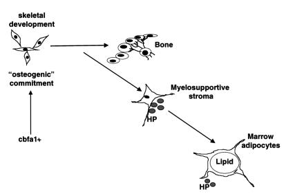
Formation of the marrow cavity and marrow stroma requires the pivotal transcription factor, cbfa1, which controls osteogenic differentiation and drives bone formation (10, 11). In development, the physical emergence of marrow stromal cells lies downstream of the physical emergence of bone and bone-forming cells, and, of course, downstream of the relevant transcriptional control (Figure 1). In postnatal organisms, cbfa1 is commonly, and perhaps consistently, expressed in clones and nontransformed lines of human or murine marrow stromal cells but does not predict their actual osteogenic capacity upon in vivo transplantation (12). Expression of cbfa1 in these same cell strains does not prevent differentiation towards nonosteoblastic phenotypes, such as adipocytes or chondrocytes. Considered along with the temporal and developmental priority of osteogenic differentiation over the physical emergence of marrow stromal cells, these observations suggest that osteogenic commitment directed by cbfa1 occurs upstream of the ontogeny of marrow stromal cells, which are the postnatal precursors of osteogenic cells. These cells retain expression of cbfa1, possibly as a legacy of their osteogenic origins, but they remain capable of entering multiple differentiation pathways and are not committed to an obligate osteogenic fate. If cbfa1 is viewed as a master gene for osteogenic commitment, then marrow stromal cells are reversibly committed and multipotential cells.
Figure 1.
During development, precursor cells become committed to skeletogenesis upon induction of the critical osteogenic transcription factor, cbfa1. The initial phenotype expressed by these cells is that of fully mature osteoblasts. Subsequently, when a threshold amount of bone has been formed, these cells form the primitive bone marrow stroma that serves as the bed upon which hematopoiesis occurs. At some point during the postnatal period, when hematopoiesis is sufficient, these same cells change phenotype yet again to become marrow adipocytes. Cells of these three phenotypes (osteoblastic, myelosupportive, and adipocytic) form a continuous network throughout the bone–bone marrow organ and maintain expression of cbfa1. These differentiated cells are able to shift from one phenotype to another, depending on the metabolic status of the organism.
Renewal versus flexibility: tissues, progenitors, molecules
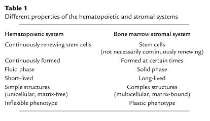
Postembryonic or postnatal differentiated cells within the stromal system can indeed adopt alternative phenotypes of other cells within this system, both in vitro and in vivo. Clonal adipocytic cell strains from postnatal rabbit marrow can be reverted to a fully osteogenic phenotype by altering the serum conditions (13). Single-cell suspensions of in vitro differentiated chick hypertrophic chondrocytes turn to fibroblastic and osteoblastic fates when allowed to adhere to appropriate substrata (14). Some evidence for direct differentiation of prehypertrophic chondrocytes to bone-forming cells in vivo has been obtained in rodents (15). Differentiated human, alkaline phosphatase–positive adventitial reticular cells, which normally function as myelosupportive elements, can rapidly accumulate fat and become adipocytes upon pharmacological myelosuppression in vivo. These cells are thus able to shift dynamically between two recognized “terminal” phenotypes (reticular and adipocytic) within the progeny of the stromal stem cell (8). These phenomena reflect the plasticity of the bone marrow stromal system and distinguish it from the hematopoietic system, in which phenotypic shifts of differentiated cells do not occur; commitment of precursor cells downstream of the HSC is generally thought to be progressive and irreversible. Plasticity of differentiated phenotypes within the stromal system implies that commitment and differentiation may not be irreversible, even in fully differentiated cells such as hypertrophic chondrocytes or myelosupportive cells. Stated another way, stromal cells downstream of a putative undifferentiated stem cell may be simultaneously differentiated and multipotential, a remarkable combination of features whose general significance was little appreciated until the current explosion of interest in somatic cell plasticity.
The plasticity of connective tissue cells extends to their functions in development and postnatal growth. These cells turn over slowly, and most are exposed to abundant extracellular matrix–directed (ECM-directed) cues, which help maintain their differentiated phenotypes. Remodeling of the ECM alters the signals that impinge on resident cells and may contribute to changes in cell morphology and patterns of gene expression. Of note, the marrow stroma is perhaps the single connective tissue characterized by a remarkable paucity of ECM, which may in part explain the ease with which stromal cells can shift from one phenotype to another.
Mesodermal, solid-phase tissues need to be plastic. The general physiological relevance of matrix remodeling events for organism growth and tissue integrity has been illustrated recently by the phenotype of membrane-type 1 matrix metalloproteinase–deficient (MT1-MMP–deficient) mice, in which connective tissue remodeling is blocked as a result of impaired matrix degradation, leading to generalized adverse changes in mesodermal tissues (16). The coordinated remodeling and adaptation of interfaced tissues (e.g., bone/tendon, bone/ligament, bone/cartilage, tendon/muscle) during organ growth demand that physical boundaries between different tissues be able to shift in space. Plasticity and multipotentiality of resident cells in mesodermal tissues may be as crucial for connective tissues and their progenitors as self-renewal is for blood and HSCs (see Table 1 for a comparison of the features of these tissues). Self-renewal and the associated patterns of cellular replication and differentiation must have evolved to serve the need for replenishing short-lived nonadherent cells in a long-lived organism, whereas phenotypic flexibility and flexibility in transcriptional control during differentiation allow for tissue adaptation.
Table 1.
Different properties of the hematopoietic and stromal systems
“Prove to me that you’re divine — turn my water into wine”
While the plasticity of the bone marrow stromal system and dependent tissues has not been acknowledged outside of the field of skeletal biology, several reports have recently revived an interest in a different order of biological plasticity, which is ascribed to “stem” cells associated with a variety of tissues. Some of these studies have implied that postnatal somatic (stem) cells can give rise to tissues normally originating from different embryonic layers. For example, it was reported that marrow stromal cells transplanted into the brain might acquire a neural fate (17), and that neural and muscle stem cells can give rise to blood (18). In the charged atmosphere that has prevailed since the birth of Dolly, these sensational claims play upon a desire for biotechnological omnipotence. “Stem cells,” seemingly, allow for extraordinary, not to say miraculous, transformations: of bone into brain, of brain or muscle into blood. If confirmed, such findings would indicate that somatic cells with a range of differentiative capabilities similar to those of embryonic stem cells remain in the postnatal organism at multiple developmentally unrelated sites, including the bone marrow stroma.
The existence of totipotent postnatal somatic stem cells would necessitate a dramatic change in our view of the biological significance of tissue stem cells, far beyond the need for tissue turnover and repair as required by nature, or even by biotechnology. Obviously, blood is not normally made in the brain or muscle, nor brain tissue in the marrow stroma. Likewise, it is unlikely that some of these unorthodox and unexpected differentiation potentials would ever be applied for clinical purposes. Still, these findings pose fascinating questions and demand a rigorous study of developmental pathways whereby the postulated somatic totipotent stem cells might arise and be retained throughout development and postnatal growth. To date, such a pathway is unknown, and the prevailing paradigms in developmental biology only account for the existence of local committed progenitors in growing tissues. A clear definition of the mechanisms by which somatic stem cells are generated and maintained would help elucidate their distinctive biological features and, ultimately, their possible uses and would undoubtedly reveal important novel aspects of pre- and postnatal development.
Marrow stromal cells and their plastic properties might thus turn out to represent a special case in a more widespread system of somatic stem cells. If so, their properties would provide insights of general relevance. Marrow stromal cells, a cell type that exhibits impressive plasticity, are in fact perivascular cells, much like retinal pericytes — perivascular cells within the central nervous system. Interestingly, bovine retinal pericytes have been found to give rise to cartilage and bone in vitro (19). Cells from the embryonic aorta can give rise to satellite cells and skeletal muscle (20). It has been proposed that microvascular districts may represent the specific niche where multipotential progenitors are retained in adult tissues (21). While accounting for the occurrence of postnatal stem cells in a variety of diverse tissues and organs, this hypothesis links this unexpected common property to a simple structural theme shared by all tissues — the existence of a vasculature and its ability to grow during organ growth. Further experimental work is needed to validate the hypothesis and to address the issue of whether the common theme to somatic postnatal progenitors is the vasculature, and whether embryonic differentiation potential, like the potential for angiogenesis, lies dormant within it.
Marrow stromal stem cells and skeletal diseases
A natural extension of the principle whereby a normal miniature ossicle can be formed by stromal stem cells would hold that stromal stem cells with intrinsic genetic defects generate miniatures of diseased bones. This principle was originally applied to human fibrous dysplasia of bone, a disease in which somatic mutations of the GNAS1 gene lead to severe crippling skeletal lesions (22). It was later extended to the skeletal abnormalities observed in mice with a targeted null mutation of the MT1-MMP gene (16). In both cases, transplanting strains of mutated stromal cells resulted in diseased ossicles with phenotypic abnormalities that directly reflected the changes observed in the intact organism. Using in vivo transplantation assays, diseases or changes of the skeleton due to intrinsic dysfunction of osteogenic cells can thus be singled out. Marrow stromal stem cells and their progeny thus emerge as the units of skeletal disease. This approach provides a handy way to generate animal models of skeletal diseases and validates the use of stromal cells in vitro for dissecting the pathophysiology of the skeletal tissues. For example, the ability to transplant progenitor cells in mice allowed us to develop a model of fibrous dysplasia and show that formation of lesional tissue depends upon somatic mosaicism (22).
Recognition of the broad growth and differentiation potential of marrow stromal cells and the ease with which they can be obtained and expanded in number (23) has opened the door to at least three classes of clinical applications, each with benefits and inherent problems. Perhaps the most readily implemented use of the osteogenic potential of marrow stromal cells involves reconstructing localized skeletal defects. The advantage provided over existing alternative methods (e.g., the use of uncultured marrow or biomaterials) lies in the theoretical full biological compatibility of a prosthetic device composed entirely of cells only, which might overcome the usual limits to the size and shape of defects to be repaired. Second, marrow stromal cells might be used for gene therapy — a more difficult challenge, since human stromal cells cannot yet be transduced with high enough efficiency to generate the required number of engineered cells. Furthermore, proper regulation of expression of a desired gene in these cells appears to be problematic, and transgenes that are expressed successfully in standard, continuous, or immortalized cell lines cannot be used directly for in vitro models using human cells, let alone for clinical applications. Finally, perhaps the most ambitious use for these cells would be to reconstitute some or all of the skeletal system to cure systemic diseases of the bone.
Are marrow stromal stem cells systemically transplantable?
The precedent of hematopoietic transplantation has led many to a simplistic view of stromal stem cells and their dependent tissues. The notion that stromal stem cells can be transplanted using the same principles and procedures used for HSCs is clearly an oversimplification. The widely known key principle of bone marrow transplantation (BMT), the seed and soil paradigm, postulates that upon ablation of a recipient marrow, progenitors infused via the circulation (the seed) can home into the nonablated marrow stroma (the soil) and can regenerate a hematopoietic tissue. The principle relies on a few established biological properties of HSC and the dependent hematopoietic lineages that do not apply to stromal progenitors and the dependent connective tissues. Furthermore, the principle of HSC transplantation depends on the remarkable radio- and chemoresistance of marrow stromal cells, traits that facilitate the replacement of hematopoietic cells in a minimally disturbed cellular environment. Clearly, this property limits the ability to remove the endogenous stroma prior to replacing it with stromal cells cultured ex vivo.
Despite claims that small numbers of donor stromal cells can be found in recipients of BMT, the bulk of the evidence indicates that marrow stromal cells are not transplanted during this procedure (24). Systemic infusion of stromal stem cells for treatment of skeletal diseases remains unlikely because of their inherent differences from HSCs. Whereas HSCs are known to circulate and negotiate the sinusoidal wall in the marrow via selective cell-cell interactions that allow them to settle in the extravascular compartment, circulating progenitors of the stromal system (25) have not been identified conclusively. Even assuming that such cells exist, there is little doubt that noncirculating, locally resident progenitors fabricate the bulk of skeletal tissues during both development and postnatal growth. Likewise, both blood and bone turn over, but the skeleton turns over at a vastly lower rate: HSCs can replenish the whole hematopoietic system in a few weeks, while building an adult skeleton requires 15 years. To generate individual cells is all that HSCs have to do to replenish a whole hematopoietic system, whereas building a skeleton entails creating a complex physical structure whose precise spatial layout reflects an equally precise timing of events over a period of years.
In the face of these concerns and the clear potential for danger to patients if systemic infusion of stromal stem cells is attempted blindly or prematurely, human studies should proceed only after animal studies have demonstrated that viable cells of donor origin can be found in the bone–bone marrow organ and that these cells are capable of homing. That is, transplanted marrow stromal cells must be detectable specifically in the appropriate macroscopic (skeletal) and microscopic (extravascular) environment. Moreover, these cells must be shown to be competent for engraftment, functioning in the recipient’s marrow to produce differentiated progeny, and these progeny must occur at high enough levels to influence tissue function. Finally, these cells must be shown to produce the desired biological effect in appropriate preclinical models.
Studies so far have generally fallen short of providing convincing evidence of engraftment of infused stromal progenitor cells, but the pioneering nature of these attempts has prevailed in some cases over stringent assessment of evidence. The bone marrow, like the spleen and the liver, normally functions as a clearing site for exogenous materials in the bloodstream, so neither the detection of reporter genes in tissue extracts nor the isolation in culture of viable cells carrying genetic markers of donor origin suffices to prove the engraftment of infused stromal progenitors. Rather, this kind of evidence may be used to assess the life-span of marked cells that have reached the marrow environment. Since stromal cells are normally mitotically quiescent and long-lived in vivo, infused stromal cells might survive for long periods after settling in the marrow but might not participate in any dynamic event of bone physiology. Much as engraftment of hematopoietic progenitors following BMT is demonstrated by the appearance of circulating blood cells of donor origin, engraftment of stromal progenitors ought to rest on evidence of various differentiated lineages of donor origin. Osteoblasts, osteocytes, adipocytes, and marrow reticular cells of donor origin must be unequivocally identified in the intact tissue and must be shown to be physically and functionally integrated, as in normal stroma.
Two studies employing animal models have sought evidence that differentiated progeny of infused stromal cells exist in the recipient’s intact tissue — undoubtedly steps in the right direction. Nilsson et al. (26) detected fully differentiated, quiescent, donor-derived osteocytes in the femoral cortex of mice receiving marrow grafts. More recently, Hou et al. (27) used marrow stromal cells carrying a reporter gene driven by the osteocalcin promoter to provide additional evidence for some engraftment of stroma-related, infused cells in mice. Because the osteocalcin gene is expressed and regulated in a tissue- and differentiation stage–specific manner, reporter gene expression in bone of host mice in this elegant study does provide evidence of osteogenic differentiation of cells of donor origin. Histologically proven donor bone cells were also reported to be present, but no quantitative assessment of their frequency was provided. Overall, these data may provide provisional evidence for some engraftment of stroma-related, infused cells in mice. However, caution is in order before concluding that systemic transplantation of osteogenic cells is feasible in principle. Quantitative aspects of engraftment and of actual rates of bone turnover need to be evaluated carefully. For example, the presence of donor osteocytes in a femoral shaft 6 months after transplant was interpreted by Nilsson et al. (26) as proof of their local origin from engrafted donor progenitors, but this conclusion relied on estimated multiple turnover cycles of an entire femur, which in reality cannot occur. At the known rates of bone remodeling in mice, it would take a mouse a lifetime to renew a mass of bone equivalent to one femur a single time. Furthermore, mouse cortical bone does not undergo Haversian remodeling (intracortical remodeling that generates osteons, as occurs in larger mammals), but rather growth-related modeling. Large areas of a mouse femur, especially in the cortex, never remodel, while other areas turn over constantly. Any osteocyte found in mouse cortical bone may therefore have been generated months before and then remained undisturbed in an unremodeled area of bone. For the same reason, osteocytes of donor origin found in cortical bone months after transplantation do not prove recruitment of functional progenitors long after engraftment, as claimed. Reliable quantitative estimates of engrafted progeny, as well as careful consideration of cell identity and function within the host environment, should also be sought. In this respect, it will be important to rely on standard means for assessing actual bone formation in vivo using fluorescent labels, and to match these data with the identity and location of any donor-derived cells that might be detected.
Despite the absence of conclusive evidence of feasibility from animal models, human BMT following a myeloablative regimen was recently attempted in children with severe osteogenesis imperfecta (OI). This study (28) indicated a rate of engraftment of 1–2% bone cells (assessed by ex vivo culture of recipient bone–bone marrow cells) and claimed improvement of clinically assessed parameters of disease over time, but it lacked appropriate clinical controls and did not provide convincing histological data. The authors also failed to reconcile the extremely low rate of observed engraftment with what was purportedly a profound, systemic effect on bone growth, affecting cartilage growth plates and sites of bone formation proper. Changes in bone mineral content, a clinical parameter used to assess treatments for OI, are unreliable estimates of donor cells’ effects. Finally, since myeloablation apparently boosts osteogenic activity in several animal models, this aspect of the treatment may complicate the analysis of donor cell function in the subject’s tissues.
Caution remains the watchword in evaluating the clinical promise of this technology. Critical basic issues require extensive animal studies, and shortcuts do not work in the interests of patients, for whom alternative therapeutic approaches are at hand. Ignoring problems in this area may well hinder the development of stem cells as therapeutic tools.
Thirty years after their first appearance on the biomedical scene, marrow stromal stem cells are more appealing than ever. The epitome of somatic cell plasticity, they feature some of the most exciting aspects of stem cell biology. As key elements of skeletal disease, they offer approaches to the study of these pathologies. More easily expanded ex vivo than stem cells in many other tissues, they lend themselves to a number of potential therapeutic applications. Turning promises into reality only rests, as always, with the quality of the forthcoming science.
Acknowledgments
The support of Telethon (grant E1029) to P. Bianco is gratefully acknowledged.
References
- 1.Friedenstein AJ, Petrakova KV, Kurolesova AI, Frolova GP. Heterotopic transplants of bone marrow. Analysis of precursor cells for osteogenic and hematopoietic tissues. Transplantation. 1968;6:230–247. [PubMed] [Google Scholar]
- 2.Friedenstein AJ, et al. Precursors for fibroblasts in different populations of hematopoietic cells as detected by the in vitro colony assay method. Exp Hematol. 1974;2:83–92. [PubMed] [Google Scholar]
- 3.Owen M. Marrow stromal stem cells. J Cell Sci Suppl. 1988;10:63–76. doi: 10.1242/jcs.1988.supplement_10.5. [DOI] [PubMed] [Google Scholar]
- 4.Friedenstein AJ, Shapiro-Piatetzky II, Petrakova KV. Osteogenesis in transplants of bone marrow cells. J Embryol Exp Morphol. 1966;16:381–390. [PubMed] [Google Scholar]
- 5.Johnstone B, Hering TM, Caplan AI, Goldberg VM, Yoo JU. In vitro chondrogenesis of bone marrow-derived mesenchymal progenitor cells. Exp Cell Res. 1998;238:265–272. doi: 10.1006/excr.1997.3858. [DOI] [PubMed] [Google Scholar]
- 6.Kuznetsov SA, et al. Single-colony derived strains of human marrow stromal fibroblasts form bone after transplantation in vivo. J Bone Miner Res. 1997;12:1335–1347. doi: 10.1359/jbmr.1997.12.9.1335. [DOI] [PubMed] [Google Scholar]
- 7.Pittenger MF, et al. Multilineage potential of adult human mesenchymal stem cells. Science. 1999;284:143–147. doi: 10.1126/science.284.5411.143. [DOI] [PubMed] [Google Scholar]
- 8.Bianco P, Costantini M, Dearden LC, Bonucci E. Alkaline phosphatase positive precursors of adipocytes in the human bone marrow. Br J Haematol. 1988;68:401–403. doi: 10.1111/j.1365-2141.1988.tb04225.x. [DOI] [PubMed] [Google Scholar]
- 9.Bianco P, Riminucci M, Kuznetsov S, Robey PG. Multipotential cells in the bone marrow stroma: regulation in the context of organ physiology. Crit Rev Eukaryot Gene Expr. 1999;9:159–173. doi: 10.1615/critreveukargeneexpr.v9.i2.30. [DOI] [PubMed] [Google Scholar]
- 10.Ducy P, Zhang R, Geoffroy V, Ridall AL, Karsenty G. Osf2/Cbfa1: a transcriptional activator of osteoblast differentiation. Cell. 1997;89:747–754. doi: 10.1016/s0092-8674(00)80257-3. [DOI] [PubMed] [Google Scholar]
- 11.Komori T, et al. Targeted disruption of Cbfa1 results in a complete lack of bone formation owing to maturational arrest of osteoblasts. Cell. 1997;89:755–764. doi: 10.1016/s0092-8674(00)80258-5. [DOI] [PubMed] [Google Scholar]
- 12.Satomura, K., Krebsbach, P.A., Bianco, P., and Gehron Robey, P. Osteogenic imprinting upstream of marrow stromal cell differentiation. J. Cell. Biochem. In press. [PubMed]
- 13.Bennett JH, Joyner CJ, Triffitt JT, Owen ME. Adipocytic cells cultured from marrow have osteogenic potential. J Cell Sci. 1991;99:131–139. doi: 10.1242/jcs.99.1.131. [DOI] [PubMed] [Google Scholar]
- 14.Gentili C, et al. Cell proliferation, extracellular matrix mineralization, and ovotransferrin transient expression during in vitro differentiation of chick hypertrophic chondrocytes into osteoblast-like cells. J Cell Biol. 1993;122:703–712. doi: 10.1083/jcb.122.3.703. [DOI] [PMC free article] [PubMed] [Google Scholar]
- 15.Riminucci M, et al. Vis-a-vis cells and the priming of bone formation. J Bone Miner Res. 1998;13:1852–1861. doi: 10.1359/jbmr.1998.13.12.1852. [DOI] [PubMed] [Google Scholar]
- 16.Holmbeck K, et al. MT1-MMP-deficient mice develop dwarfism, osteopenia, arthritis, and connective tissue disease due to inadequate collagen turnover. Cell. 1999;99:81–92. doi: 10.1016/s0092-8674(00)80064-1. [DOI] [PubMed] [Google Scholar]
- 17.Azizi SA, Stokes D, Augelli BJ, DiGirolamo C, Prockop DJ. Engraftment and migration of human bone marrow stromal cells implanted in the brains of albino rats: similarities to astrocyte grafts. Proc Natl Acad Sci USA. 1998;95:3908–3913. doi: 10.1073/pnas.95.7.3908. [DOI] [PMC free article] [PubMed] [Google Scholar]
- 18.Bjornson CR, et al. Turning brain into blood: a hematopoietic fate adopted by adult neural stem cells in vivo. Science. 1999;283:534–537. doi: 10.1126/science.283.5401.534. [DOI] [PubMed] [Google Scholar]
- 19.Doherty MJ, et al. Vascular pericytes express osteogenic potential in vitro and in vivo. J Bone Miner Res. 1998;13:828–838. doi: 10.1359/jbmr.1998.13.5.828. [DOI] [PubMed] [Google Scholar]
- 20.De Angelis L, et al. Skeletal myogenic progenitors originating from embryonic dorsal aorta coexpress endothelial and myogenic markers and contribute to postnatal muscle growth and regeneration. J Cell Biol. 1999;147:869–878. doi: 10.1083/jcb.147.4.869. [DOI] [PMC free article] [PubMed] [Google Scholar]
- 21.Bianco P, Cossu G. Uno, nessuno e centomila: searching for the identity of mesodermal progenitors. Exp Cell Res. 1999;251:257–263. doi: 10.1006/excr.1999.4592. [DOI] [PubMed] [Google Scholar]
- 22.Bianco P, et al. Reproduction of human fibrous dysplasia of bone in immunocompromised mice by transplanted mosaics of normal and Gsalpha-mutated skeletal progenitor cells. J Clin Invest. 1998;101:1737–1744. doi: 10.1172/JCI2361. [DOI] [PMC free article] [PubMed] [Google Scholar]
- 23.Krebsbach PH, Kuznetsov SA, Bianco P, Gehron Robey P. Bone marrow stromal cells: characterization and clinical application. Crit Rev Oral Biol Med. 1998;10:165–181. doi: 10.1177/10454411990100020401. [DOI] [PubMed] [Google Scholar]
- 24.Simmons PJ, Przepiorka D, Thomas ED, Torok-Storb B. Host origin of marrow stromal cells following allogeneic bone marrow transplantation. Nature. 1987;328:429–432. doi: 10.1038/328429a0. [DOI] [PubMed] [Google Scholar]
- 25.Luria EA, Panasyuk AF, Friedenstein AY. Fibroblast colony formation from monolayer cultures of blood cells. Transfusion. 1971;11:345–349. doi: 10.1111/j.1537-2995.1971.tb04426.x. [DOI] [PubMed] [Google Scholar]
- 26.Nilsson SK, et al. Cells capable of bone production engraft from whole bone marrow transplants in nonablated mice. J Exp Med. 1999;189:729–734. doi: 10.1084/jem.189.4.729. [DOI] [PMC free article] [PubMed] [Google Scholar]
- 27.Hou Z, et al. Osteoblast-specific gene expression after transplantation of marrow cells: implications for skeletal gene therapy. Proc Natl Acad Sci USA. 1999;96:7294–7299. doi: 10.1073/pnas.96.13.7294. [DOI] [PMC free article] [PubMed] [Google Scholar]
- 28.Horwitz EM, et al. Transplantability and therapeutic effects of bone marrow-derived mesenchymal cells in children with osteogenesis imperfecta. Nat Med. 1999;5:309–313. doi: 10.1038/6529. [DOI] [PubMed] [Google Scholar]


