Contrary to Smaers’s claim (1) that all recent studies find “allometric scaling of the human frontal lobe is not larger than expected,” one study (2) clearly claimed otherwise. Smaers’s own studies (3) are somewhat more ambiguous on this point, and he contradicts himself here by stating that “the conclusion that no aspects of the human frontal lobe are larger than expected (controlling for allometry) is not upheld.” It was such ambiguity that we sought to resolve in our report (4).
We brought available datasets together within a uniform and statistically valid analytical framework to remove doubts resulting from analytical differences (4). Smaers (1) claims that phylogenetic analysis is unnecessary because values of λ were <0.01. However, in two of the datasets we found larger values. Despite Smaers’s (1) concern, his own analyses (1, 3) of the same data also use phylogenetic generalized least-squares regressions, yet fail to report λ values. Smaers misinterprets our variable-rates analysis, which does not concern trait values in extant species, but evolutionary rates of change along branches of the primate phylogeny. No such analysis of frontal lobe evolution had previously been published.
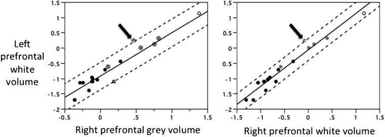
Smaers’s (1) comment that “larger than expected human (left) prefrontal white to grey matter is suggested to be a result of smaller than expected nonprefrontal white matter or (left) prefrontal grey matter” conflates our reexamination of two different studies. First, we reanalyzed the claim that human prefrontal white matter is large relative to nonprefrontal white matter (2). We found this effect was a result of “smaller than expected values of nonprefrontal white matter volume in this particular data set” (4) (emphasis added). We made no claim that this effect would be found in other data sets, instead urging caution in interpreting “deviation in a single species based on analyses of single data sets” (4). Second, we noted Smaers’s much more specific claim that (i) human left (but not right) prefrontal cortex (PFC) white matter is large relative to left PFC gray matter (3). Smaers’s study nevertheless found (ii) no significant deviation of human left PFC white matter relative to the rest of the brain (figure 4B in ref. 3), leaving our overall conclusion unchallenged and rendering the interpretation of result (i) problematic. In fact, figure 5C in ref. 3 suggests that small values of left PFC gray matter (relative to brain volume) contribute to result (i). In the same data set, human left PFC white matter is not large relative to either white or gray matter in the right hemisphere PFC (Fig. 1), further undermining the interpretation that left PFC white matter is unexpectedly large.
Fig. 1.
Reanalysis of data from Smaers et al. (3). Regressions fitted by phylogenetic generalized least-squares regression, as in ref. 4. Humans are indicated by an open circle, apes by gray circles, and other anthropoid primates by black circles. Dashed lines are 95% prediction intervals for values of y relative to x. Arrows indicate orangutan, which, unlike humans, has a significant positive residual in both regressions. Instead of interpreting this finding as indicative of orangutan cognitive attributes, we reiterate our caution about overinterpretation of such outliers in single studies, compared with finding consistent patterns across independent datasets.
Smaers states that his study (3) did not claim a significant ape-monkey difference, but merely “a trend in which apes, contrary to monkeys, consistently have positive residuals” (1). Thus, the “trend” is acknowledged to be statistically nonsignificant, yet we are asked to believe that it is biologically meaningful. In Smaers’s report (3) this nonsignificant result is referred to under the heading “Ape Uniqueness.” We cannot see why any faith should be placed in claims of quantitative uniqueness, or why “larger than expected” is an acceptable term in the absence of a statistically significant difference.
Footnotes
The authors declare no conflict of interest.
References
- 1.Smaers JB. How humans stand out in frontal lobe scaling. Proc Natl Acad Sci USA. 2013;110:E3682. doi: 10.1073/pnas.1308850110. [DOI] [PMC free article] [PubMed] [Google Scholar]
- 2.Schoenemann PT, Sheehan MJ, Glotzer LD. Prefrontal white matter volume is disproportionately larger in humans than in other primates. Nat Neurosci. 2005;8(2):242–252. doi: 10.1038/nn1394. [DOI] [PubMed] [Google Scholar]
- 3.Smaers JB, et al. Primate prefrontal cortex evolution: Human brains are the extreme of a lateralized ape trend. Brain Behav Evol. 2011;77(2):67–78. doi: 10.1159/000323671. [DOI] [PubMed] [Google Scholar]
- 4.Barton RA, Venditti C. Human frontal lobes are not relatively large. Proc Natl Acad Sci USA. 2013;110(22):9001–9006. doi: 10.1073/pnas.1215723110. [DOI] [PMC free article] [PubMed] [Google Scholar]


