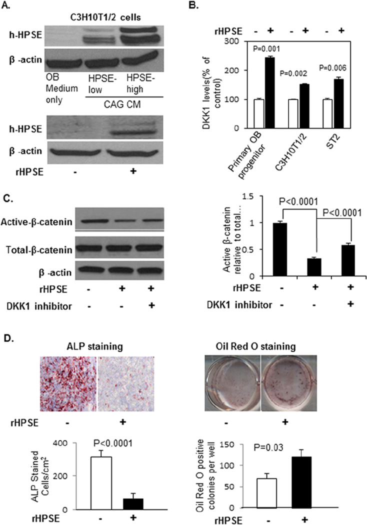
Figure 5. rHPSE stimulates DKK1 secretion from osteoblast progenitor and stromal cells, inhibits osteoblastogenesis, and promotes adipogenesis by inhibiting Wnt signaling pathway in these cells.
(A). Human heparanase is detectable in cell lysates of C3H10T1/2 cells after the cells were cultured in CM of HPSE-low and HPSE-high CAG cells (upper panel) or in the presence of human rHPSE (100 ng/ml) (lower panel) for 3 days. (B). Primary murine osteoblast progenitor cells, C3H10T1/2 pre-osteoblastic cells, and ST2 stromal cells were cultured in absence or presence of rHPSE(100 ng/ml) for 3 days, and the level of murine DKK1 in the CM measured by ELISA. Each bar represents the Mean ± SD of 2 measurements. Significance is indicated in the panel. (C). Primary osteoblast progenitor cells cultured in osteogenic medium in absence or presence of rHPSE (100 ng/ml) with or without DKKinhibitor (3.0 mM) for 10 days (medium changed every 3 days). Cells were harvested for Western blot and stained with antibodies against active-β-catenin, total-β-catenin and β-actin (left). The active β-catenin bands from Western blot were quantified by ImageJ from two independent experiments (right). (D). ALP and Oil Red O staining were performed at day 10 for evaluation of osteoblast and adipocyte differentiation. ALP positive osteoblast cells and Oil Red O stained adipocyte colonies were enumerated from two independent experiments (Mean ± SEM). Significance is indicated in each panel.

