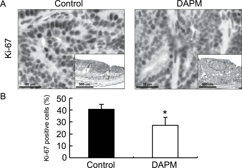
Fig. 4.
Ki-67 immunostaining of tumors from control and DAPM-treated mice. Thirty mice were injected with AOM as described in Materials and methods. Ten weeks after the last injection, mice were subjected to colonoscopic imaging to verify the presence of colon tumors. Mice were then administered vehicle (control) or DAPM and killed 4 weeks later. Tissue sections were prepared from the colon of control (n = 15) and DAPM-treated mice (n = 15) and processed for immunohistochemical analysis of Ki-67 as described in Materials and methods. (A) Representative images for Ki-67 staining of the tumors from control and DAPM-treated mice (The inset depicts a lower magnification of the tissue and the circled area is shown at the high magnification.) (B) The relative percentage of Ki-67-positive cells in the tumor of control and DAPM-treated mice. The positive cells were counted as described in Materials and methods. Columns, mean percent positive cells of 15 samples per group; bars, standard deviation. *P < 0.05 compared with control mice (Student’s t-test).

