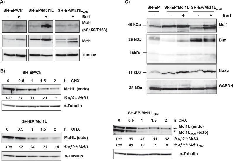
Figure 3. Mcl1LJAM rescues Mcl1L from degradation and cooperates with Bim.
SH-EP/Ctr, SH-EP/Mcl1L and SH-EP/ Mcl1LJAM cell lysates were analysed for phosphorylated Mcl1 (Ser159/Thr163) and Mcl1 after treatment with bortezomib for four hours (a). α-Tubulin served as loading control. Protein stability was analysed by treating SH-EP/Ctr, SH-EP/Mcl1L or SH-EP/Mcl1LJAM cells with 10 μg/ml CHX for the times indicated (b). α-Tubulin served as loading control. SH-EP/Ctr, SH-EP/Mcl1L and SH-EP/Mcl1LJAM cells were treated for four hours with 200 nM bortezomib. Cell lysates were analysed for the expression of Noxa and Bim. GAPDH served as loading control (c).

