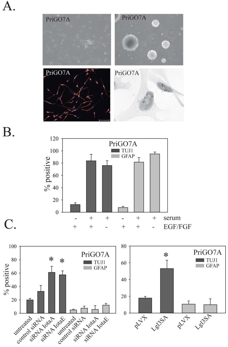
Figure 10. Effects of PKC and Lgl1 on PriGO7A differentiation.
Morphology under phase contrast microscopy (top left panel), neurosphere formation (top right panel), nestin immunofluorescence (lower left panel) and EGFR chromogenic in situ hybridization for PriGO7A cells (lower right panel). B. Differentiation of PriGO7A cells after exposure to serum or growth factor withdrawal. C. Bar graphs show the effects of PKCι knockdown (left panel) and Lgl3SA transduction (right panel) on PriGO7A differentiation.

