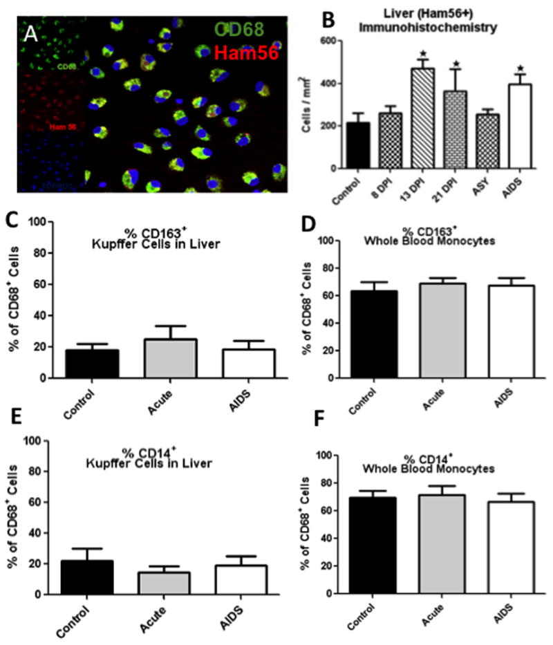
Figure 1.

A: Triple-label confocal microscopy of liver macrophages (Kupffer cells) in vitro showing co-localization of CD68 (green) and Ham56 (red), indicating Kupffer cells co-express CD68 & Ham56 (CD68 with Alexa 488, green; Ham56 with Alexa 568, red; and cell nuclei with Topro3). B: Absolute numbers of Ham56+ Kupffer cells per/mm2 of liver in uninfected (control) and various stages of SIV infection as determined by immunohistochemistry for Ham56. Note significant increases in Ham56+ Kupffer cells per mm2 are detected after early SIV infection and in macaques with AIDS. *Indicates significant differences from controls (P<0.05). C–F: Percentages of CD68+ cells in the liver (C and E) and blood (D and F) co-expressing CD163 (C and D) or CD14 (E and F) in acute and chronic infection compared to controls. No significant differences in CD14 or CD163 expression were detected on CD68+ cells in liver or blood due to SIV infection (C–F).
