Abstract
Filoviruses (viruses in the genus Ebolavirus and Marburgvirus in the family Filoviridae) cause severe haemorrhagic fever in humans and nonhuman primates. Rapid, highly sensitive, and reliable filovirus-specific assays are required for diagnostics and outbreak control. Characterisation of antigenic sites in viral proteins can aid in the development of viral antigen detection assays such immunochromatography-based rapid diagnosis. We generated a panel of mouse monoclonal antibodies (mAbs) to the nucleoprotein (NP) of Ebola virus belonging to the species Zaire ebolavirus. The mAbs were divided into seven groups based on the profiles of their specificity and cross-reactivity to other species in the Ebolavirus genus. Using synthetic peptides corresponding to the Ebola virus NP sequence, the mAb binding sites were mapped to seven antigenic regions in the C-terminal half of the NP, including two highly conserved regions among all five Ebolavirus species currently known. Furthermore, we successfully produced species-specific rabbit antisera to synthetic peptides predicted to represent unique filovirus B-cell epitopes. Our data provide useful information for the development of Ebola virus antigen detection assays.
Keywords: Ebola virus, Nucleoprotein, Antibody epitope, Monoclonal antibody, Synthetic peptide
1. Introduction
Filoviruses are among the most lethal human pathogens recognised to date with case fatality rates up to 90%, depending on the virus species and strain (Pittalis et al, 2009; Bente et al., 2009). Filoviruses are grouped into two genera, Ebolavirus and Marburgvirus. There is one known species of Marburgvirus, Marburg marburgvirus, consisting of two viruses, Marburg virus (MARV) and Ravn virus (RAVV). In contrast, the genus Ebolavirus has five known species, Zaire ebolavirus, Sudan ebolavirus, Taï Forest ebolavirus, Bundibugyo ebolavirus and Reston ebolavirus, represented by Ebola virus (EBOV), Sudan virus (SUDV), Taï Forest virus (TAFV), Bundibugyo virus (BDBV) and Reston virus (RESTV), respectively. Furthermore, there is a newly discovered filovirus named Lloviu virus (LLOV) assigned to the proposed genus Cuevavirus, with one species, Lloviu cuevavirus (Negredo et al., 2011; Kuhn et al., 2010). The genome of filoviruses is approximately 19kb long, and contains seven genes arranged sequentially in the order: nucleoprotein (NP), viral protein (VP) 35, VP40, glycoprotein (GP), VP30, VP24 and polymerase (L) genes (Sanchez et al, 2007).
The lack of therapeutics and vaccines for filovirus infections and the fact that other pathogens cause clinical symptoms comparable to those of Ebola and Marburg haemorrhagic fever highlights the need for rapid, sensitive, reliable and virus-specific diagnostic tests to control the spread of these viruses (Qiu et al., 2011; Sanchez et al., 2007). Rapid antigen-detection tests with filovirus-specific monoclonal antibodies (mAb) are likely one of the best ways for early diagnosis of filovirus infections in the field setting. NP may be the ideal target antigen because of its abundance in filovirus particles and its strong antigenicity (Niikura et al., 2001, 2003). The average EBOV virion, which is up to 1028nm in length, contains about 3200 NP molecules (Bharat et al., 2012). EBOV NP consists of 739 amino acid residues, with a conserved hydrophobic N-terminus and a variable hydrophilic C-terminal part (Niikura et al., 2001; Sanchez et al, 2007). NP plays an important role in the replication of the viral genome and is essential for formation of the nucleocapsid (Watanabe et al., 2006). The C-terminus of EBOV NP binds to VP40 while the N-terminus forms a condensed helix with the same diameter as the inner nucleocapsid helix of an EBOV particle (Bharat et al., 2012). Following expression of VP40 in cultured cells, virus-like particles (VLPs) are produced and, upon co-expression of NP, the VLP contains NP as its core (Bharat et al., 2012; Noda et al, 2007). It has been demonstrated that the C-terminal half of the filovirus NP has strong antigenicity (Saijo et al, 2001). Multiple studies have identified conformational and linear epitopes for antibodies in this NP region for several viruses within the genus Ebolavirus (Ikegami et al., 2003; Niikura et al., 2001, 2003).
In general, characterisation of antigenic sites in a viral protein can aid in the development of diagnostic tools, therapeutics and vaccines (Gershoni et al., 2007; Toyoda et al., 2000). Here, we identified antigenic regions within the NP molecule using mouse NP-specific mAbs and rabbit antisera to synthetic NP peptides representing viruses from all known filovirus species. Some of the identified antigenic regions are shared among multiple virus species within the Ebolavirus genus, whereas others are species-specific. Our data provide useful information for future development of antigen-based detection assays for the diagnosis of filovirus infections.
2. Materials and methods
2.1. Plasmid construction
Plasmids expressing GP, VP40 and NP were constructed as described previously (Nakayama et al, 2010; Nidom et al, 2012). Briefly, viral RNAs were extracted from the supernatant of Vero E6 cells infected with EBOV (Mayinga), SUDV (Boniface), TAFV (Côte d'Ivoire), BDBV (Bundibugyo), RESTV (Pennsylvania) or MARV (Angola). Full length NP, VP40 and GP cDNA were amplified by RT-PCR using KOD-plus-Neo polymerase (Toyobo) and cloned into TOPO® vector using the Zero Blunt® TOPO® PCR Cloning Kit (Invitrogen). After sequence confirmation, the cloned genes were inserted into the mammalian expression vector pCAGGS.
2.2. Preparation of purified VLPs and NP
Human epithelial kidney 293T cells were grown in Dulbeco's modified Eagle's medium (DMEM), supplemented with 10% FCS, penicillin (100 unit/ml) and streptomycin (100 μg/ml). VLPs were produced by transfection of 293T cells with plasmids expressing NP and VP40 together with or without the plasmid expressing GP as described previously (Licata et al., 2004; Urata et al., 2007). Forty-eight hours after transfection, VLPs in the supernatant were purified by centrifugation through a 25% sucrose cushion at 28,000 × g and 4 °C for 1.5 h. The pelleted VLPs were resuspended in PBS and stored at −80 °C. For the preparation of purified NP, 293 T cells transfected with the plasmid encoding EBOV NP were lysed, and the NP fraction was collected by discontinuous CsCl gradient centrifugation of the lysates as described previously (Bharat et al., 2012; Noda et al., 2010).
2.3. Mouse mAb production
On day 0, six-week-old female Balb/c mice were immunised intramuscularly with 100 μg of EBOV VLPs consisting of NP and VP40 with complete Freund's adjuvant (Difco). The animals were boosted intramuscularly on day 14 with 100 μg of the same EBOV VLPs and incomplete Freund's adjuvant. After a final intravenous boost with 100 μg of the same EBOV VLPs without adjuvant on day 39, spleen cells were harvested on day 42 and fused to P3-U1 myeloma cells according to standard procedures (Shahhosseini et al., 2007). Hybridomas were maintained in Roswell Park Memorial Institute medium 1640 containing 20% FCS, penicillin (100 unit/ml), streptomycin (100 μg/ml), l-glutamine (4mM) and 2-mercaptoethanol (55 μM). Hybridoma supernatants were screened by an enzyme linked immunosorbent assay (ELISA) for secretion of NP-specific antibodies using purified EBOVNP and VLP as antigens. Specificity and cross-reactivity of mAbs were also confirmed by Western blotting. Selected hybridoma cells were then cloned twice performing limiting dilution.
2.4. Production of rabbit antisera
Genetyx ver6.0 for Windows (GENETYX CORPORATION) was used to predict B-cell epitopes in the NPs of EBOV, SUDV, TAFV, BDBV, RESTV and MARV, and the amino acid (aa) positions around 630–650 were selected. Synthetic peptides corresponding to this aa region in NP were produced (Sigma). Rabbits were then immunised with keyhole limpet haemocyanin-conjugated synthetic peptides by the standard procedure, and antisera were obtained on day 49.
2.5. ELISA
Ninety six-well ELISA plates (Nunc®, Maxisorp) were coated with 50 μl PBS containing purified EBOV NP (2 μg/ml), VLPs (2-5 μg/ml) or synthetic peptides (100 μg/ml) per well overnight at 4 °C. ELISA was carried out as described previously (Nakayama et al., 2011), using mouse antisera, hybridoma supernatants, purified mAbs or rabbit antisera as primary antibodies and goat anti-mouse IgG (H + L) or donkey anti-rabbit IgG (H + L) conjugated with peroxidase (Jackson ImmunoResearch) as secondary antibodies.
2.6. Western blotting
Vero E6 cells cultured in DMEM supplemented with 10% FBS, penicillin (100 unit/ml), streptomycin (100 μg/ml) and l-glutamine (4 mM) were infected with EBOV (Mayinga), SUDV (Boniface), TAFV (Cote d'Ivoire), BDBV (Bundibugyo), RESTV (Pennsylvania), MARV (Angola, Musoke, Ozolin and Ci67) or RAVV (Ravn) at a multiplicity of infection of 1 and maintained for 72h. Cell culture supernatants were subjected to SDS-PAGE. For the screening of hybridoma supernatants (see above), VLPs were used instead of authentic virus lysates. After electrophoresis, separated proteins were blotted on a polyvinylidene difluoride membrane (Millipore) or Immobilon-P transfer membrane (Millipore). Mouse mAbs and rabbit antisera were used as primary antibodies. The bound antibodies were detected with peroxidase-conjugated goat anti-mouse IgG (H + L) or donkey anti-rabbit IgG (H + L) (Jackson ImmunoResearch), followed by visualisation with Immobilon Western (Millipore).
2.7. Ethics and biocontainment statements
Animal studies were carried out in strict accordance with the Guidelines for Proper Conduct of Animal Experiments of the Science Council of Japan. The protocol was approved by the Hokkaido University Animal Care and Use Committee. All efforts were made to minimise the suffering of animals. All infectious work with filoviruses was performed under high containment complying with standard operating procedures approved by the Institutional Biosafety Committee in the BSL4 Laboratories of the Integrated Research Facility at the Rocky Mountain Laboratories, Division of Intramural Research, National Institute of Allergy and Infectious Diseases, National Institutes of Health, Hamilton, Montana, USA.
3. Results
3.1. Specificity and cross-reactivity of NP-specific mAbs
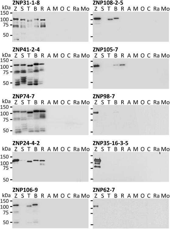
In the first screening process, we obtained 126 hybridomas producing mAbs reactive to the recombinant EBOV NP. None of them showed cross-reactivity to MARV NP. These mAbs were further assessed by ELISA for their cross-reactivity with the recombinant NPs of the other known Ebolavirus species (SUDV, TAFV, BDBV and RESTV). We found several different profiles for the cross-reactivities of these mAbs. Representative clones for each obtained cross-reactivity profile showing the highest OD450 values were selected and further cloned by limiting dilution. We then established 10 clones of NP-specific mAbs (ZNP31-1-8, ZNP41-2-4, ZNP74-7, ZNP24-4-2, ZNP106-9, ZNP108-2-5, ZNP105-7, ZNP98-7, ZNP35-16-3-5 and ZNP62-7) which were divided into 7 groups based on their cross-reactivity profiles in ELISA (Table 1). Four mAbs (ZNP31-1-8, ZNP41-2-4, ZNP74-7 and ZNP24-4-2) reacted with all known viruses of Ebolavirus species, with one (ZNP24-4-2) having relatively weak reactivity with SUDV. Four mAbs (ZNP106-9, ZNP108-2-5, ZNP105-7 and ZNP98-7) bound to NPs of some viruses in addition to EBOV, and 2 mAbs (ZNP35-16-3-5 and ZNP62-7) reacted only to EBOV. Importantly, these different reactivity profiles enabled us to distinguish the known Ebolavirus species by using combinations of these MAbs: EBOV was recognised by all the mAbs, SUDV by ZNP31-1-8, ZNP41-2-4, ZNP74-7, ZNP24-4-2 and ZNP106-9; TAFV by ZNP31-1-8, ZNP41-2-4, ZNP74-7, ZNP24-4-2, ZNP106-9 and ZNP108-2-5; BDBV by ZNP31-1-8, ZNP41-2-4, ZNP74-7, ZNP24-4-2, ZNP106-9, ZNP108-2-5, ZNP105-7 and ZNP98-7; and RESTV by ZNP31-1-8, ZNP41-2-4, ZNP74-7, ZNP24-4-2 and ZNP105-7. The reactivities of these NP-specific mAbs were further tested by Western blotting using lysates of actual filovirus particles grown in Vero E6 cells (Fig. 1). We found that the mAbs predominantly bound to proteins of approximately 100 kD and some smaller proteins, representing full-length NP and likely degraded NP molecules, respectively (Watanabe et al., 2006). The cross-reactivity profiles and virus specificities were similar to those obtained by ELISA, thus confirming the utility of these mAbs to detect not only recombinant but also virus-derived NPs.
Table 1.
Cross reactivity profiles of mAbs.
| mAb (group) | Isotype | EBOVa | SUDV | TAFV | BDBV | RESTV | MARV |
|---|---|---|---|---|---|---|---|
| ZNP31-1-8 (I) | IgG1 | ++b | ++ | ++ | ++ | ++ | − |
| ZNP41-2-4 (I) | IgG1 | ++ | ++ | ++ | ++ | ++ | − |
| ZNP74-7 (I) | IgG1 | ++ | ++ | ++ | ++ | ++ | − |
| ZNP24-4-2 (II) | IgG1 | ++ | + | ++ | ++ | ++ | − |
| ZNP106-9 (III) | IgG1 | ++ | + | ++ | ++ | − | − |
| ZNP108-2-5 (IV) | IgG1 | ++ | − | ++ | ++ | − | − |
| ZNP105-7 (V) | IgG1 | ++ | − | − | ++ | ++ | − |
| ZNP98-7 (VI) | IgG2a | ++ | − | − | ++ | − | − |
| ZNP35-16-3-5 (VII) | IgG1 | ++ | − | − | − | − | − |
| ZNP62-7 (VII) | IgG2b | ++ | − | − | − | − | − |
VLPs of each virus species were used as antigens.
Antibody reactivity was evaluated based on ELISAOD450 values at a mAb concentration of 2.5 μg/ml. ++, OD≥ 1.0, +, 0.5<OD<1; −, OD≤0.5.
Fig. 1.
Reactivity of mouse mAbs in Western blotting. Vero E6 cells were infected with EBOV (Z), SUDV (S), TAFV (T), BDBV (B), RESTV (R), MARV Angola (A), MARV Musoke (M), MARVOzolin(O), MARV Ci67(C) or RAVV (Ra). Cell culture supernatants containing virus particles were collected, inactivated and subjected to SDS-PAGE under reducing conditions. Mo, mock–infected.
3.2. Synthetic peptide-based scanning to determine linear epitopes recognised by mAbs
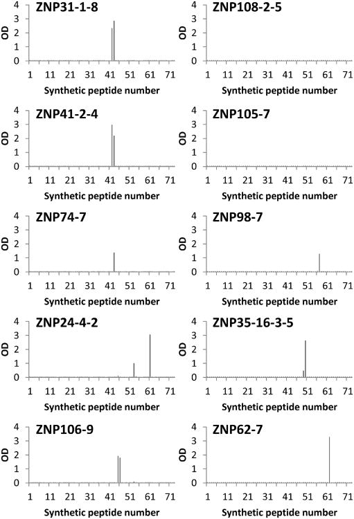
To determine the epitopes recognised by the mAbs, their reactivities to synthetic peptides (20 amino acids in length) were analysed by ELISA. The antigen peptides corresponded to 73 overlapping peptide sequences (10 amino acids overlapped between consecutive peptides) derived from EBOV NP and covered the entire amino acid sequence of this protein. This synthetic peptide-based scanning enabled us to determine some linear antigenic peptide sequences on EBOV NP. Of the 10 mAbs described above, 8 (ZNP31-1-8, ZNP41-2-4, ZNP74-7, ZNP24-4-2, ZNP106-9, ZNP98-7, ZNP35-16-3-5 and ZNP62-7) bound to at least one peptide, whereas 2 (ZNP108-2-5 and ZNP105-7) had no positive reaction (Fig. 2). The amino acid sequences recognised by these 8 mAbs are summarised in Table 2 and Fig. 3. Three highly cross-reactive mAbs, ZNP41-2-4, ZNP31-1-8 and ZNP74-7, strongly reacted to the peptide corresponding to aa positions 421–440. ZNP41-2-4 and ZNP31-1-8 reacted further with the consecutive peptides corresponding to aa positions 411–430, restricting the recognised epitope to 10 amino acids (aa positions 421–430). Another cross-reactive mAb, ZNP24-4-2, bound to two peptides corresponding to very different regions in NP. ZNP106-9 reacted with 2 consecutive peptides with overlapping aa sequences corresponding to aa positions 441–460 and 451–470, sharing the 10 aa at positions 451–460. ZNP98-7, ZNP35-16-3-5 and ZNP62-7 each recognised a single peptide derived from different regions of NP (aa 561–580, aa 491–510 and aa 611–630, respectively).
Fig. 2.
Reactivities of mAbs to EBOV NP-derived synthetic peptides. Seventy-three overlapping peptide sequences (20 aa in length with a 10 aa overlap) covering the entire amino acid sequence of NPof EBOV Mayinga were coated on ELISA plates at a concentration of 100 μg/ml. Purified mAbs were used as primary antibodies at a concentration of 1 μg/ml. OD measurements were determined at 450 nm.
Table 2.
Amino acid sequences important for epitope formation.
| mAb | Peptide sequences recognised by mAb | Amino acid positions |
|---|---|---|
| ZNP31-1-8 ZNP41-2-4 | YDDDDDIPFPa | 421–430a |
| ZNP74-7 | YDDDDDIPFPGPINDDDNPG | 421–440 |
| ZNP24-4-2 | QTQFRPIQNVPGPHRTIHHA | 521–540 |
| TPTVAPPAPVYRDHSEKKEL | 601–620 | |
| ZNP106-9 | DTTIPDVVVDa | 451–460a |
| ZNP98-7 | MLTPINEEADPLDDADDETS | 561–580 |
| ZNP35-16-3-5 | DDEDTKPVPNRSTKGGQQKN | 491–510 |
| ZNP62-7 | YRDHSEKKELPQDEQQDQDH | 611–630 |
Overlapping sequence of 2 consecutive peptides to which the antibodies bound.
Fig. 3.
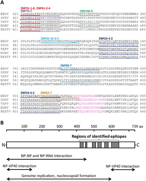
Epitope sequences in amino acid sequence alignment and known functional regions NPs. (A) Amino acid sequences of EBOV, SUDV, TAFV, BDBV and RESTV were obtained from GenBank under accession numbers AF272001, AF173836, FJ217162, FJ217161 and AF522874, respectively. Amino acid sequences at positions 421–660 of each virus are shown. EBOV NP peptides recognised by the mAbs are highlighted with solid lines. Corresponding regions of the other NPs to which each mAb showed strong cross-reactivity are underlined (dashed lines). Amino acid sequences used for producing species-specific rabbit antisera are shown in pink. (B) Locations of the identified epitopes are shown in the schematic diagram of NP. Functional domains (Bharat et al., 2012; Noda et al., 2007; Watanabe et al., 2006) are also shown. (For interpretation of the references to colour in this figure legend, the reader is referred to the web version of the article.)
3.3. Reactivity of rabbit antisera produced by immunisation with synthetic peptides
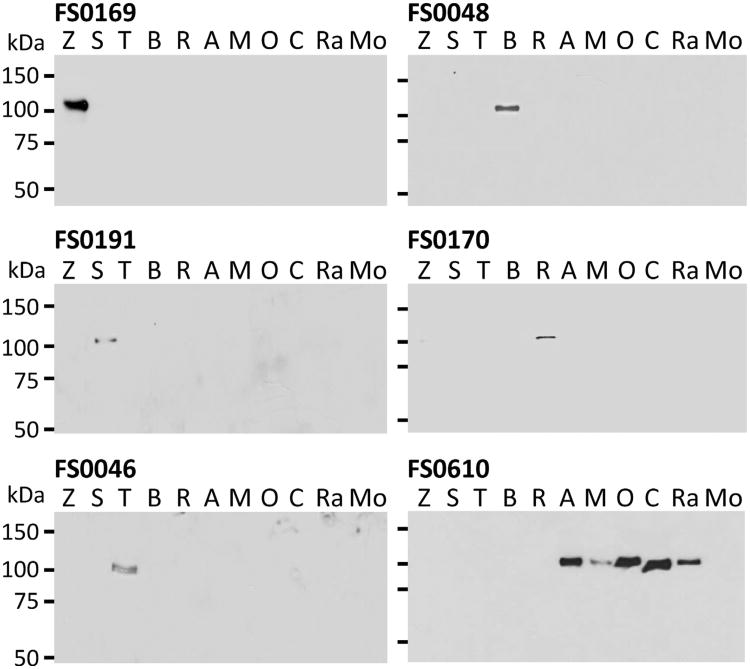
We then sought to determine epitopes distinctive among the NPs of each Ebolavirus and Marburgvirus species. Based on a programme used to predict B-cell epitopes, we selected region around aa positions 630–650 from viruses representing each filovirus species (Fig. 3) (EBOV, SUDV, TAFV, BDBV, RESTV and MARV), and generated rabbit antisera to the respective synthetic peptides as described in Materials and Methods. The reactivity of each antiserum (FS0169, FS0191, FS0046, FS0048, FS0170 and FS0610) was analysed by ELISA (Table 3). According to the high sequence variation in this region among these viruses, the antisera reacted specifically with the homologous NPs, although FS0046 and FS0048 (antisera to TAFV and BDBV, respectively), showed limited cross-reactivity to RESTV NP. The virus specificity was further confirmed using filovirus lysates in Western blotting (Fig. 4). Notably, all the virus strains tested within the genus Marburgvirus (including RAVV) were recognised by antiserum FS0610. These results indicated that the region around aa 630–650 in filovirus NP served as a filovirus species-specific epitope.
Table 3.
Reactivity of rabbit antisera produced by immunisation with synthetic peptides.
| Antiserum | Synthetic peptide used for immunisation (amino acid sequence) | EBOVa | SUDV | TAFV | BDBV | RESTV | MARV |
|---|---|---|---|---|---|---|---|
| FS0169 | EBOV NP 628-638 (QDHTQEARNQD) | ++b | − | ||||
| FS0191 | SUDV NP 631-644 (QGSESEALPINSKK) | − | ++ | − | − | − | − |
| FS0046 | TAFV NP 630-643 (NQVSGSENTDNKPH) | − | − | ++ | − | + | − |
| FS0048 | BDBV NP 628-641 (QSNQTNNEDNVRNN) | − | − | − | ++ | + | − |
| FS0170 | RESTV NP 630-643 (TSQLNEDPDIGQSK) | − | − | − | − | + | − |
| FS0610 | MARV NP 635-652 (RVVTKKGRTFLYPNDLLQ) | − | − | − | − | − | ++ |
VLPs of each virus species were used as antigens.
Antibody reactivity was evaluated based on ELISA OD450 values at a serum dilution of 1:2000. ++, OD> 1.0; +, 0.5<OD<1; −, OD<0.5.
Fig. 4.
Reactivity of rabbit antisera in Western blotting. Rabbit antisera (FS0169, FS0191, FS0046, FS0048, FS0170 and FS0610) were produced using synthetic peptides derived from EBOV, SUDV, TAFV, BDBV, RESTV and MARV, respectively. Experimental conditions were the same as in Fig. 1. EBOV (Z), SUDV (S), TAFV (T), BDBV (B), RESTV (R), MARV Angola (A), MARV Musoke (M), MARV Ozolin (O), MARV Ci67 (C), RAVV (Ra). Mo, mock-infected.
4. Discussion
Using mouse mAbs and synthetic peptide-based scanning, we determined 2 highly conserved antigenic regions (aa 421–440 and aa 601–620) serving as linear epitopes in the filovirus NP (Fig. 3). In addition, a stretch of 10 amino acids at aa 421–430 (YDDDDDIPFP) was found to be important for 3 mAbs (ZNP31-1-8, ZNP41-2-4 and ZNP74-7), which strongly recognised all known Ebolavirus species. This finding is consistent with a previous study demonstrating that mAbs reactive to EBOV, RESTV and SUDV recognised the sequence at aa 424–430 (Niikura et al., 2003). In this specific region, the amino acid sequence IPFP is completely conserved among all analysed viruses in the Ebolavirus genus, suggesting that these aa residues are crucial for conformation of this common epitope.
ZNP24-4-2 was highly cross-reactive to all known viruses of the genus Ebolavirus, with weaker reactivity to SUDV (Table 1 and Fig. 1). This mAb reacted with two different peptides corresponding to aa 521–540 and aa 601–620 (Fig. 2).These two peptide sequences may be parts of a conformational epitope. However, there is no conserved sequence in the region at aa 521-540 among all the analysed viruses, whereas the sequence at aa 601–620 shows some conservation. Although SUDV was only weakly recognised by this mAb, this conserved region might be required for recognition as a conserved epitope.
ZNP106-9 and ZNP108-2-5 were strongly reactive to EBOV, TAFV and BDBV, but only weakly reactive or nonreactive to SUDV and RESTV, respectively. This reactivity pattern is consistent with the phylogenetic relationship among the viruses (Towner et al., 2008). Only ZNP106-9 reacted with the peptide sequence D451TTIPDVVVD460, demonstrating that ZNP108-2-5 recognises a different epitope. The amino acid sequence alignment of this region suggests that D456 in EBOV, TAFV and BDBV is critical for the ZNP106-9 specificity to these viruses, since SUDV and RESTV have G or N at this aa position, respectively (Fig. 3).
ZNP35-16-3-5 and ZNP62-7 recognised EBOV only, and bound to aa 611–630 and aa 491–510, respectively. According to the sequence variation among the analysed viruses, these aa likely form EBOV-specific epitopes. It can be speculated that the same region of NP of the other viruses in the Ebolaviruse genus forms species-specific epitopes. In addition to these two regions, the success of the production of antisera to the synthetic peptides with the predicted sequences around aa 630–650 provided further information on the filovirus species-specific epitopes. The antigenic region of EBOV NP was previously shown to be located in the C-terminal half of the protein (Saijo et al., 2001). The N-terminal aa 1-451 of the EBOV NP assemble into a condensed helix, which forms the inner structure of the viral nucleocapsid (Bharat et al., 2012). The amino acid residues in this region are highly conserved among the known viruses in the genus Ebolavirus. It is likely that this region forms functionally important structures inside the NP molecule, and as a result, has limited antigenic properties. This is consistent with our results in which most antigenic regions were found in the highly variable C-terminal region starting at aa 451 (Fig. 3). The only epitope found on the condensed helix structure was the one recognised by ZNP31-1-8, ZNP41-2-4 and ZNP74-7, mAbs cross-reactive to all known Ebolavirus species.
In this study, we established a panel of NP-specific mAbs divided into 7 groups based on their cross-reactivity profiles to all known viruses of the genus Ebolavirus. Using synthetic peptide-based screening, 8 antigenic regions in the EBOV NP molecule, each consisting of roughly 10-20 aa residues, were determined. These well-characterised mAbs with detailed epitope information should be useful for the development of filovirus antigen detection assays such as immunochromatography-based rapid antigen diagnosis.
Acknowledgments
We thank Dr. Hideki Ebihara (National Institute of Allergy and Infectious Diseases, National Institutes of Health, Rocky Mountain Laboratories) for valuable advice and Kim Barrymore for editing the manuscript. This work was supported by the Japan Science and Technology Agency within the framework of the Science and Technology Research Partnership for Sustainable Development (SATREPS) and by the Japan Initiative for Global Research Network on Infectious Diseases (J-GRID). Funding was also provided by a Grant-in-Aid from the Ministry of Health, Labour and Welfare of Japan, and in part by the Intramural Research Program of the National Institute of Allergy and Infectious Diseases (NIAID), National Institutes of Health (NIH).
Abbreviations
- mAb
monoclonal antibodies
- EBOV
Ebola virus
- SUDV
Sudan virus
- TAFV
Taï Forest virus
- BDBV
Bundibugyo virus
- RESTV
Reston virus
- MARV
Marburg virus
- RAVV
Ravn virus
- NP
nucleoprotein
- VP
viral protein
- GP
glycoprotein
- VLP
virus-like particle
References
- Bente D, Gren J, Strong JE, Feldmann H. Disease modeling for Ebola and Marburg viruses. Disease Models and Mechanisms. 2009;2:12–17. doi: 10.1242/dmm.000471. [DOI] [PMC free article] [PubMed] [Google Scholar]
- Bharat TA, Noda T, Riches JD, Kraehling V, Kolesnikova L, Becker S, Kawaoka Y, Briggs JA. Structural dissection of Ebola virus and its assembly determinants using cryo-electron tomography. Proceedings of the National Academy of Sciences of the United States of America. 2012;109:4275–4280. doi: 10.1073/pnas.1120453109. [DOI] [PMC free article] [PubMed] [Google Scholar]
- Gershoni JM, Roitburd-Berman A, Siman-Tov DD, Tarnovitski FN, Weiss Y. Epitope mapping: the first step in developing epitope-based vaccines. BioDrugs. 2007;21:145–156. doi: 10.2165/00063030-200721030-00002. [DOI] [PMC free article] [PubMed] [Google Scholar]
- Ikegami T, Niikura M, Saijo M, Miranda ME, Calaor AB, Hernandez M, Acosta LP, Manalo DL, Kurane I, Yoshikawa Y, Morikawa S. Antigen capture enzyme-linked immunosorbent assay fors pecific detection of Reston Ebolavirus nucleoprotein. Clinical and Diagnostic Laboratory Immunology. 2003;10:552–557. doi: 10.1128/CDLI.10.4.552-557.2003. [DOI] [PMC free article] [PubMed] [Google Scholar]
- Kuhn JH, Becker S, Ebihara H, Geisbert TW, Johnson KM, Kawaoka Y, Lipkin WI, Negredo AI, Netesov SV, Nichol ST, Palacios G, Peters CJ, Tenorio A, Volchkov VE, Jahrling PB. Proposal for a revised taxonomy of the family Filoviridae: classification, names of taxa and viruses, and virus abbreviations. Archives of Virology. 2010;155:2083–2103. doi: 10.1007/s00705-010-0814-x. [DOI] [PMC free article] [PubMed] [Google Scholar]
- Licata JM, Johnson RF, Han Z, Harty RN. Contribution of ebola virus glycoprotein, nucleoprotein, and VP24 to budding of VP40 virus-like particles. Journal of Virology. 2004;78:7344–7351. doi: 10.1128/JVI.78.14.7344-7351.2004. [DOI] [PMC free article] [PubMed] [Google Scholar]
- Nakayama E, Tomabechi D, Matsuno K, Kishida N, Yoshida R, Feldmann H, Takada A. Antibody-dependent enhancement of Marburg virus infection. Journal of Infectious Diseases. 2011;204(Suppl. 3):S978L S985. doi: 10.1093/infdis/jir334. [DOI] [PMC free article] [PubMed] [Google Scholar]
- Nakayama E, Yokoyama A, Miyamoto H, Igarashi M, Kishida N, Matsuno K, Marzi A, Feldmann H, Ito K, Saijo M, Takada A. Enzyme-linked immunosorbent assay for detection of filovirus species-specific antibodies. Clinical and Vaccine Immunology. 2010;17:1723–1728. doi: 10.1128/CVI.00170-10. [DOI] [PMC free article] [PubMed] [Google Scholar]
- Negredo A, Palacios G, Vazquez-Moron S, Gonzalez F, Dopazo H, Molero F, Juste J, Quetglas J, Savji N, de la Cruz MM, Herrera JE, Pizarro M, Hutchison SK, Echevarria JE, Lipkin WI, Tenorio A. Discovery of an ebolavirus-like filovirus in europe. PLOS Pathogens. 2011;7:e1002304. doi: 10.1371/journal.ppat.1002304. [DOI] [PMC free article] [PubMed] [Google Scholar]
- Nidom CA, Nakayama E, Nidom RV, Alamudi MY, Daulay S, Dharmayanti IN, Dachlan YP, Amin M, Igarashi M, Miyamoto H, Yoshida R, Takada A. Serological evidence of ebola virus infection in Indonesian orangutans. PLoS ONE. 2012;7:e40740. doi: 10.1371/journal.pone.0040740. [DOI] [PMC free article] [PubMed] [Google Scholar]
- Niikura M, Ikegami T, Saijo M, Kurane I, Miranda ME, Morikawa S. Detection of Ebola viral antigen by enzyme-linked immunosorbent assay using a novel monoclonal antibody to nucleoprotein. Journal of Clinical Microbiology. 2001;39:3267–3271. doi: 10.1128/JCM.39.9.3267-3271.2001. [DOI] [PMC free article] [PubMed] [Google Scholar]
- Niikura M, Ikegami T, Saijo M, Kurata T, Kurane I, Morikawa S. Analysis of linear B-cell epitopes of the nucleoprotein of ebola virus that distinguish ebola virus subtypes. Clinical and Diagnostic Laboratory Immunology. 2003;10:83–87. doi: 10.1128/CDLI.10.1.83-87.2003. [DOI] [PMC free article] [PubMed] [Google Scholar]
- Noda T, Hagiwara K, Sagara H, Kawaoka Y. Characterization of the Ebola virus nucleoprotein-RNA complex. Journal of General Virology. 2010;91:1478–1483. doi: 10.1099/vir.0.019794-0. [DOI] [PMC free article] [PubMed] [Google Scholar]
- Noda T, Watanabe S, Sagara H, Kawaoka Y. Mapping of the VP40-binding regions of the nucleoprotein of Ebola virus. Journal of Virology. 2007;81:3554–3562. doi: 10.1128/JVI.02183-06. [DOI] [PMC free article] [PubMed] [Google Scholar]
- Pittalis S, Fusco FM, Lanini S, Nisii C, Puro V, Lauria FN, Ippolito G. Case definition for Ebola and Marburg haemorrhagic fevers: a complex challenge for epidemiologists and clinicians. New Microbiologica. 2009;32:359–367. [PubMed] [Google Scholar]
- Qiu X, Alimonti JB, Melito PL, Fernando L, Stroher U, Jones SM. Characterization of Zaire ebolavirus glycoprotein-specific monoclonal antibodies. Clinical Immunology. 2011;141:218–227. doi: 10.1016/j.clim.2011.08.008. [DOI] [PubMed] [Google Scholar]
- Saijo M, Niikura M, Morikawa S, Ksiazek TG, Meyer RF, Peters CJ, Kurane I. Enzyme-linked immunosorbent assays for detection of antibodies to Ebola and Marburg viruses using recombinant nucleoproteins. Journal of Clinical Microbiology. 2001;39:1–7. doi: 10.1128/JCM.39.1.1-7.2001. [DOI] [PMC free article] [PubMed] [Google Scholar]
- Sanchez A, Geisbert TW, Feldmann H. Filoviridae: Marburg and Ebola Viruses. In: Knipe DM, Howley PM, Griffin DE, Lamb RA, Martin MA, Roizman B, Straus SE, editors. Fields Virology. Lippincott Williams & Wilkins; Philadelphia: 2007. pp. 1409–1448. [Google Scholar]
- Shahhosseini S, Das D, Qiu X, Feldmann H, Jones SM, Suresh MR. Production and characterization of monoclonal antibodies against different epitopes of Ebola virus antigens. Journal of Virological Methods. 2007;143:29–37. doi: 10.1016/j.jviromet.2007.02.004. [DOI] [PubMed] [Google Scholar]
- Towner JS, Sealy TK, Khristova ML, Albarino CG, Conlan S, Reeder SA, Quan PL, Lipkin WI, Downing R, Tappero JW, Okware S, Lutwama J, Bakamutumaho B, Kayiwa J, Comer JA, Rollin PE, Ksiazek TG, Nichol ST. Newly discovered ebola virus associated with hemorrhagic fever outbreak in Uganda. PLoS Pathogens. 2008;4:e1000212. doi: 10.1371/journal.ppat.1000212. [DOI] [PMC free article] [PubMed] [Google Scholar]
- Toyoda T, Masunaga K, Ohtsu Y, Hara K, Hamada N, Kashiwagi T, Iwahashi J. Antibody-scanning and epitope-tagging methods; molecular mapping of proteins using antibodies. Current Protein and Peptide Science. 2000;1:303–308. doi: 10.2174/1389203003381360. [DOI] [PubMed] [Google Scholar]
- Urata S, Noda T, Kawaoka Y, Morikawa S, Yokosawa H, Yasuda J. Interaction of Tsg101 with Marburg virus VP40 depends on the PPPY motif, but not the PT/SAP motif as in the case of Ebola virus, and Tsg101 plays a critical role in the budding of Marburg virus-like particles induced by VP40, NP, and GP. Journal of Virology. 2007;81:4895–4899. doi: 10.1128/JVI.02829-06. [DOI] [PMC free article] [PubMed] [Google Scholar]
- Watanabe S, Noda T, Kawaoka Y. Functional mapping of the nucleoprotein of Ebola virus. Journal of Virology. 2006;80:3743–3751. doi: 10.1128/JVI.80.8.3743-3751.2006. [DOI] [PMC free article] [PubMed] [Google Scholar]


