Abstract
We previously discovered that nuclear import of high risk HPV16 E7 is mediated by a cNLS located within the zinc-binding domain via a pathway that is independent of karyopherins/importins (Angeline et al., 2003; Knapp et al., 2009). In this study we continued our characterization of the cNLS and nuclear import pathway of HPV16 E7. We find that an intact zinc-binding domain is essential for the cNLS function in mediating nuclear import of HPV16 E7. Mutagenesis of cysteine residues to alanine in each of the two CysXXCys motifs involved in zinc-binding changes the nuclear localization of the EGFP-16E7 and 2xEGFP-16E7 mutants. We further discover that a patch of hydrophobic residues, 65LRLCV69, within the zinc-binding domain of HPV16 E7 mediates its nuclear import via hydrophobic interactions with the FG domain of the central channel nucleoporin Nup62.
Introduction
Human Papillomaviruses (HPVs) are estimated to be the most common sexually transmitted infection in the United States. The largest group of HPVs are the alpha HPVs, and consists of both cutaneous viruses causing common warts, as well as the approximately 40 mucosal types known to infect the cervical epithelium. Mucosal HPVs are further divided into high and low risk groups, dependent upon the frequency with which they have been linked to the malignant progression of their resultant lesions (Doorbar, 2006; zur Hausen, 2000, 2009). Fifteen of the sexually transmitted genital HPVs can be classified as high risk, notably HPV16, HPV18, HPV31, HPV33 and HPV45, which may result in squamous intraepithelial lesions capable of progressing to invasive carcinomas (Doorbar, 2006; Munger et al., 2004; zur Hausen, 2000, 2009). Nearly 99% of cervical cancers and 20% of oropharynx cancers are positive for high risk HPV DNA, with HPV16 found in nearly 63% of the cervical cancers (Doorbar, 2006).
HPVs are dependent on the replication machinery of the host cells and consequently they have evolved the E7 oncoproteins to induce reentry into the S phase of the differentiated epithelial cells and establish the appropriate environment required to support viral DNA amplification. To accomplish this, the high risk HPV16/18 E7 oncoproteins bind and destabilize the retinoblastoma protein (pRB) and the RB-related pocket proteins, p130 and p107. E7 oncoproteins interact also with other components of the cell cycle machinery, such as cyclin A, cyclin E and the cyclin-dependent kinase inhibitors p27 and p21 (Jones and Munger,1996; Zwerschke and Jansen-Durr, 2000; McLaughlin-Drubin and Munger, 2009). In addition to these nuclear target proteins, HPV16 E7 binds to targets in the cytoplasm, such as the microtubule-associated N-end rule ubiqutin ligase p600 (Huh et al., 2005) and the nuclear mitotic apparatus protein (NuMA) (Nguyen and Munger, 2009).
Structurally, the E7 proteins consist of three biochemically distinct domains. Both NMR and X-ray crystal structures have been solved for the C terminal domain of E7, and the three-dimensional structure has been shown to organize into a tightly packed zinc-binding fold (McLaughlin-Drubin and Munger, 2009). The N terminus conserved regions (CR) CR1 (aa1-15) and CR2 (aa16-37) of HPV16 E7 possess significant sequence similarity to a portion of CR1 and the entirety of CR2 of adenovirus E1A and related sequences of SV40 large tumor antigen, as well as functional similarity, contributing to the transforming ability of HPV16 E7. The CR2 domain contains both the Leu-X-Cys-XGlu (LxCxE) pRB binding motif as well as a consensus casein kinase II (CKII) phosphorylation site. The C-terminal CR3 domain (aa38-98) contains a zinc-binding domain with two Cys-X-X-Cys motifs separated by 29 amino acids (McLaughlin-Drubin and Munger, 2009).
High risk HPV16 E7 is predominantly nuclear in the CaSki cervical carcinoma cell line, when expressed transiently in HaCaT and U2OS cells, and in invasive cervical carcinoma (Guccione et al., 2002; Fiedler et al., 2004; Cid-Arregui et al., 2003). We have previously discovered that nuclear import of HPV16 E7 and HPV11 E7 is mediated by a Ran-dependent pathway that is independent of karyopherins/importins and it is mediated by their cNLS located within the unique zinc-binding domain (Angeline et al., 2003; Knapp et al., 2009; Piccioli et al., 2010). Both HPV16 E7 and HPV11 E7 proteins have also a leucine-rich nuclear export signal (NES) located within the zinc-binding domain that mediates their nuclear export via a CRM1 pathway (Knapp et al., 2009; McKee et al., 2013).
Nuclear import and export of different cargoes take place through nuclear pore complexes (NPCs) embedded at the nuclear envelope. There are some 30 nucleoporins (Nups) that assemble to form the NPC and they can be classified into: transmembrane Nups (Poms) that anchor the NPCs at the nuclear envelope, structural Nups, and FG-Nups containing phenylalanine-glycine (FG) repeats, and involved in nuclear import and export (Terry and Wente, 2009). Transport through the NPCs can be via passive diffusion for cargoes up to approximately 40 kDa, active transport mediated by karyopherins (importins and exportins) interacting with FG-Nups for cargoes over 40 kDa, and direct interaction of some cargoes with FG-Nups bypassing the requirement for karyopherins (Terry and Wente, 2009). The central channel for nucleocytoplasmic transport consists of three nucleoporins, Nup62, Nup58 and Nup54 and the structures of the Nup62-Nup54 and Nup54-Nup58 interacting domains has been recently determined revealing the architecture of the mammalian transport channel (Solmaz et al., 2011).
In this study we continue our characterization of the cNLS and nuclear import pathway of high risk HPV16 E7 oncoprotein. We determine that an intact zinc-binding domain within the CR3 domain is essential for the cNLS function in mediating nuclear import of HPV16 E7. Mutagenesis of Cys residues in each of the two CysXXCys motifs involved in zinc binding changes the nuclear localization of the resultant EGFP-16E7 and 2xEGFP-16E7 mutants. We further discover that a patch of hydrophobic residues, 65LRLCV69, within the zinc-binding domain of HPV16 E7 mediates its nuclear import via direct hydrophobic interactions with the FG domain of the channel nucleoporin, Nup62.
Results
The zinc-binding domain of HPV16 E7 is essential for its nuclear localization
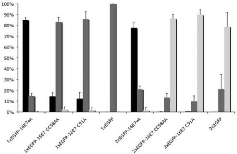
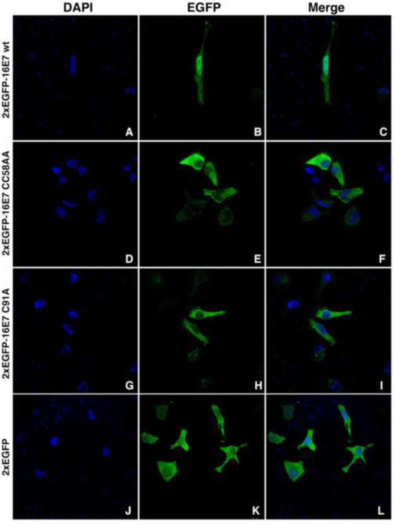
We have previously established the presence of a C-terminal NLS in the CR3 domain of HPV16 E7 containing the zinc-binding fold that consists of two copies of a Cys-X-X-Cys motif separated by 29 amino acids (Knapp et al., 2009). Moreover, we have determined that the zinc-binding domain of HPV11 E7 is essential for its nuclear import and localization (Piccioli et al., 2010). To examine the possibility that the intact zinc-binding domain of HPV16 E7 is essential for the nuclear import activity of the cNLS of HPV16 E7 we have now performed site-directed mutagenesis of cysteine residues in each of the two copies of Cys-X-X-Cys in the context of EGFP-16E7 and 2xEGFP-16E7 generating the CC58/59AA and C91A mutants. The CC58/59AA mutant is designated CC58AA for simplicity. HeLa cells were transiently transfected with the wild type EGFP-16E7, the EGFP-16E7CC58AA and EGFP-16E7C91A cysteine mutants and EGFP control, and the cellular localization of the expressed proteins was examined via confocal fluorescence microscopy. As previously reported, the wild type EGFP-16E7 had a nuclear localization in the majority of transfected cells with the rest of cells having a pancellular phenotype (Fig. 1); EGFP had a pancellular localization in all the transfected cells (Fig. 1). Both the EGFP-16E7CC58AA and EGFP-16E7C91A cysteine mutants exhibited a pancellular localization, similar with that of EGFP, in the majority of transfected cells, 83.2% +/− 4.1%, and 85.8% +/− 6.9% respectively (Fig. 1). These data indicate that, while disruption of the zinc-binding domain in the context of EGFP-16E7 led to a change in the localization from mostly nuclear to pancellular, it did not completely prevent the EGFP-16E7CC58AA or EGFP-16E7C91A mutants from gaining some entry to the nucleus. As the cysteine mutations can affect the dimerization of 16E7, and in the absence of dimerization the EGFP-16E7CC58AA and EGFP-16E7C91A mutants could enter by passive diffusion (as the EGFP itself), we generated the same mutants in the context of 2xEGFP-16E7 (containing a 2xEGFP module) to exceed the limit for passive diffusion across the nuclear pore complex. HeLa cells were transiently transfected with the 2xEGFP-16E7CC58AA and 2xEGFP-16E7C91A mutants, along with the 2xEGFP-16E7 wild type and 2xEGFP control. As previously reported the 2xEGFP-16E7 wild type had a predominantly nuclear localization in the majority of cells (Fig. 1 and Fig. 2, panels B and C) and 2xEGFP had a cytoplasmic localization (Fig. 1 and Fig. 2, panels K and L). Significantly, the 2xEGFP-16E7CC58AA and 2xEGFP-16E7C91A, exhibited predominantly cytoplasmic localization in the majority of transfected cells, 86.4% +/− 3.6% and 89.8% +/− 5.1%, respectively (Fig. 1 and Fig. 2, panels E, F, H and I). Together these data support the conclusion that the nuclear import activity of the cNLS of HPV16 E7 is dependent upon an intact zinc-binding domain. The loss of zinc coordination in the cysteine mutants can lead to structural changes in the zinc-binding domain that would make unavailable the critical residues of the cNLS.
Fig. 1. Quantitative analysis of the effect of mutations of the zinc coordinating cysteine residues to alanine on the nuclear localization of EGFP-16E7 and 2xEGFP-16E7.
Data from five experiments using EGFP-16E7, EGFP-16E7CC58AA, EGFP-16E7C91A, EGFP, 2xEGFP-16E7, 2xEGFP-16E7CC58AA, 2xEGFP-16E7C91A, and 2xEGFP plasmids were used for quantitative analysis and graphic representation of the localization distribution of transfected HeLa cells. Black bars represent cells exhibiting predominant nuclear localization; gray bars represent cells exhibiting pancellular localization; white bars represent cells exhibiting predominant cytoplasmic localization.
Fig. 2. Mutation of the zinc coordinating cysteine residues to alanine changes the nuclear localization of 2xEGFP-16E7.
HeLa cells were transfected with either 2xEGFP-16E7 wild type (panels A-C), 2xEGFP-16E7CC58AA (panels D-F), 2xEGFP-16E7C91A (panels G-I), or 2xEGFP (panels J-L) plasmids and examined by confocal fluorescence microscopy at 24 h post transfection. Panels A, D, G and J represent the DAPI staining of the nuclei, panels B, E, H and K represent the fluorescence of the EGFP and panels C, F, I, and L represent the merge.
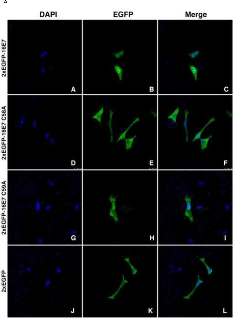
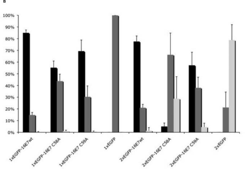
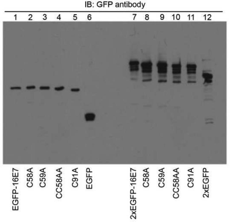
We have previously showed that a cysteine residue at position 59 - conserved between the high risk and low risk HPVs, and not involved in coordinating the zinc ion - plays some role in the cNLS function of HPV11 E7 (Piccioli et al., 2010). To examine if this conserved cysteine residue performs a similar function in HPV16 E7, site directed mutagenesis was used to generate a pair of single amino acid substitutions of the N-terminal cysteines within the zinc-binding domain in the context of 2xEGFP-16E7. The 2xEGFP-16E7 wild type, 2xEGFP-16E7 single cysteine mutants and 2xEGFP, were transiently transfected into HeLa cells and their resultant subcellular localizations were examined via confocal fluorescence microscopy. Both 2xEGFP-16E7 wild type and 2xEGFP-16E7C59A exhibited predominantly nuclear localization (Fig. 3A, panels B and C; and panels H and I), although at reduced levels for the 2xEGFP-16E7C59A mutant (57.3% +/− 11.1%) (Fig. 3B). In contrast, the 2xEGFP-16E7C58A exhibited predominantly pancellular localization (Fig. 3A, panels E, and F) in 66.3% +/− 18.6% cells and also cytoplasmic localization in 28.8% +/−18.8% cells (Fig. 3B). In the context of 1xEGFP, the reductions in nuclear localization caused by the C58A and C59A mutations were more modest. EGFP-16E7C58A and EGFP-16E7C59A were predominantly nuclear in 55.3% +/− 5.7% and 69.3% +/−9.5% transfected cells with the rest of cells exhibiting a pancellular phenotype (Fig. 3B); this is in comparison with 85% +/− 2.4% cells showing nuclear localization for the EGFP-16E7 wild type (Fig. 3B). Immunoblot analyses of the expressed EGFP-16E7, 2xEGFP-16E7 and the different cysteine mutants performed with a GFP antibody indicated that the wild type and mutant proteins were expressed properly at similar levels (Fig. 4). These data suggest that cysteine 59 can partially compensate for the loss of cysteine 58 in coordinating the zinc ion and maintaining the zinc-binding domain and nuclear localization, and also that cysteine 59 may play a very modest role in the cNLS function.
Fig. 3.
A. Single cysteine substitution to alanine within the zinc finger alters the nuclear localization of 2xEGFP-16E7. HeLa cells were transfected with either 2xEGFP-16E7 wild type (panels A-C), 2xEGFP-16E7C58A (panels D-F), 2xEGFP-16E7C59A (panels G-I), or 2xEGFP (panels J-L) plasmids and examined by confocal fluorescence microscopy at 24 h post-transfection. Panels A, D, G and J show the DAPI staining of the nuclei, panels B, E, H and K represent the fluorescence of the EGFP and panels C, F, I, and L represent the merge. B. Quantitative analysis of the effect of single cysteine substitution within the zinc finger on the localization of EGFP-16E7 and 2xEGFP-16E7. Data from five experiments using EGFP-16E7, EGFP-16E7C58A, EGFP-16E7C59A, EGFP, 2xEGFP-16E7, 2xEGFP-16E7C58A, 2xEGFP-16E7C59A, and 2xEGFP plasmids were used for quantitative analysis and graphic representation of the localization distribution of transfected HeLa cells. Black bars represent cells exhibiting predominant nuclear localization; gray bars represent cells exhibiting pancellular localization; white bars represent cells exhibiting predominant cytoplasmic localization.
Fig. 4. The EGFP-16E7 and 2xEGFP-16E7 cysteine mutants are properly expressed in HeLa cells.
HeLa cells were transfected with EGFP-16E7 (lane 1), EGFP-16E7C58A (lane 2), EGFP-16E7C59A (lane 3), EGFP-16E7CC58AA (lane 4), EGFP-16E7C91A (lane 5), EGFP (lane 6), 2xEGFP-16E7 (lane 7), 2xEGFP-16E7C58A (lane 8), 2xEGFP-16E7C59A (lane 9), 2xEGFP-16E7CC58AA (lane 10), 2xEGFP-16E7C91A (lane 11), or 2xEGFP (lane 12) plasmids. Cell lysates were prepared 24 h post transfection and probed with a GFP antibody.
A hydrophobic patch within the zinc-binding domain is essential for the nuclear localization of HPV16 E7
The surface hydrophobicity of karyopherins/importins is sufficient to provide access to the NPC and translocation is achieved through a series of non-specific interactions between short hydrophobic patches on the surface of the karyopherins and the FG-Nups within the NPC (Naim et al., 2009). We have previously identified a small hydrophobic patch of amino acids, 65VRLVV69, within the zinc-binding domain of HPV11 E7 that was required for its nuclear localization (McKee et al., 2013). A small hydrophobic patch within the zinc-binding domain of HPV16 E7, 65LRLCV69, has significant homology to the patch previously characterized in HPV11 E7, 65VRLVV69 (McKee et al., 2013). To examine the possibility that this hydrophobic patch of amino acids within the zinc-binding domain of HPV16 E7 is required for mediating its nuclear import we used site-directed mutagenesis to replace all five amino acids with alanine and generated the LRLCV65-69AAAAA mutation in the context of 2xEGFP-16E7 and GST-16cE7. The LRLCV65-69AAAAA mutation is designated LRLCV65AAAAA, again for simplicity. We also generated single mutants by substituting the arginine at position 66 with alanine in the context of 2xEGFP-16E7 and GST-16cE7.
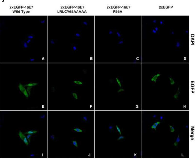
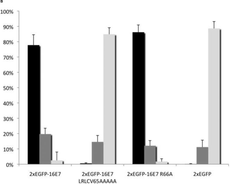
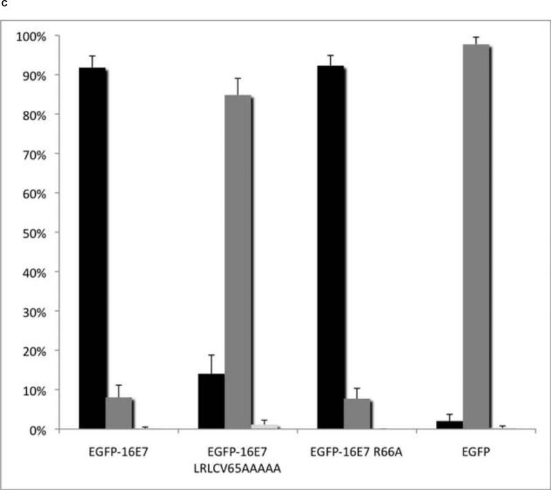
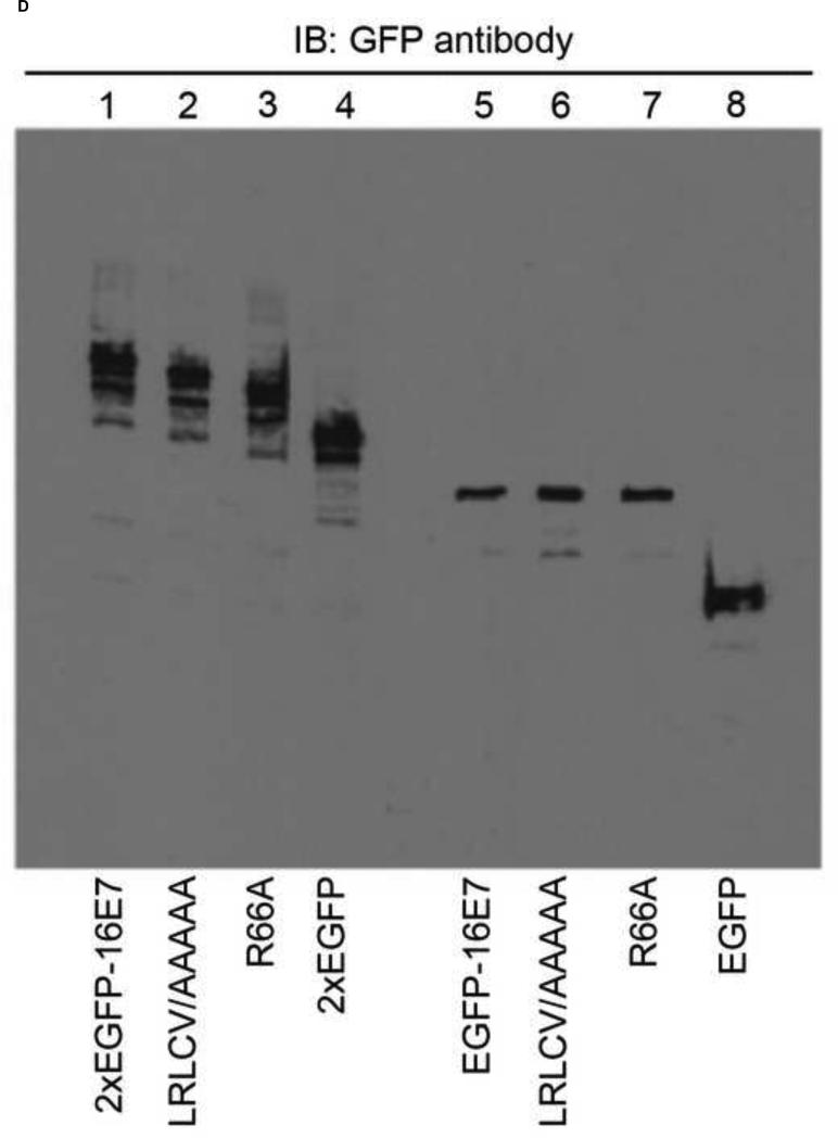
HeLa cells were transiently transfected with the 2xEGFP-16E7LRLCV65AAAAA and 2xEGFP-16E7R66A mutants, along with the 2xEGFP-16E7 wild type and 2xEGFP and the localization of the resultant proteins was examined via confocal fluorescence microscopy. As previously shown the 2xEGFP-16E7 wild type was nuclear in the majority of cells (Fig. 5A, panels E and I; and Fig. 5B) whereas the 2xEGFP was cytoplasmic in all the transfected cells (Fig. 5A, panels H and L; and Fig. 5B). In contrast with the nuclear localization of the wild type, the 2xEGFP-16E7LRLCV65AAAAA mutant was cytoplasmic in 84.9% +/− 4.3% of transfected cells, with the rest of cells having a pancellular localization (Fig. 5A, panels F and J; and Fig. 5B). The 2xEGFP-16E7R66A mutant had a similar nuclear localization as the wild type in the majority of cells (86.2% +/− 4.2%) (Fig. 5A, panels G and K; and Fig. 5B). In the context of EGFP-16E7, 84.8% +/− 4.2% of EGFP-16E7LRLCV65AAAAA expressing cells exhibited a pancellular phenotype similarly as the EGFP itself (Fig. 5C), whereas 92.3% +/− 2.6% of EGFP-16E7R66A expressing cells exhibited a predominantly nuclear phenotype (Fig. 5C). Together these data suggest that these hydrophobic residues, 65LRLCV69, are responsible for the nuclear localization of 2xEGFP-16E7 and EGFP-16E7 whereas the positively charged arginine 66 residue plays no significant role in the function of the cNLS. To ensure that the results observed were not due to degradation of the mutants, immunoblot analysis with an anti-GFP antibody was performed. The data indicate that all the 2xEGFP-16E7 and EGFP-16E7 mutant proteins were expressed at the correct molecular weight and at similar levels with their corresponding wild type proteins; although the 2xEGFP-16E7 mutants show a certain level of proteolytic degradation, it is at the same level as for the 2xEGFP-16E7 wild type (Fig. 5D).
Fig. 5.
A. Mutation of hydrophobic residues within the zinc-binding domain disrupts the nuclear localization of 2xEGFP-16E7. HeLa cells were transfected with either 2xEGFP-16E7 wild type (panels A, E, and I), 2xEGFP-16E7LRLCV65AAAAA (panels B, F, and J), 2xEGFP-16E7R66A (panels C, G, and K), or 2xEGFP (panels D, H, and L) plasmids and examined by confocal fluorescence microscopy at 24 h post-transfection. Panels A-D represent DAPI staining of the nuclei, panels E-H represent EGFP fluorescence, and panels I-L represent the merge. B. Quantitative analysis of the effect of mutation of hydrophobic residues within the zinc-binding domain on the localization of 2xEGFP-16E7 in HeLa cells. Data from seven experiments using 2xEGFP-16E7 wild type, 2xEGFP-16E7LRLCV65AAAAA, 2xEGFP-16E7R66A, and 2xEGFP plasmids were used for quantitative analysis and graphic representation of the localization distribution of transfected HeLa cells. Black bars represent cells exhibiting predominant nuclear localization; gray bars represent cells exhibiting pancellular localization; white bars represent cells exhibiting predominant cytoplasmic localization. C. Quantitative analysis of the effect of mutation of hydrophobic residues within the zinc-binding domain on the localization of EGFP-16E7 in HeLa cells. Data from five experiments using EGFP-16E7 wild type, EGFP-16E7LRLCV65AAAAA, EGFP-16E7R66A, and EGFP plasmids were used for quantitative analysis and graphic representation of the localization distribution of transfected HeLa cells. Black bars represent cells exhibiting predominant nuclear localization; gray bars represent cells exhibiting pancellular localization; white bars represent cells exhibiting predominant cytoplasmic localization. D. EGFP-16E7 and 2xEGFP-16E7 hydrophobic residues and R66A mutants are properly expressed in HeLa cells. HeLa cells were transfected with 2xEGFP-16E7 (lane 1), 2xEGFP-16E7LRLCV65AAAAA (lane 2), 2xEGFP-16E7R66A (lane 3), 2xEGFP (lane 4), EGFP-16E7 (lane 5), EGFP-16E7LRLCV65AAAAA (lane 6), EGFP-16E7R66A (lane 7), or EGFP (lane 8) plasmids. Cell lysates were prepared 24 h post transfection and probed with a GFP antibody.
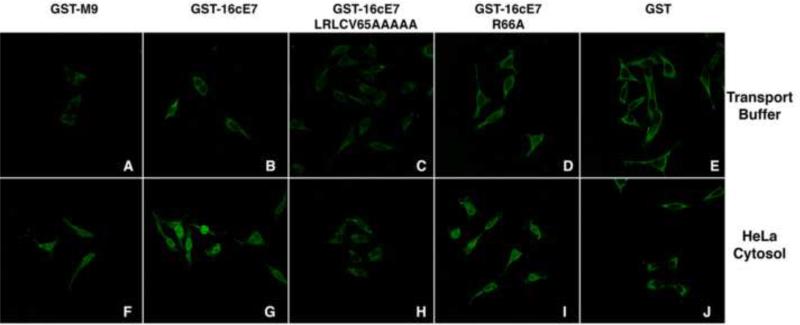
To further confirm this conclusion drawn from the transient transfection experiments in HeLa cells, in vitro nuclear import assays were performed using the same mutants in the context of GST-16cE7. In these experiments, digitonin permeablized HeLa cells were incubated with GST-16cE7, GST-16cE7LRLCV65AAAAA, GST-16cE7R66A, GST-M9 as a positive control, or GST as a negative control, in the presence of either transport buffer or HeLa cytosol. Detection of the GST fusion proteins was via immunofluorescence staining with a specific GST antibody and subsequent examination by confocal fluorescence microscopy. As expected, both GST-16cE7 and GST-M9 were imported into the nucleus in the presence of HeLa cytosol (Fig. 6, panels G and F). In agreement with the previous transfection data the GST-16cE7LRLCV65AAAAA hydrophobic mutant was no longer imported into the nucleus, whereas the GST-16cE7R66A was imported into the nucleus in the presence of cytosol (Fig. 6, panels H and I). As expected, no nuclear import was observed for the GST negative control (Fig. 6, panel J). Together these data support the previous conclusion that the hydrophobic residues, 65LRLCV69, within the zinc-binding domain of HPV16 E7 are essential for the cNLS function and that the charged arginine residue at position 66 has no significant role in nuclear import.
Fig. 6. Mutation of hydrophobic residues within the zinc-binding domain inhibits nuclear import of GST-16cE7.
Digitonin-permeabilized HeLa cells were incubated with GST-M9 (panels A and F), GST-16cE7 (panels B and G), GST-16cE7LRLCV65AAAAA (panels C and H), GST-16cE7R66A (panels D and I), or GST (panels E and J) in the presence of only transport buffer (panels A-E) or HeLa cytosol and energy mix (panels F-J).
HPV16 E7 interacts via its zinc-binding domain with the FG domain of Nup62
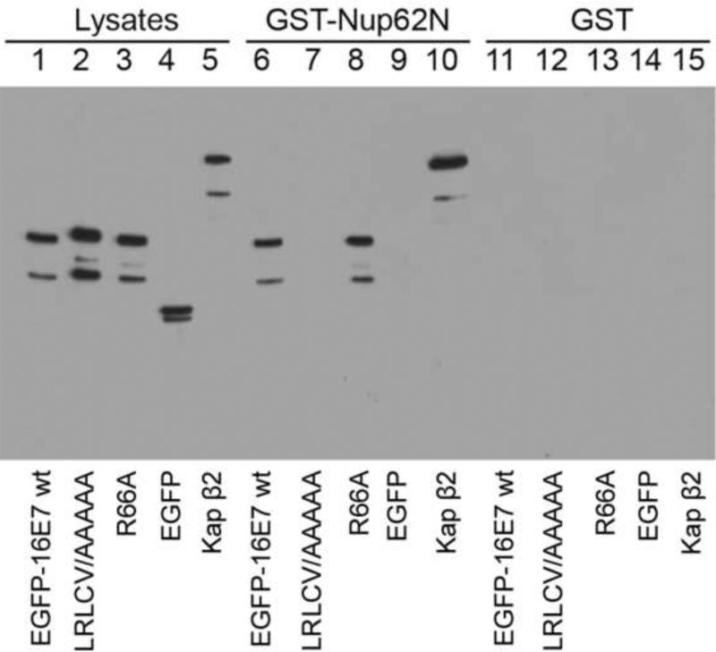
The mechanism by which hydrophobic residue patches of karyopherins mediate nuclear import is through non-specific hydrophobic interactions with the FG repeats in nucleoporins (Terry and Wente, 2009). To examine the possibility that the hydrophobic patch of HPV16 E7, 65LRLCV69, mediates nuclear import through direct non-specific interactions with FG-Nups we performed isolation assays. For these experiments we chose the FG nucleoporin Nup62 because it is located at the central transport channel of the NPC and interacts with karyopherins/importins mediating nucleocytoplasmic traffic of cargoes (Solmaz et al., 2011). Nup62 forms triple helices with Nup54 that project alternatively up and down from either side of the midplane Nup54-Nup58 ring, and constitute cytoplasmic and nucleoplasmic entries for nuclear import and export complexes via their N-terminal FG domains (Solmaz et al., 2011). In these experiments we used GST-Nup62N, containing all six FG repeats of Nup62, and GST, as a negative control. These GST fusion proteins were immobilized on glutathione-Sepharose beads and incubated with HeLa cell lysates expressing EGFP-16E7 wild type, EGFP-16E7LRLCV65AAAAA, EGFP-16E7R66A, or EGFP. The bound proteins were then eluted and analyzed via immunoblotting with a GFP antibody. As a positive control for binding to Nup62N we used Kap β2/transportin. The positive control, Kap β2 bound to GSTNup62N (Fig. 7, lane 10) but not to GST itself (Fig. 7, lane 15), whereas the negative control EGFP did not bind to either one (Fig. 7, lanes 9 and 14). Significantly, the results show that both EGFP-16E7 wild type and EGFP-16E7R66A interacted with GST-Nup62N (Fig. 7, lanes 6 and 8), but not with GST (Fig. 7, lanes 11 and 13, respectively). This correlates well with the previously observed nuclear localization of both EGFP-16E7 and EGFP-16E7R66A. Significantly, the EGFP-16E7LRLCV65AAAAA hydrophobic residue mutant failed to interact with GST-Nup62N (Fig. 7, lane 7). We also performed isolation assays with GST-Nup62C, representing the C- terminal domain of Nup62 lacking any FG repeats, and neither Kap β2 nor EGFP-16E7 bound to GST-Nup62C (data not shown). Taken together, these data strongly suggest that the patch of hydrophobic residues, 65LRLCV69, within the zinc-binding domain of HPV16 E7 is essential for the cNLS function through the interaction with the FG repeats in Nup62, and that the charged arginine residue at position 66 has no role in this process.
Fig. 7. HPV16 E7 interacts with the FG domain of Nup62 and mutations of hydrophobic residues within the zinc-binding domain disrupt this interaction.
HeLa cells were transfected with EGFP-16E7, EGFP-16E7LRLCV65AAAAA, EGFP-16E7R66A, and EGFP plasmids. Cell lysates were prepared 24 h post transfection and probed with either a GFP antibody (lane 1, EGFP-16E7; lane 2, EGFP-16E7LRLCV65AAAAA; lane 3, EGFP-16E7R66A; lane 4, EGFP) or Kap 2 antibody (lane 5). GST-Nup62N (lanes 6-10) and GST (lanes 11-15) immobilized on glutathione-Sepharose were incubated with the cell lysates and the bound proteins were eluted and analyzed via immunoblotting with a GFP antibody (lanes 6 and 11, EGFP-16E7; lanes 7 and 12, EGFP-16E7 LRLCV65AAAAA; lanes 8 and 13, EGFP-16E7R66A; and lanes 9 and 14, EGFP). Binding of Kap 2 to GSTNup62N and GST was detected with a Kap 2 antibody (lanes 10 and 15).
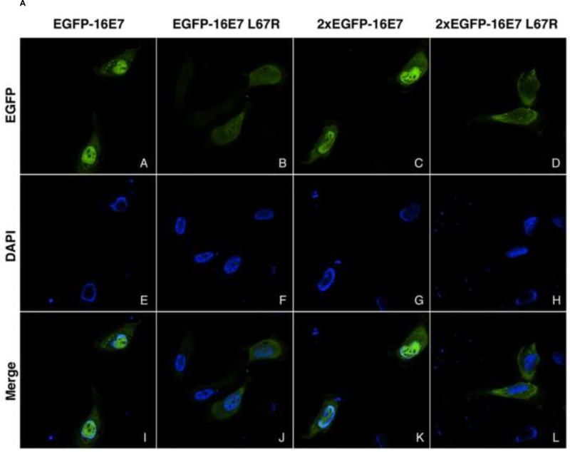
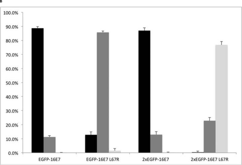
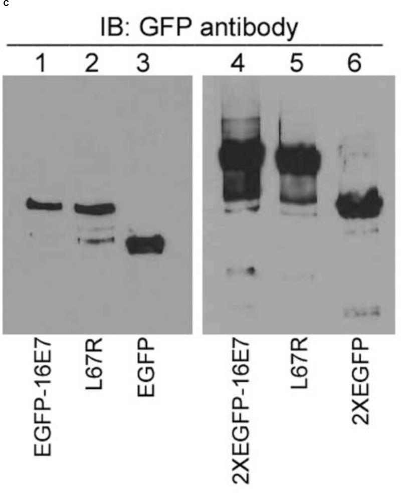
Interestingly, an HPV16 E7 mutant containing a mutation in this mostly hydrophobic patch L67R, although retaining its ability to bind pRb, is defective in abrogating pRB-mediated cell cycle arrest and fails to bind HDAC (Avvakumov et al., 2003; Brehm et al., 1999). To examine if this L67R mutation within 65LRLCV69 interferes with the nuclear localization of HPV16 E7 we introduced this mutation both in the context of EGFP-16E7 and 2xEGFP-16E7 and analyzed their localization in comparison with the wild type proteins. In contrast with the nuclear localization of EGFP-16E7 and 2xEGFP-16E7 (Fig. 8A, panels A and I; panels C and K; and Fig. 8B), the EGFP-16E7L67R had a pancellular localization in 85.8 +/− 0.9 % of transfected cells (Fig. 8A, panels B and J; and Fig. 8B), whereas the 2xEGFP-16E7L67R had a cytoplasmic localization in 76.8 +/− 2.5% of cells and a pancellular localization in the rest of cells (Fig. 8A, panels D and L; and Fig. 8B). Immunoblot analysis with an anti-GFP antibody indicated that EGFP-16E7L67R and 2xEGFP-16E7L67R mutant proteins were expressed at the correct molecular weight and at similar levels with their corresponding wild type proteins (Fig. 8C). These results indicate that the hydrophobic leucine 67 residue is essential for the nuclear localization of 16E7 and its substitution with the positively charged arginine residue leads to a loss of nuclear import function of the cNLS.
Fig. 8. The L67R mutation disrupts the nuclear localization of EGFP-16E7 and 2xEGFP-16E7.
A. HeLa cells were transfected with EGFP-16E7 (panels A, E and I), EGFP-16E7L67R (panels B, F and J), 2xEGFP-16E7 (panels C, G and K), or 2xEGFP-16E7L67R (panels D, H and L) plasmids, and the localization of the expressed proteins was analyzed by confocal fluorescence microscopy. Panels A-D represent the fluorescence of the EGFP, panels E-H the DAPI staining of the nuclei, and panels I-L the merge. B. Quantitative analysis of the effect of L67R mutation on the localization of EGFP-16E7 and 2xEGFP-16E7. Data from four experiments using EGFP-16E7 wild type, EGFP-16E7L67R, 2xEGFP-16E7 wild type and 2xEGFP-16E7L67R plasmids were used for the quantitative analysis and graphic representation of the localization distribution of transfected HeLa cells. Black bars represent cells showing predominant nuclear localization; gray bars represent cells showing pancellular localization; white bars represent cells showing predominant cytoplasmic localization. C. EGFP-16E7L67R and 2xEGFP-16E7L67R mutants are properly expressed in HeLa cells. HeLa cells were transfected with EGFP-16E7 (lane 1), EGFP-16E7L67R (lane 2), EGFP (lane 3), 2xEGFP-16E7 (lane 4), 2xEGFP-16E7L67R (lane 5) and 2xEGFP (lane 6). Cell lysates were prepared 24 h post transfection and probed with a GFP antibody.
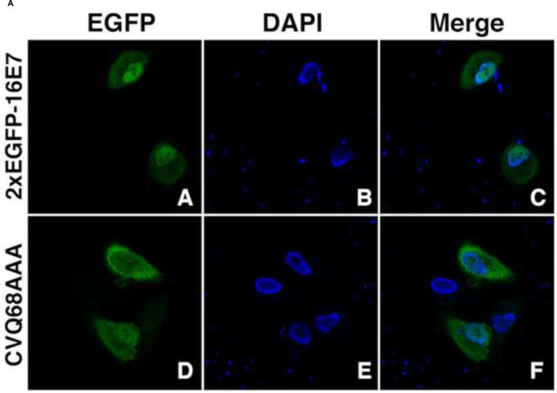
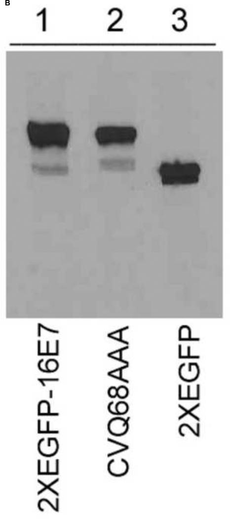
There is another HPV16 E7 mutant that partially overlaps with the 65LRLCV69 hydrophobic patch, CVQ68-70AAA, that has a mixed phenotype: it has wild type activity regarding the ability to block binding of pRb to E2F and to destabilize pRb (Helt and Galloway, 2001), and to bind to p600 and E2F6 (Huh et al., 2005; McLaughlin-Drubin et al., 2008); however, the CVQ68-70AAA mutant fails to bypass DNA damage checkpoints, to extend HFK lifespan, to inactivate p21 and to bind to cullin 2 ubiquitin ligase complex (Helt and Galloway, 2001; Helt et al., 2002; Huh et al., 2007; Roman and Munger, 2013). We generated the 2xEGFP-16E7CVQ68AAA mutant and analyzed its localization in comparison with the wild type and found that the CVQ68-70AAA mutation caused a change in the localization from nuclear in the majority of cells (85.65% +/− 7.2%) for the wild type to pancellular in 61.83% +/− 4.4% of cells and cytoplasmic in 35.73% +/− 4.17% cells for the mutant (Fig. 9A). These data indicate that substitution of the last two residues in the hydrophobic patch, cysteine and valine, to alanine leads to significant weakening of the cNLS nuclear import function.
Fig. 9.
A. Analysis of the effect of CVQ68AAA mutation on the nuclear localization of 2xEGFP-16E7. HeLa cells were transfected with 2xEGFP-16E7 (panels A, B and C) or 2xEGFP-16E7CVQ68AAA (panels D, E and F) plasmids, and the localization of the expressed proteins was analyzed by confocal fluorescence microscopy. Panels A and D represent the fluorescence of the EGFP, panels B and E the DAPI staining of the nuclei, and panels C and F the merge. B. The 2xEGFP-16E7CVQ68AAA mutant is expressed properly in HeLa cells. HeLa cells were transfected with 2xEGFP-16E7 (lane 1), 2xEGFP-16E7CVQ68AAA (lane 2) and 2xEGFP (lane 3). Cell lysates were prepared 24 h post transfection and probed with a GFP antibody.
Discussion
In this study we continued our analysis of the nuclear import pathway and characterization of the cNLS of high risk HPV16 E7 using both transfection assays in HeLa cells with EGFP fusions containing 16E7 and different mutants and nuclear import assays in digitonin-permeabilized HeLa cells with GST fusions to the CR3 of 16E7. The CR3 domain contains a zinc-binding domain that consists of two Cys-X-X-Cys motifs separated by 29 amino acids that is conserved between the high and low risk HPV types (McLaughlin-Drubin and Munger, 2009). The integrity of this zinc-binding domain has been shown to be necessary for dimerization and, in high risk HPV types, the transformative capacity of E7 (McLaughlin-Drubin and Munger, 2009). Here we presented data showing that mutations of Cys residues in the two Cys-X-X-Cys motifs that affect the zinc binding clearly disrupt the nuclear localization of the resultant EGFP-16E7 and 2xEGFP-16E7 mutants. These data strongly suggest that the integrity of the zinc-binding domain of HPV16 E7 is critical for the nuclear import activity of the cNLS, in addition to the requirement for dimerization and transformation potential. The cysteine residues involved in zinc coordination within the CR3 domain of E7 proteins are the most highly conserved cysteine residues among the different HPV types and we have previously established for the low risk HPV11 E7 that the integrity of its zinc-binding domain is essential for the nuclear import activity of the cNLS (Piccioli et al., 2010). The loss of zinc coordination can lead to structural changes of the CR3 domain that would make unavailable the critical hydrophobic residues of the cNLS. In addition, another viral protein, the TAX protein of human T lymphotropic virus type 1 (HTLV-1), also enters the nucleus via a pathway mediated by its zinc-binding domain (Tsuji et al., 2007).
Our previous studies have shown that nuclear entry of HPV16 E7 is mediated by its unique cNLS using a pathway that it is independent of karyopherins/importins (Angeline et al., 2003; Knapp et al., 2009). One possible mechanism that bypasses the requirement of nuclear import receptors is via direct low affinity hydrophobic interactions between E7 and the phenylalanine-glycine (FG) repeats in FG nucleoporins in a manner similar to that used by traditional receptors/karyopherins. Significantly, the zinc-binding domain of HPV16 E7 is very rich in nonpolar hydrophobic residues, with some of them arranged in small hydrophobic patches, suggesting a similar mechanism for how the zinc-binding domain of HPV16 E7 is able to mediate nuclear import. In this study we determined that HPV16 E7 interacts via its zinc-binding domain with the FG repeats domain of Nup62, a nucleoporin located at the central channel of the NPC and involved in nuclear import and export of cargoes (Solmaz et al., 2011). Significantly, we found a mostly hydrophobic patch, 65LRLCV69, within the zinc-binding domain that is essential for the nuclear localization of EGFP-16E7 and 2xEGFP-16E7. Mutation of the hydrophobic residues to alanine was sufficient to disrupt the predominantly nuclear localization of EGFP-16E7 and 2xEGFP-16E7 and inhibit the nuclear import of GST-16cE7 in digitoninpermeabilized cells. In contrast, no change in localization was observed in either the transfection assays, or the nuclear import assays by mutating the arginine 66 residue to alanine, suggesting that this positively charged residue has no role in the nuclear import of HPV16 E7. Significantly, mutation of the hydrophobic residues to alanine inhibited the interaction of HPV16 E7 with the FG domain of Nup62, suggesting that the hydrophobic residues of this sequence play a critical role in facilitating the interaction of the zinc-binding domain of HPV16 E7 with Nup62. Based on the structure of the zinc-binding domain dimer of HPV1A E7 and the sequence alignment with other E7 proteins, the 65LRLCV69 patch is located on the β2 strand and L65, L67 and V69 are highly conserved hydrophobic residues and may participate in hydrophobic interactions that stabilize the dimer (Liu et al., 2006). Interestingly, an HPV16 E7 L67R mutant, although it binds pRb in vitro, fails to overcome cell cycle arrest in osteosarcoma cells in vivo and to transactivate E2F-dependent promoters (Avvakumov et al., 2003; Brehm et al., 1999). As this L67R mutation within the hydrophobic patch involved in nuclear import of HPV16 E7 disrupts 16E7 nuclear localization, it can be speculated that this may contribute to the phenotype of the mutant in vivo. Another HPV16 E7 mutant, that partially overlaps with the hydrophobic patch, CVQ68-70AAA, fails to bypass DNA damage checkpoints, to extend HFK lifespan and to inactivate p21 (Helt and Galloway, 2001; Helt et al., 2002). The CVQ68-70AAA mutation decreases the nuclear localization and increases the cytoplasmic localization of 16E7, which may contribute to some of its loss of function(s). The R66A mutant, that retains the nuclear localization of the wild type, also retains the wild type phenotype, the ability to destabilize pRb and to bypass DNA damage checkpoints (Helt and Galloway, 2001).
Overall, the data strongly suggest that the cNLS mediated nuclear import of HPV16 E7 is via non-specific hydrophobic interactions of its zinc-binding domain with the FG repeats domain of Nup62 at the NPC. The mechanism of nuclear import of high risk HPV16 E7 oncoprotein is conserved with that of low risk HPV11 E7 protein (McKee et al., 2013). Nuclear import of HPV11 E7 is mediated by direct interactions between a homologous hydrophobic patch, 65VRLVV69, within the zinc-binding domain with the FG domain of Nup62 (McKee et al., 2013). Also, the cutaneous high risk HPV8 E7 enters the nucleus via hydrophobic interactions between a homologous hydrophobic patch, 65LRLFV69, with the FG domain of Nup62 (manuscript in preparation). It is likely that HPV E7 proteins might interact with other FG nucleoporins like Nup153 and Nup54 during their translocation through the NPC into the nucleus.
While the mechanism of nuclear import of HPV E7 oncoproteins is conserved between different HPV types, it is unique in comparison to other HPV proteins, including the L1 and L2 capsid proteins and the E6 oncoprotein that use different karyopherins to enter the nucleus (Bordeaux et al., 2006; Darshan et al., 2004; Klucevsek et al., 2006; Le Roux and Moroianu, 2003; Nelson et al., 2002). We can only speculate at this time why the HPV E7 proteins have evolved this nuclear import mechanism independent of karyopherins, but the relatively small size, the richness in hydrophobic residues and the relative low concentration may favor direct interaction of the E7 proteins with FG nucleoporins and bypassing the competition for karyopherins. An interesting possibility, that remains to be explored, is that E7 may also function as a carrier facilitating the nuclear import of a cellular target protein and changing its nucleocytoplasmic distribution.
Another viral oncoprotein, the TAX protein of HTLV-1 also enters the nucleus via direct hydrophobic interactions of its zinc-binding domain with the FG nucleoporin Nup62 at the NPC (Tsuji et al., 2007). Also, a cellular protein, like PU.1 transcription factor, enters the nucleus independent of karyopherins via direct binding to the FG nucleoporins, Nup62 and Nup153 (Zhong et al., 2005).
Materials and methods
Mutagenesis to generate different EGFP-16E7 and 2xEGFP-16E7 mutants
EGFP-16E7 and 2xEGFP-16E7 plasmids were obtained previously (Knapp et al., 2009). The cysteine mutants EGFP-16E7C58A, 2xEGFP-16E7C58A, EGFP-16E7CC58AA, 2xEGFP-16E7CC58AA, EGFP-16E7C91A, 2xEGFP-16E7C91A, EGFP-16E7C59A, and 2xEGFP-16E7C59A mutants were generated using the QuikChange™ Site-Directed Mutagenesis Kit (Stratagene) with either EGFP-16E7 or 2xEGFP-16E7 as templates, and the corresponding mutagenesis primer pairs.
The EGFP-16E7LRLCV65AAAAA, 2xEGFP-16E7LRLCV65AAAAA, EGFP-16E7R66A, 2xEGFP-16E7R66A, EGFP-16E7L67R, 2xEGFP-16E7L67R and 2xEGFP-16E7CVQ68AAA mutants were generated using the QuikChange™ Site-Directed Mutagenesis Kit with either EGFP-16E7 or 2xEGFP-16E7 as templates, and the corresponding mutagenesis primer pairs.
All mutant plasmids were transformed into XL1-Blue competent cells (Agilent Technologies), purified and sequenced for verification (Eurofins MWG).
Transient expression of EGFP fusion proteins and confocal microscopy analysis
HeLa cells (ATCC) were plated on 12 mm poly-L-lysine-coated glass coverslips to 50 % confluency 24 h prior to transfection. Cells in each well were transfected with the corresponding EGFP fusion plasmid (as indicated in the figure legends) and the FuGENE 6 reagent (Roche Applied Science, IN). Media was changed to DMEM with 10% FBS and 1% penicillin-streptomycin after 6 h and the cells were fixed 24 h after the initial transfection with 3.7% formaldehyde in PBS (10 min). Coverslips were mounted using Vectashield-DAPI mounting medium (Vector Labs, CA) to visualize the nuclei by DAPI staining. The slides were examined by confocal fluorescence microscopy using a Leica TCS Sp5 broadband confocal microscope and pictures were taken using the Leica LAS AF software (Leica Microsystems).
Intracellular localization phenotypes of the different EGFP fusion proteins were scored as predominantly nuclear, pancellular, or predominantly cytoplasmic, as visually observed via confocal fluorescence microscopy for each of the transfection experiments. Data from four to seven experiments (as indicated in the figure legends) were used for quantitative analysis. The graphical representations for each quantitative analysis display average values with standard deviations. Immunoblot analyses of the expressed EGFP-16E7, 2xEGFP-16E7 and different mutants performed with a GFP antibody indicated that the proteins were expressed at similar levels and not degraded.
Preparation of GST-16cE7 Mutant Proteins
The GST-16cE7 plasmid was obtained previously (Knapp et al., 2009). The GST-16cE7R66A and GST-16cE7LRLCV65AAAAA mutants were generated using the QuikChange™ Site-Directed Mutagenesis Kit with GST-16cE7 as the template, and the mutagenesis primer pairs as indicated above. The mutant plasmids were transformed into XL1-Blue competent cells and extracted using Quantum Prep® Plasmid MidiPrep kit. All purified plasmids were sequenced for verification (Eurofins MWG).
For protein expression and purification, each GST-16cE7 plasmid was used to transform E. coli BL21 CodonPlus (Agilent Technologies). Bacteria were then induced with 1mM IPTG for 3 h at 37°C, and each mutant protein was purified in its native state on glutathione-Sepharose beads (GE Healthcare) using a standard procedure. The GST-M9 positive control containing the NLS of hnRNPA1 was also prepared on glutathione-Sepharose beads using a standard procedure. All purified GST fusion proteins were dialyzed in transport buffer (20 mM HEPES-KOH pH 7.3, 110mM potassium acetate, 2 mM magnesium acetate, 1 mM EGTA, and 2 mM DTT) plus the protease inhibitors leupeptin and aprotinin. Purified and dialyzed proteins were aliquoted and stored at −80°C. Prior to use, all proteins were checked for purity and lack of proteolytic degradation by SDS-PAGE and Coomassie Blue staining.
In Vitro Nuclear Import Assays
Nuclear import assays were performed according to previously established protocols (Angeline et al., 2003; Knapp et al., 2009; Piccioli et al., 2010). Subconfluent HeLa cells, grown on 12 mm poly-L-lysine coated glass coverslips for 24 h, were permeablized with 70 g/ l digitonin for 5 min on ice to selectively permeabilize the plasma membrane while leaving the nuclear envelope intact. As a result, these digitoninpermeabilized cells will retain intact import-competent nuclei while being largely depleted of cytosolic transport factors. After three washes with cold transport buffer, the permeabilized cells were incubated with the import mixture for 30 min. Each import mixture contained an energy regenerating system (1 mM GTP, 1 mM ATP, 5 mM phosphocreatine, and 0.4U creatine phosphokinase) and the different GST fusion proteins (as indicated in the figure legend). In addition, each import contained either 3 ng/ l BSA in transport buffer, or HeLa cytosol. Final import reaction volumes were adjusted to 20 l with transport buffer.
For visualization of nuclear import, the GST fusion proteins were detected by immunofluorescence with a goat anti-GST antibody. Briefly, cells were washed, then fixed with 3.7% formaldehyde in PBS for 10 min on ice followed by methanol treatment for 3 min at −20°C. To reduce non-specific antibody binding the fixed cells were blocked for 1 h with 3% BSA/0.1% Tween in PBS. Cells were then incubated for 1 h with a 1:200 dilution of goat anti-GST primary antibody and after three washes with 3% BSA/0.1% Tween, cells were incubated with a 1:100 dilution of FITC conjugated rabbit anti-goat secondary antibody for 30 min. Following a final three washes, the coverslips were mounted using Vectashield-DAPI mounting medium to identify the nuclei by DAPI staining. Nuclear import was assessed by confocal fluorescence microscopy using a Leica TCS Sp5 broadband confocal microscope and representative images were taken using the Leica LAS AF software.
Preparation of GST-Nup62N and GST-Nup62C
The GST-Nup62N (containing aa 1-265 of Nup62), and GST-Nup62C (containing aa 178-522 of Nup62) plasmids (Zhong et al., 2005) were kindly provided by Dr. Nabeel Yaseen. For protein expression and purification the GST-Nup62N, GST-Nup62C and GST plasmids were used to transform E. coli BL21 CodonPlus. After induction of the bacteria with 1mM IPTG for 3 h, GST-Nup62N, GST-Nup62C and GST were purified in their native state on Glutathione-Sepharose beads using a standard procedure.
Isolation assays
Before performing the binding assays, GST-Nup62N, GST-Nup62C and GST immobilized on glutathione- Sepharose were analyzed by SDS-PAGE and Coomassie staining. This analysis revealed for GST-Nup62N and GST-Nup62C the presence of the intact protein, and also some degradation. HeLa cell lysates containing the expressed EGFP-16E7, EGFP-16E7LRLCV65AAAAA, EGFP-16E7R66A, and EGFP (negative control) were incubated with GST-Nup62N (containing the FG domain of Nup62), GST-Nup62C (lacking any FG repeats) or with GST immobilized on glutathione-Sepharose for 1 h at 4°C. After washing the beads to remove nonspecific binding the bound proteins were analyzed by immunobloting with a GFP antibody. As a positive control for binding to Nup62N, we analyzed the binding of Kap β2 nuclear import receptor to GST-Nup62N by incubating HeLa cell lysate with GST-Nup62N, GST-Nup62C, or GST and the bound proteins were analyzed by immunobloting with a Kap β2 antibody.
Highlights.
➢ An intact zinc-binding domain is essential for the nuclear localization of HPV16 E7
➢ Identification of a hydrophobic patch that is critical for the nuclear import of HPV16 E7
➢ HPV16 E7 interacts via its zinc-binding domain with the FG domain of Nup62
Acknowledgments
We thank Dr. Nabeel Yaseen for the GST-Nup62N and GST-Nup62C plasmids. We thank Rebeca Cardoso, Courtney McKee and Aditya Ashok for help with purification of GST-fusion proteins. This work was supported by a grant from the National Institutes of Health (R01 CA94898) to Junona Moroianu.
Abbreviations
- HPV
human papillomavirus
- NLS
nuclear localization signal
- NES
nuclear export signal
- NPC
nuclear pore complex
- EGFP
enhanced green fluorescent protein
- GST
Glutathione-S-transferase
Footnotes
Publisher's Disclaimer: This is a PDF file of an unedited manuscript that has been accepted for publication. As a service to our customers we are providing this early version of the manuscript. The manuscript will undergo copyediting, typesetting, and review of the resulting proof before it is published in its final citable form. Please note that during the production process errors may be discovered which could affect the content, and all legal disclaimers that apply to the journal pertain.
References
- Angeline M, Merle E, Moroianu J. The E7 oncoprotein of high-risk human papillomavirus type 16 enters the nucleus via a nonclassical Ran-dependent pathway. Virology. 2003;317:13–23. doi: 10.1016/j.virol.2003.08.024. [DOI] [PubMed] [Google Scholar]
- Avvakumov N, Torchia J, Mymryk JS. Interaction of the HPV E7 proteins with the pCAF acetyltransferase. Oncogene. 2003;22:3833–3841. doi: 10.1038/sj.onc.1206562. [DOI] [PubMed] [Google Scholar]
- Bordeaux J, Forte S, Harding E, Darshan MS, Klucevsek K, Moroianu J. The l2 minor capsid protein of low-risk human papillomavirus type 11 interacts with host nuclear import receptors and viral DNA. J Virol. 2006;80:8259–8262. doi: 10.1128/JVI.00776-06. [DOI] [PMC free article] [PubMed] [Google Scholar]
- Brehm A, Nielsen SJ, Miska EA, McCance DJ, Reid JL, Bannister AJ, Kouzarides T. The E7 oncoprotein associates with Mi2 and histone deacetylase activity to promote cell growth. The EMBO journal. 1999;18:2449–2458. doi: 10.1093/emboj/18.9.2449. [DOI] [PMC free article] [PubMed] [Google Scholar]
- Darshan MS, Lucchi J, Harding E, Moroianu J. The l2 minor capsid protein of human papillomavirus type 16 interacts with a network of nuclear import receptors. J Virol. 2004;78:12179–12188. doi: 10.1128/JVI.78.22.12179-12188.2004. [DOI] [PMC free article] [PubMed] [Google Scholar]
- Doorbar J. Molecular biology of human papillomavirus infection and cervical cancer. Clin Sci (Lond) 2006;110:525–541. doi: 10.1042/CS20050369. [DOI] [PubMed] [Google Scholar]
- Helt AM, Funk JO, Galloway DA. Inactivation of both the retinoblastoma tumor suppressor and p21 by the human papillomavirus type 16 E7 oncoprotein is necessary to inhibit cell cycle arrest in human epithelial cells. Journal of virology. 2002;76:10559–10568. doi: 10.1128/JVI.76.20.10559-10568.2002. [DOI] [PMC free article] [PubMed] [Google Scholar]
- Helt AM, Galloway DA. Destabilization of the retinoblastoma tumor suppressor by human papillomavirus type 16 E7 is not sufficient to overcome cell cycle arrest in human keratinocytes. Journal of virology. 2001;75:6737–6747. doi: 10.1128/JVI.75.15.6737-6747.2001. [DOI] [PMC free article] [PubMed] [Google Scholar]
- Huh K, Zhou X, Hayakawa H, Cho JY, Libermann TA, Jin J, Harper JW, Munger K. Human papillomavirus type 16 E7 oncoprotein associates with the cullin 2 ubiquitin ligase complex, which contributes to degradation of the retinoblastoma tumor suppressor. Journal of virology. 2007;81:9737–9747. doi: 10.1128/JVI.00881-07. [DOI] [PMC free article] [PubMed] [Google Scholar]
- Huh KW, DeMasi J, Ogawa H, Nakatani Y, Howley PM, Munger K. Association of the human papillomavirus type 16 E7 oncoprotein with the 600-kDa retinoblastoma protein-associated factor, p600. Proc Natl Acad Sci U S A. 2005;102:11492–11497. doi: 10.1073/pnas.0505337102. [DOI] [PMC free article] [PubMed] [Google Scholar]
- Klucevsek K, Daley J, Darshan MS, Bordeaux J, Moroianu J. Nuclear import strategies of high-risk HPV18 L2 minor capsid protein. Virology. 2006;352:200–208. doi: 10.1016/j.virol.2006.04.007. [DOI] [PubMed] [Google Scholar]
- Knapp AA, McManus PM, Bockstall K, Moroianu J. Identification of the nuclear localization and export signals of high risk HPV16 E7 oncoprotein. Virology. 2009;383:60–68. doi: 10.1016/j.virol.2008.09.037. [DOI] [PMC free article] [PubMed] [Google Scholar]
- Le Roux LG, Moroianu J. Nuclear entry of high-risk human papillomavirus type 16 E6 oncoprotein occurs via several pathways. J Virol. 2003;77:2330–2337. doi: 10.1128/JVI.77.4.2330-2337.2003. [DOI] [PMC free article] [PubMed] [Google Scholar]
- Liu X, Clements A, Zhao K, Marmorstein R. Structure of the human Papillomavirus E7 oncoprotein and its mechanism for inactivation of the retinoblastoma tumor suppressor. J Biol Chem. 2006;281:578–586. doi: 10.1074/jbc.M508455200. [DOI] [PubMed] [Google Scholar]
- McKee CH, Onder Z, Ashok A, Cardoso R, Moroianu J. Characterization of the transport signals that mediate the nucleocytoplasmic traffic of low risk HPV11 E7. Virology. 2013 doi: 10.1016/j.virol.2013.04.031. [DOI] [PMC free article] [PubMed] [Google Scholar]
- McLaughlin-Drubin ME, Huh KW, Munger K. Human papillomavirus type 16 E7 oncoprotein associates with E2F6. Journal of virology. 2008;82:8695–8705. doi: 10.1128/JVI.00579-08. [DOI] [PMC free article] [PubMed] [Google Scholar]
- McLaughlin-Drubin ME, Munger K. The human papillomavirus E7 oncoprotein. Virology. 2009;384:335–344. doi: 10.1016/j.virol.2008.10.006. [DOI] [PMC free article] [PubMed] [Google Scholar]
- Munger K, Baldwin A, Edwards KM, Hayakawa H, Nguyen CL, Owens M, Grace M, Huh K. Mechanisms of human papillomavirus-induced oncogenesis. J Virol. 2004;78:11451–11460. doi: 10.1128/JVI.78.21.11451-11460.2004. [DOI] [PMC free article] [PubMed] [Google Scholar]
- Naim B, Zbaida D, Dagan S, Kapon R, Reich Z. Cargo surface hydrophobicity is sufficient to overcome the nuclear pore complex selectivity barrier. Embo J. 2009;28:2697–2705. doi: 10.1038/emboj.2009.225. [DOI] [PMC free article] [PubMed] [Google Scholar]
- Nelson LM, Rose RC, Moroianu J. Nuclear import strategies of high risk HPV16 L1 major capsid protein. J Biol Chem. 2002;23:23. doi: 10.1074/jbc.M200724200. [DOI] [PubMed] [Google Scholar]
- Nguyen CL, Munger K. Human papillomavirus E7 protein deregulates mitosis via an association with nuclear mitotic apparatus protein 1. J Virol. 2009;83:1700–1707. doi: 10.1128/JVI.01971-08. [DOI] [PMC free article] [PubMed] [Google Scholar]
- Piccioli Z, McKee CH, Leszczynski A, Onder Z, Hannah EC, Mamoor S, Crosby L, Moroianu J. The nuclear localization of low risk HPV11 E7 protein mediated by its zinc binding domain is independent of nuclear import receptors. Virology. 2010;407:100–109. doi: 10.1016/j.virol.2010.07.045. [DOI] [PMC free article] [PubMed] [Google Scholar]
- Roman A, Munger K. The papillomavirus E7 proteins. Virology. 2013 doi: 10.1016/j.virol.2013.04.013. [DOI] [PMC free article] [PubMed] [Google Scholar]
- Solmaz SR, Chauhan R, Blobel G, Melcak I. Molecular architecture of the transport channel of the nuclear pore complex. Cell. 2011;147:590–602. doi: 10.1016/j.cell.2011.09.034. [DOI] [PMC free article] [PubMed] [Google Scholar]
- Terry LJ, Wente SR. Flexible gates: dynamic topologies and functions for FG nucleoporins in nucleocytoplasmic transport. Eukaryot Cell. 2009;8:1814–1827. doi: 10.1128/EC.00225-09. [DOI] [PMC free article] [PubMed] [Google Scholar]
- Tsuji T, Sheehy N, Gautier VW, Hayakawa H, Sawa H, Hall WW. The nuclear import of the human T lymphotropic virus type I (HTLV-1) tax protein is carrier and energy-independent. J Biol Chem. 2007;282:13875–13883. doi: 10.1074/jbc.M611629200. [DOI] [PubMed] [Google Scholar]
- Zhong H, Takeda A, Nazari R, Shio H, Blobel G, Yaseen NR. Carrier-independent nuclear import of the transcription factor PU.1 via RanGTP-stimulated binding to Nup153. J Biol Chem. 2005;280:10675–10682. doi: 10.1074/jbc.M412878200. [DOI] [PubMed] [Google Scholar]
- zur Hausen H. Papillomaviruses causing cancer: evasion from host-cell control in early events in carcinogenesis. J Natl Cancer Inst. 2000;92:690–698. doi: 10.1093/jnci/92.9.690. [DOI] [PubMed] [Google Scholar]
- zur Hausen H. Papillomaviruses in the causation of human cancers - a brief historical account. Virology. 2009;384:260–265. doi: 10.1016/j.virol.2008.11.046. [DOI] [PubMed] [Google Scholar]


