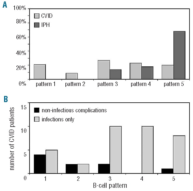
Figure 3.

Pathophysiological B-cell patterns in CVID and IPH and relation to clinical phenotypes. (A) Comparison of B-cell patterns between CVID (n=44) and IPH (n=21). B-cell patterns 1 and 2 are exclusively observed in CVID (P =0.02). (B) B-cell patterns and clinical phenotypes in CVID (n=44). Non-infectious complications (auto-immunity and polyclonal lymphocytic proliferation) were more often observed in B-cell pattern 1–2 compared to B-cell pattern 3–5 (P =0.003).
