Abstract
Background. Worldwide, ocular toxoplasmosis (OT) is the principal cause of posterior uveitis, a severe, life-altering disease. A Toxoplasma gondii enzyme-linked immunoassay that detects strain-specific antibodies present in serum was used to correlate serotype with disease.
Methods. Toxoplasma serotypes in consecutive serum samples from German uveitis patients with OT were compared with non-OT seropositive patients with noninfectious autoimmune posterior uveitis. OT patients were tested for association of parasite serotype with age, gender, location, clinical onset, size, visual acuity, or number of lesions (mean follow-up, 3.8 years) to determine association with recurrences.
Results. A novel, nonreactive (NR) serotype was detected more frequently in serum samples of OT patients (50/114, 44%) than in non-OT patients (4/56, 7%) (odds ratio, 10.0; 95% confidence interval 3.4–40.8; P < .0001). Non-OT patients were predominantly infected with Type II strains (39/56; 70%), consistent with expected frequencies in Central Europe. Among OT patients, those with NR serotypes experienced more frequent recurrences (P = .037). Polymerase chain reaction detected parasite DNA in 8/60 OT aqueous humor specimens but failed to identify Type II strain alleles.
Conclusions. Toxoplasma NR and Type II serotypes predominate in German OT patients. The NR serotype is associated with OT recurrences, underscoring the value of screening for management of disease.
Keywords: Diagnosis, ocular inflammatory disease, serotype, toxoplasmosis, uveitis
Toxoplasma gondii is a globally distributed protozoan parasite. Prevalence of infection varies based on geography, and is estimated to be about 50%–85% in Europe and Central and South America [1]. Progression and severity of disease is variable, ranging from asymptomatic to causing lymphadenopathy, encephalitis, and infectious retinochoroiditis, which accounts for 30%–50% of all cases of posterior uveitis globally [2–5]. The risk of developing eye lesions among T. gondii–infected patients varies geographically. In the United States and Europe, approximately 2% of patients present with OT versus 18% in southern Brazil [6, 7]; permanent visual loss is observed in up to 25% of affected patients [8].
In Europe and North America, T. gondii possesses a simple population genetic structure; 3 clonal lineages (referred to as Type I, II, or III) dominate the majority of human infections. Animal infections have established Type I (but not Type II or III) strains as highly virulent in mice due primarily to their proliferative capacity and ability to inactivate host immune responses [9, 10]. Development of OT in people is multifactorial and variable in onset, recurrence rate, clinical presentation, and severity [2]. Disease is thought to be dependent on a variety of factors, including host genetics, immune status, parasite genotype, and when infection is acquired (ie, congenital or postnatally) [11]. Recently, Toxoplasma strain type was identified as a significant factor determining prematurity and severity of congenital toxoplasmosis in the United States [12], and a diversity of recombinant and atypical strains, including a recently described Type × (Haplogroup 12) strain commonly infecting US wildlife, have previously been found associated with severe disease in patients experiencing acquired immunodeficiency syndrome (AIDS) and OT in North, Central, and South America [13–20]. In Europe, however, Type II strains account for 70%–80% of human infections [1, 21], the majority of congenital cases (85%) among pregnant women [22, 23], and OT cases in France [24, 25].
To assess whether strain type is a contributing factor determining the severity and frequency of recurrence of OT, we utilized a previously established enzyme-linked immunosorbent assay (ELISA)–based serotyping assay that detects strain-specific antibodies circulating in human serum samples, to distinguish infection caused by Type II strains from non-Type II strains, both in prospectively as well as retrospectively collected samples [12, 23, 26, 27]. We applied this technique to serum samples obtained from a cohort of 114 consecutive OT and 56 T. gondii–seropositive control patients with (non T. gondii–related) uveitis in Germany. Parasite DNA amplified from aqueous humor specimens was used to validate some of the obtained serological results. We show that a novel Toxoplasma serotype is associated with recurrent OT, and our results have the potential to personalize disease management protocols to improve the treatment and control of OT.
METHODS
Subjects with Ocular Toxoplasmosis
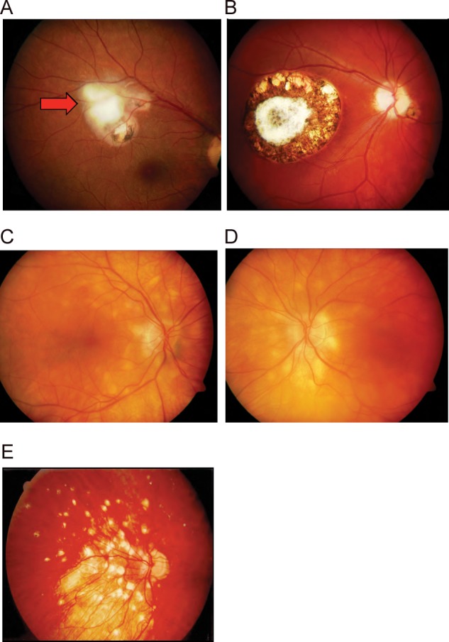
Serum and aqueous humor specimens were collected from March 1999 to June 2003 from 170 patients with inflammation due to uveitis who tested seropositive for Toxoplasma infection using a commercial immunofluorescence assay (Enzygnost, Siemens, Marburg, Germany). All patients lived in Germany at the time of clinical disease and were evaluated at the Charité eye clinic in Berlin. Patients were human immunodeficiency virus (HIV) negative and underwent a standardized testing program, including chest X-ray, and serology for angiotensin-converting enzyme, human leukocyte antigen (HLA)–B27, Borrelia burgdorferi, and Treponema pallidum to investigate other potential causes for their uveitis. One hundred fourteen patients were symptomatic and presented with active disease, the majority had observable OT lesions characterized by typical focal necrotizing retinochoroiditis accompanied by a vitreous inflammatory reaction (Figure 1A). Twenty-five percent of all acute OT patients were 0–15 years old, 49% were 15–35, and 26% were >35 years of age. Intraocular infection was confirmed by a positive Goldmann-Witmer coefficient; intraocular anti–T. gondii antibody levels were assessed in all patients. The Goldmann-Witmer coefficient (C) was calculated using paired serum and aqueous humor samples and the formula C = C1/C2, where C1 = anti–T. gondii IgG titer/total IgG concentration within ocular fluid, and C2 represents the same ratio within serum. A Goldmann-Witmer coefficient of 3.0 or greater was considered positive, indicating OT [28].
Figure 1.

Fundoscopic examination plates of German uveitis patients with or without OT. A, Retinochoroidal lesions in a patient with recurrent OT. Active inflammation is seen at the upper temporal border of 2 pigmented, older lesions (scars). Serum and intraocular antibodies were indicative of acute infection with T. gondii. B, Central retinochoroidal scar in a congenital OT patient with inactive disease. C, Typical clinical presentation of birdshot retinochoriodopathy in control patients. D, Typical autoimmune uveitis associated with HLA-A 29 in control non-OT patients. Fundus photographs show bilateral nonpigmented, cream-white scattered lesions nasally and superior to the optic nerve. The patients were persistently IgM negative and only positive for serum IgG antibodies (no intraocular IgG), indicative of latent infection with T. gondii. E, Posterior uveitis associated with sarcoidosis in a seropositive non-OT control patient. This patient had serum but no intraocular IgG antibodies indicative of latent infection with T. gondii. Abbreviations: HLA, human leukocyte antigen; IgG, immunoglobin G; IgM, immunoglobin M; OT, ocular toxoplasmosis.
Acute OT patients were examined at weeks 1, 2, and 6 after initiation of therapy. Each visit, patients underwent an ophthalmologic examination, including measurement of best-corrected visual acuity, applanation tonometry, undilated and dilated slit-lamp biomicroscopy, and indirect fundus examination. Toxoplasmic retinochoroiditis scars were mapped by automated visual field test in patients with central OT lesions (central 30-2-threshold protocol; Humphrey Visual Field Analyzer equipment, Carl Zeiss Meditec Inc, Dublin, CA). Fluorescein angiography was performed at the examiner's discretion. Fundus photographs were performed in patients before and after treatment.
During the same time period, 56 control patients presented with noninfectious autoimmune posterior uveitis, including birdshot choroiditis, Behcet's disease, or sympathetic ophthalmia [29]. Only patients who (1) presented with clinical signs other than those characteristic for OT, and (2) showed signs of autoimmune or other eye diseases supported by laboratory findings as indicated above (eg, HLA typing A29) were included as controls. None of the 56 control patients had lesions or scars characteristic of OT (Figure 1C–E). The majority of control and OT subjects were white, with the following exceptions: (1) 8 Behcet's patients were of Turkish ancestry (second-generation German offspring), 2 of 4 Vogt–Koyanagi–Harada patients were Asian (both Vietnamese). All patients filled out medical history questionnaires and only 1 female OT patient documented a long-term stay (>6 months) in an “unusual/exotic” destination: South America and Africa. No control patients reported a positive travel history related to their active intraocular inflammation. The 170 cases and controls were followed passively (by routine exams and as necessary when symptomatic) from their entry in the study until 2005; mean follow up time: 3.8 years. Fifty serum samples from T. gondii–seronegative uveitis patients were tested to establish serotyping assay thresholds. Ophthalmic examinations were recorded at time of specimen collection.
This study was conducted in accordance with tenets of the Declaration of Helsinki. All specimens were collected from volunteers who gave informed consent. Serum samples were anonymized prior to transfer to the testing laboratory; the protocol used in this study was approved by the local Committee on Human Research, Charité-Universitätsmedizin Berlin. Testing of anonymized serum samples was exempt from institutional review board approval at the National Institutes of Health.
T. gondii Serotyping ELISA
The ELISA assay and peptides used to determine T. gondii serotype have been described previously [23, 26, 27]. Serum samples from uveitis patients with or without OT were tested for their antibody reactivity against GRA6 and GRA7 allelic peptide motifs derived from distinct parasite types. ELISA results are presented as an optical density (OD) index by dividing the OD value obtained at 405 nm for each of the 5 serotyping peptides by the mean of the OD readings for 2 control peptides; results are expressed as arbitrary units. Threshold values (above which normalized assay values were considered positive) were determined by averaging the normalized OD ratio from 50 seronegative samples and adding 2 standard deviations (SDs).
Polymerase Chain Reaction Genotyping
Aqueous humor specimens were stored at −20°C until assayed. DNA was extracted as described previously [15]. Multilocus polymerase chain reaction (PCR) DNA sequencing was performed at B1, NTS2, SAG3, and GRA6 loci as described previously [30]. PCR products were treated with ExoSAP-IT (USB Corp, Cleveland, OH) and DNA sequenced at National Institute of Allergy and Infectious Diseases Rocky Mountain Laboratories Genomics, Hamilton, MT.
Statistical Analysis
Fisher's exact test was used to determine whether a specific T. gondii serotype was associated with OT or non-OT controls. We repeated this after collapsing the serotypes to compare NR to all other types using a standard 2-sided Fisher's exact test, estimating the effect with conditional maximum likelihood estimates of odds ratios, and using the matching 95% confidence intervals (CIs) [31]. Fisher's exact test comparing NR to all other types was used for the analyses in Table 1, except the test for age effects, which used the Mann–Whitney U test. Visual acuity was scored using a LogMAR chart. No corrections were made for multiple testing. P values < .05 were considered statistically significant.
Table 1.
Clinical Characteristics of Ocular Toxoplasmosis Caused by Different Genotypes of T. gondii
| Type I/III |
Type II |
Atypical |
Nonreactive |
P Value** |
|
|---|---|---|---|---|---|
| N (%) | N (%) | N (%) | N (%) | N (%) | |
| Number of patients | |||||
| Total (n = 104) | 6 (5.8) | 44 (42) | 9 (8.7) | 45 (43) | |
| Mean patient age at first manifestation (years ± SD) | 21 (±13) | 34 (±21) | 24 (±17) | 27 (±18) | .294 |
| Clinical occurrence | .037* | ||||
| No recurrent OT observed | 1 (16.7) | 16 (43.2) | 3 (37.5) | 7 (17.9) | |
| Recurrent OT observed | 5 (83.3) | 21 (56.8) | 5 (62.5) | 32 (82.1) | |
| Undetermineda (n = 14) | |||||
| Affected side*** | .321 | ||||
| Unilateral | 3 (50.0) | 27 (75.0) | 6 (75.0) | 32 (82.1) | |
| Bilateral | 3 (50.0) | 9 (25.0) | 2 (25.0) | 7 (17.9) | |
| Variable not recorded (n = 15) | |||||
| Disease localization*** | .445 | ||||
| Central | 1 (16.7) | 9 (28.1) | 3 (37.5) | 7 (20.0) | |
| Peripheral | 5 (83.3) | 23 (71.9) | 5 (62.5) | 28 (80.0) | |
| Variable not recorded (n = 23) | |||||
| Size of lesion*** | |||||
| 1 PD | 3 (60.0) | 18 (66.7) | 3 (50.0) | 13 (46.4) | .214 |
| >2 PD | 2 (40.0) | 9 (33.3) | 3 (50.0) | 15 (53.6) | |
| Variable not recorded (n = 38) | |||||
Abbreviations: OT, ocular toxoplasmosis; PD, papillar diameter.
a Undetermined refers to those patients who reported recurrent symptoms (ie, floaters) but upon clinical examination, new lesions were not confirmed. These patients were not included as “recurrent OT observed” for the statistical analysis.
* Statistically significant (P < .05).
** P values refer to comparison of all other serotypes against Nonreactive (NR).
*** For OT at first manifestation.
RESULTS
Type II Serotype Predominates in German Uveitis Patients without OT
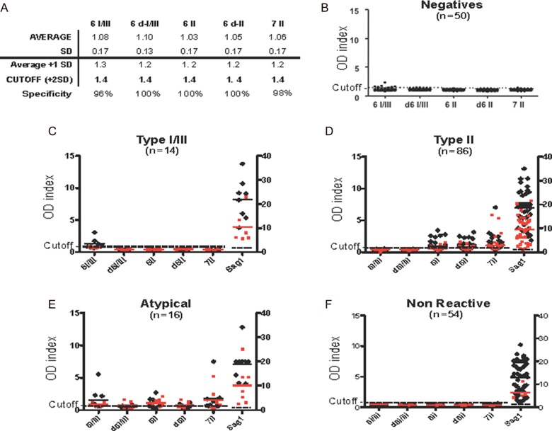
Serum samples from 56 consecutive Toxoplasma-seropositive autoimmune uveitis patients who were symptomatic but without OT were tested for their antibody reactivity against GRA6 and GRA7 allelic peptide motifs to determine their serotype. The Type II serotype was predominant (39/56; 70%), with all serotypes detected in the uveitis control group (Figure 2). This incidence rate is consistent with expected frequencies for Central Europe [1, 23]. Fifty T. gondii–seronegative patients that tested negative for reactivity against the 5 diagnostic peptides, 2 control peptides, and rSAG1 antigen were used to define a threshold value of 1.4 for the serotyping assay (Figure 2A).
Figure 2.
Serotypes associated with German OT uveitis patients. A and B, Toxoplasma dye-test-negative serum samples (n = 50) were tested using a panel of 5 strain-specific peptides to determine the specificity and empiric threshold values for negative serum samples in the serotyping assay. Peptide names were abbreviated as follows: “6” denoting peptides from GRA6; “7” from GRA7; “I/III” or “II” indicating the archetypal strain from which the peptide allele was derived; and “d” indicating a truncated diagnostic peptide. Reactivity at SAG1 indicates the presence of anti-Toxoplasma antibodies. Type I/III strain infections produce allele-specific antibodies that react with 1 or both of the 6-I/III and d6-I/III peptide epitopes. Type II infections produce antibodies that react with at least 1 of the 6-II, d6-II, and 7-II peptide epitopes. “Atypical” serotypes identified strain-specific antibodies that reacted with both the Type I/III and Type II strain-typing peptides or did not react (“Nonreactive” [NR]) with any of the allele-specific peptides. Red circles depict individual serum sample results for uveitis patients without OT; black diamonds depict individual sera results for OT patients. Strong reactivity against single-diagnostic peptides (with no reactivity against any other peptides, including recombinant SAG1 [rSAG1]) was considered false-positive. False reactivity generally occurs in <2% of samples [27]. C, Fourteen patients had specific reactivity against the 6-I/III peptide and rSAG1 protein, consistent with infection by a Type I or III strain. D, The majority of serum samples (86/170 or 51%) reacted with both rSAG1 and 1 or more of the Type II allelic peptides designated 6-II, d6-II, and 7-II, consistent with infection by Type II strains. E, Sixteen serum samples possessed Atypical serotypes that reacted strongly with both Type II and Type I or III peptides as well as rSAG1. F, 54/170 (32%) serum samples only reacted with rSAG1 and were peptide-NR. Abbreviation: OT, ocular toxoplasmosis.
Non–Type II Serotypes Predominate in German Uveitis Patients with OT
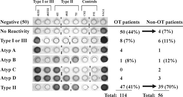
All parasite serotypes were detected among 114 consecutive uveitis patients experiencing OT (Figure 2). In contrast, non-Type II serotypes were predominant (67/114; 59%) and significantly associated with development of OT (P < .0001). The incidence of detecting the Type II serotype among German OT patients (47/114; 41%) was substantially lower than that of France, which is almost exclusively Type II [24, 25]. The relative proportion of Atypical and Type I or III serotypes did not vary between the 2 cohorts. Among “Atypical” serotypes, 4 serologic signatures were identified, designated Atypical A through D, distinguished by their differing reactivities against the allele-specific peptides (Figure 3).
Figure 3.
ELISA showing 7 distinct serotype patterns. The majority of OT samples possessed no peptide reactivity (labeled “NR”), although infection with T. gondii was confirmed by positive reactivity against recombinant SAG1 antigen. Four patterns (labeled “Atypic”) were suggestive of infection by nonarchetypal strains, as evidenced by high reactivity to more than 1 of the polymorphic peptides (labeled Atypical A–D). Reaction with any or all of peptides 6II, d6II or 7II confirmed infection with Type II strains, whereas reaction with 6/I/III and/or d6I/III confirmed infection by a Type I or III strain. Abbreviations: Atypic, atypical; ELISA, enzyme-linked immunosorbent assay; OT, ocular toxoplasmosis.
Figure 4.
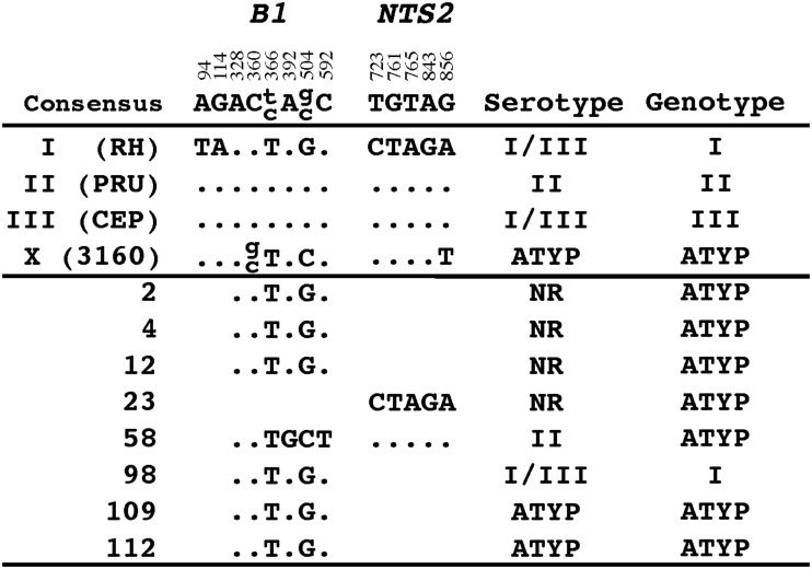
DNA sequence analysis at the B1 and NTS2 loci. Polymorphic sites that exist between the archetypal Type I, II, and III lineages were compared with sequences obtained from PCR-amplified parasite DNA from the aqueous humor of 8 OT patients. The consensus sequence was defined as the nucleotide common to at least 2 of the 3 archetypes. Periods (.) indicate identity with the consensus sequence. The numerical positions annotated refer to the numbered sites in the published GenBank sequences AF179871 for B1 and AF158092 for NTS2. The serotype for each sample was recorded beside the allele identified and multilocus genotypes established. Abbreviations: ATYP; atypical (nonarchetypal) genotype; NR, nonreactive; OT, ocular toxoplasmosis; PCR, polymerase chain reaction.
A Novel NR Serotype is Common among German OT Patients
A substantial number of OT patient serum samples (50/114; 44%) showed no reactivity with any serotyping peptides, but did react strongly for T. gondii by both commercial and rSAG1 antigen ELISA. Failure to react to the allelic peptides could not be explained by waning titers of anti-Toxoplasma antibodies (Figure 2F). The novel serotype was referred to as peptide “Nonreactive” (NR). In contrast, among seropositive uveitis patients without OT, only 4 of 56 (7%) patients possessed NR serotype (Supplementary Tables 1 and 2). The odds of identifying an NR serotype in German OT patients was approximately 10 times greater than the odds of finding an NR serotype in non-OT control patients with autoimmune uveitis (odds ratio 10.0 [95% CI, 3.4, 40.8; P < .0001]) (Figure 2; Supplementary Tables 1 and 2).
No Type II DNA Is Found in the Aqueous Humor of German OT Patients with Uveitis
DNA extracted from archived aqueous humor specimens obtained from 60 of the serotyped OT patients was available for multilocus PCR-DNA sequence analysis. Fifty-two samples tested PCR negative at 5 genotyping markers: B1, NTS2, SAG1, SAG3, and GRA6 [15, 30, 32]. Eight of 60 samples (13%) tested PCR positive, consistent with expected detection sensitivities for aqueous humor [24, 25, 33, 40]. No Type II lineage alleles were detected (Figure 4). Type I alleles were amplified from 7 out of 8 samples, similar to a previous study from US OT patients [15]. The eighth sample possessed alleles consistent with infection by a recombinant genotype, indicating that only non–Type II infections had occurred among the PCR-positive German OT uveitis patients. Four of the PCR-positive patients (samples 2, 4, 12, and 23) possessed NR serotypes, contraindicating Type II genotypes being associated with NR serotype.
Recurrent OT Is More Common in Patients with NR Serotype
Presentation of OT varies widely from subclinical to severe [8] and depends on location, size, number of lesions, extent of retinal inflammation, and number of recurrences. Because parasite genotype and/or additional factors likely correlate with incidence, severity, or type of OT, we examined whether Toxoplasma serotype correlated with 7 variables: age, gender, distribution (affected side, disease localization), visual acuity, and size and number of lesions. No significant association of serotype with visual acuity and gender was detected (data not shown), nor with age, pattern of retinal lesion (unilateral vs bilateral; peripheral vs central), or lesion size (Table 1). However, OT patients with NR serotypes were more likely to have recurrent OT when compared against all infected patients (odds ratio [OR] = 2.92; 95% CI, 1.05–8.89; P = .037). When comparing NR serotypes specifically against Type II–infected patients at presentation, we observed an even stronger effect for recurrence (OR = 3.42; 95% CI, 1.21–10.41; P = .024). Intraocular inflammation was severe in patients with large choroidal lesions, and vitreous haze was substantial. Extensive retinal lesions defined as equal or more than 2 disc diameters were observed in 29 patients. There was, however, no significant correlation of centrally localized disease, lesion size, or visual outcome with T. gondii serotype (Table 1).
DISCUSSION
A novel, nonreactive (NR) serotype was commonly identified among Toxoplasma-infected German uveitis patients who developed OT (44%), but was found only infrequently among seropositive control patients with autoimmune uveitis (7%) attending the same eye clinic. As in France, a significant number of OT patients possessed Type II serotypes (41%), but this patient group had fewer recurrences of OT. Patients who presented with OT were more likely to have recurrent OT if they had NR serotypes (P = .037), indicating that the serum-based strain typing assay is capable of predicting latently infected individuals who are at risk of developing recurrent OT.
This paper focuses on the NR serotype. Previous work identified a strong association of non–Type II serotypes (NE-II) with the development of more serious congenital disease in the United States [12]. Similar effects are shown for non–Type II serotypes in our study. For example, the odds of identifying a non–Type II serotype in German OT patients was approximately 3.2 times greater than the odds of finding a non–Type II serotype in non-OT control patients with autoimmune uveitis (OR, 3.2 [95% CI, 1.6, 6.9; P = .0008]), and non-Type II serotype is correlated with recurrence (P = .035). Although NR serotype appears more strongly associated with OT occurrence than non–Type II serotype, NR serotype is not sufficient to predict all individuals who will develop recurrent OT; hence, additional factors likely contribute to disease etiology. Significant risk factors influencing disease progression and clinical outcome of OT include parasite strain, ethnic origin, and host immune status [24, 34, 35]. Previous work in France linked Type II parasites with the development of OT [24]. However, the patient group examined consisted primarily of HIV-infected individuals and was conceivably biased toward strain types associated with chronic infection in France, which is predominantly the Type II lineage [21–23]. The study by Fekkar et al [24] did not assess whether immunosuppression or some other factor associated with HIV infection led to increased recrudescence of disease and the development of OT. When only immunocompetent, non-HIV–infected French OT patients are considered, both Type II and non–Type II parasites associated with mild to severe disease in France, not unlike our study [24]. Studies in North America among healthy adult patients who presented with clinically severe OT identified only non–Type II (ie, Type I or recombinant) genotypes [15]. Serum samples available from 2 of these patients possessed NR serotypes [26]. Why the majority of T. gondii–infected OT-susceptible individuals do not produce antibodies that react with archetypal allelic peptide motifs is enigmatic. Regardless of mechanism, the NR serotype predominates among non-HIV–infected individuals who experience recurrent OT, which is a more severe form of disease. Consistent with this, patients with NR serotype trended toward larger retinochoroidal lesions when compared to patients with any other serotype (see Table 1), although the finding did not reach statistical significance. Whether disease in the NR patient group is the result of (1) a failure to mount an appropriate immune response capable of controlling infection, (2) infection by a nonarchetypal parasite genotype, or (3) some combination of host factors and parasite strain, remains to be established.
Ultimately, to determine the significance of finding NR serotypes in patients who develop more serious, recurrent OT will require information on the genotype(s) of strain(s) associated with disease and the contributing roles of immune mediators induced during active episodes of OT. Infections in mice show that different strains of Toxoplasma differentially secrete polymorphic effector proteins (eg, ROP5, ROP16, ROP18, GRA15) that target host proinflammatory responses that alter protective immunity. For example, strains expressing the GRA15II allele activate nuclear factor κB (NF-κB) and produce protective levels of interleukin-12 and interferon-γ [36], whereas strains expressing ROP16I and/or ROP5I/ROP18I cause prolonged activation of signal transducer and activation of transcriptions 3 and 6 (STAT3 and STAT6) signaling [10, 37] or inactivate host GTPases required for efficient parasite killing [38, 39], respectively. It is likely that parasite proteins also regulate the induction of host effector responses in people, and recent work examining congenital infections in the United States has shown that NE-II correlate with prematurity and severity of disease [12]. In our study, insufficient parasite DNA was recovered from Type II–infected OT patient aqueous humor, indicating that, relative to other active, non–Type II OT infections, parasite load was lower, or at least below the detection limits of 1–10 parasites (B1, NTS2 loci) [23]. Lack of detectable parasites among Type II–infected patients may positively correlate with the decreased numbers of recurrences among this patient cohort. Among patients possessing sufficient parasite DNA to permit genotyping (8/60), no Type II alleles were identified. A strong bias toward strains bearing Type I–lineage alleles (7/8) or recombinant genotypes (1/8) was observed, implicating infection by non–Type II, and conceivably, novel genotype(s), as the most likely explanation for the observed lack of reactivity against the serotyping peptides. Nonarchetypal strains typically possess entirely different polymorphic epitopes at GRA6 or GRA7, and these epitopes do not necessarily produce antibodies in serum samples of infected subjects capable of reacting with the diagnostic allelic peptide motifs. Examples of this have been observed previously among people infected during an outbreak of toxoplasmosis in Brazil [27]. Alternatively, it is also possible that host genetic factors preclude the ability of OT patients to produce allele-specific antibodies at GRA6 and GRA7, or that the polymorphic epitopes are not immunogenic in this patient group.
The possibility that some complex combination of host resistance and/or susceptibility alleles is likewise influencing the outcome and severity of OT cannot be excluded. Patients bearing specific HLA-DQ3 alleles or polymorphisms at ABCA4 and COL2A1 are more susceptible to congenital toxoplasmosis [35]. Recent follow-up work has shown that Type II serotypes associate with severe congenital disease more frequently among patients bearing susceptibility alleles at ABCA4 and COL2A1 [12]. This might explain how Type II parasites cause fulminant disease only when associated with a host genetic risk factor, and prospective studies are underway to test whether host toxoplasmosis susceptibility alleles or any skewing in HLA type is associated with the development and severity of OT among patients with Type II serotypes.
In Europe, infections with strains harboring Type I–lineage alleles or recombinant genotypes are considered rare. Toxoplasma genotypes associated with AIDS and congenital toxoplasmosis in France have established Type II as the dominant strain causing the majority of infections [21, 22]. Our data, however, demonstrate the necessity of analyzing patient groups in combination with appropriate control groups (ie, Toxoplasma-infected subjects without OT from the same institution). Because OT is thought to develop among only 2% of infected individuals in North America and Europe [7, 41], previous studies perhaps failed to identify the significance of finding non–Type II strains and the NR serotype associated with recurrent OT. It is also unlikely that the NR serotype is the result of an outbreak by a limited set of genotypes because the samples were collected over a 4-year period.
To test whether NR serotype predicts those patients who go on to develop recurrent OT [42], we measured odds ratios related to OT in 2 ways. In our OT patient cohort followed passively for recurrence, we found the odds for developing recurrent OT was 2.92 (95% CI, 1.05–8.89) times higher for NR serotypes than for all other serotypes. From the case-control part of the study, we found the odds were 10.0 (95% CI, 3.4–40.8) times higher for those with the NR serotype to present with OT compared to all other serotypes. The case-control data do not allow for estimating the positive predictive value for predicting OT over a defined time period because there was not complete cohort follow-up with precise time periods. However, we can interpret the 10.0 odds ratio measured from the case-control data as a hazard ratio if we assume that the proportion of people with the NR serotype was constant over time [43]. In order to determine the positive predictive value of the NR serotype for predicting initial OT over a prespecified time period, a cohort study may be done.
In conclusion, results presented here may have marked implications for future management of patients with OT. On the one hand, serotyping may allow identification of patients without OT but at increased risk to develop OT, thereby allowing changes in their management (ie, raising patients’ awareness of signs and symptoms, more frequent eye exams, or even prophylactic treatment). On the other hand, NR-serotype patients with active OT who have a higher risk of recurrences will receive appropriate management (ie, preventive treatment). Long-term intermittent treatment with trimethoprim/sulfamethoxazole has previously been shown to reduce the rate of recurrent OT in Brazil [43]. These specific measures may also be implemented in patients with perimacular lesions and monocular patients, or patients with markedly diminished visual acuity (eg, <20/400) regardless of serotype. It is important to make healthcare providers aware of these findings because current consensus among physicians is that Toxoplasma infection among healthy adults is self-limiting and relatively benign.
Supplementary Data
Supplementary materials are available at The Journal of Infectious Diseases online (http://jid.oxfordjournals.org/). Supplementary materials consist of data provided by the author that are published to benefit the reader. The posted materials are not copyedited. The contents of all supplementary data are the sole responsibility of the authors. Questions or messages regarding errors should be addressed to the author.
Notes
Author Contributions. Conceived and designed experiments: L. S., E. R. J., O. L., U. P., and M. E. G. Performed the experiments: L. S., E. R. J., and M. E. G. Analyzed the data: L. S., J. J., M. P. F., U. P., and M. E. G. Contributed reagents/materials: S. M., N. T., U. P. Wrote the paper: L. S., O. L., M. P. F., U. P., and M. E. G.
Acknowledgments. The authors thank Drs Siddhartha Mahanty and Tom Nutman for critically reviewing the manuscript.
Financial support. This work was supported by the Intramural Research Program of the National Institutes of Health (NIH) and National Institute of Allergy and Infectious Diseases (NIAID; M. E. G.). M. E. G. is a scholar of the Canadian Institute for Advanced Research (CIFAR) Program for Integrated Microbial Biodiversity. U. P., N. T., and O. L. were supported by ToxoNet, a program of the German Bundesministerium für Bildung und Forschung (BMBF).
Potential Conflict of Interest. All authors: No reported conflicts.
All authors have submitted the ICMJE Form for Disclosure of Potential Conflicts of Interest. Conflicts that the editors consider relevant to the content of the manuscript have been disclosed.
References
- 1.Dubey JP, Beattie CP. Toxoplasmosis of animals and man. Boca Raton, FL: CRC Press; 1988. pp 1-220. [Google Scholar]
- 2.Holland GN. Reconsidering the pathogenesis of ocular toxoplasmosis. Am J Ophthalmol. 1999;128:502–5. doi: 10.1016/s0002-9394(99)00263-9. [DOI] [PubMed] [Google Scholar]
- 3.McCannel CA, Holland GN, Helm CJ, Cornell PJ, Winston JV, Rimmer TG. Causes of uveitis in the general practice of ophthalmology. UCLA Community-Based Uveitis Study Group. Am J Ophthalmol. 1996;121:35–46. doi: 10.1016/s0002-9394(14)70532-x. [DOI] [PubMed] [Google Scholar]
- 4.Montoya JG, Liesenfeld O. Toxoplasmosis. Lancet. 2004;363:1965–76. doi: 10.1016/S0140-6736(04)16412-X. [DOI] [PubMed] [Google Scholar]
- 5.Remington JS. Toxoplasmosis in the adult. Bull N Y Acad Med. 1974;50:211–27. [PMC free article] [PubMed] [Google Scholar]
- 6.Glasner PD, Silveira C, Kruszon-Moran D, et al. An unusually high prevalence of ocular toxoplasmosis in Southern Brazil. Am J Ophthalmol. 1992;114:136–44. doi: 10.1016/s0002-9394(14)73976-5. [DOI] [PubMed] [Google Scholar]
- 7.Holland GN. Ocular toxoplasmosis: a global reassessment. Part I: Epidemiology and course of disease. Am J Ophthalmol. 2003;136:973–88. doi: 10.1016/j.ajo.2003.09.040. [DOI] [PubMed] [Google Scholar]
- 8.Bosch-Driessen LH, Verbraak FD, Suttorp-Schulten MSA, et al. A prospective, randomized trial of pyrimethamine and azithromycin vs pyrimethamine and sulfadiazine for the treatment of ocular toxoplasmosis. Am J Ophthalmol. 2002;134:34–40. doi: 10.1016/s0002-9394(02)01537-4. [DOI] [PubMed] [Google Scholar]
- 9.Saeij JP, Boyle JP, Coller S, et al. Polymorphic secreted kinases are key virulence factors in toxoplasmosis. Science. 2006;314:1780–3. doi: 10.1126/science.1133690. [DOI] [PMC free article] [PubMed] [Google Scholar]
- 10.Saeij JP, Coller S, Boyle JP, Jerome ME, White MW, Boothroyd JC. Toxoplasma co-opts host gene expression by injection of a polymorphic kinase homologue. Nature. 2007;445:324–7. doi: 10.1038/nature05395. [DOI] [PMC free article] [PubMed] [Google Scholar]
- 11.Holland GN. Ocular toxoplasmosis: A global reassessment. Part II: Disease manifestations and management. Am J Ophthalmol. 2004;137:1–17. [PubMed] [Google Scholar]
- 12.McLeod R, Boyer KM, Lee MA, et al. Prematurity and severity associate with T. gondii alleles (NCCCTS, 1981–2009) Clin Infect Dis. 2012;54:1595–605. doi: 10.1093/cid/cis258. [DOI] [PMC free article] [PubMed] [Google Scholar]
- 13.Ajzenberg D, Banuls AL, Su C, et al. Genetic diversity, clonality and sexuality in Toxoplasma gondii. Int J Parasitol. 2004;34:1185–96. doi: 10.1016/j.ijpara.2004.06.007. [DOI] [PubMed] [Google Scholar]
- 14.Grigg ME, Bonnefoy S, Hehl AB, Suzuki Y, Boothroyd JC. Success and virulence in Toxoplasma as the result of sexual recombination between two disctinct ancestries. Science. 2001;294:161–5. doi: 10.1126/science.1061888. [DOI] [PubMed] [Google Scholar]
- 15.Grigg ME, Ganatra J, Boothroyd JC, Margolis TP. Unusual abundance of atypical strains associated with human ocular toxoplasmosis. J Infect Dis. 2001;184:633–9. doi: 10.1086/322800. [DOI] [PubMed] [Google Scholar]
- 16.Khan A, Jordan C, Muccioli C, et al. Genetic divergence of Toxoplasma gondii strains associated with ocular disease, Brazil. Emerg Infect Dis. 2006;12:942–9. doi: 10.3201/eid1206.060025. [DOI] [PMC free article] [PubMed] [Google Scholar]
- 17.Khan A, Su C, German M, Storch GA, Clifford DB, Sibley LD. Genotyping of Toxoplasma gondii strains from immunocompromised patients reveals high prevalence of type I strains. J Clin Microbiol. 2005;43:5881–7. doi: 10.1128/JCM.43.12.5881-5887.2005. [DOI] [PMC free article] [PubMed] [Google Scholar]
- 18.Lehmann T, Marcet PL, Graham DH, Dahl ER, Dubey JP. Globalization and the population structure of Toxoplasma gondii. Proc Natl Acad Sci USA. 2006;103:11423–8. doi: 10.1073/pnas.0601438103. [DOI] [PMC free article] [PubMed] [Google Scholar]
- 19.Pena HF, Gennari SM, Dubey JP, Su C. Population structure and mouse-virulence of Toxoplasma gondii in Brazil. Int J Parasitol. 2008;38:561–9. doi: 10.1016/j.ijpara.2007.09.004. [DOI] [PubMed] [Google Scholar]
- 20.Dubey JP, Velmurugan GV, Rajendran C, et al. Genetic characterisation of Toxoplasma gondii in wildlife from North America revealed widespread and high prevalence of the fourth clonal type. Int J Parasitol. 2011;41:1139–47. doi: 10.1016/j.ijpara.2011.06.005. [DOI] [PubMed] [Google Scholar]
- 21.Howe DK, Honoré S, Derouin F, Sibley LD. Determination of genotypes of Toxoplasma gondii strains isolated from patients with toxoplasmosis. J Clin Microbiol. 1997;35:1411–4. doi: 10.1128/jcm.35.6.1411-1414.1997. [DOI] [PMC free article] [PubMed] [Google Scholar]
- 22.Ajzenberg D, Cogne N, Paris L, et al. Genotype of 86 Toxoplasma gondii isolates associated with human congenital toxoplasmosis, and correlation with clinical findings. J Infect Dis. 2002;186:684–9. doi: 10.1086/342663. [DOI] [PubMed] [Google Scholar]
- 23.Nowakowska D, Colon I, Remington JS, et al. Genotyping of Toxoplasma gondii by multiplex PCR and peptide-based serological testing of samples from infants in Poland diagnosed with congenital toxoplasmosis. J Clin Microbiol. 2006;44:1382–9. doi: 10.1128/JCM.44.4.1382-1389.2006. [DOI] [PMC free article] [PubMed] [Google Scholar]
- 24.Fekkar A, Ajzenberg D, Bodaghi B, et al. Direct genotyping of Toxoplasma gondii in ocular fluid samples from 20 patients with ocular toxoplasmosis: predominance of type II in France. J Clin Microbiol. 2011;49:1513–7. doi: 10.1128/JCM.02196-10. [DOI] [PMC free article] [PubMed] [Google Scholar]
- 25.Fekkar A, Bodaghi B, Touafek F, Le Hoang P, Mazier D, Paris L. Comparison of immunoblotting, calculation of the Goldmann-Witmer coefficient, and real-time PCR using aqueous humor samples for diagnosis of ocular toxoplasmosis. J Clin Microbiol. 2008;46:1965–7. doi: 10.1128/JCM.01900-07. [DOI] [PMC free article] [PubMed] [Google Scholar]
- 26.Kong J-T, Grigg ME, Uyetake L, Parmley S, Boothroyd JC. Serotyping of Toxoplasma gondii infections in humans using synthetic peptides. J Infect Dis. 2003;187:1484–95. doi: 10.1086/374647. [DOI] [PubMed] [Google Scholar]
- 27.Vaudaux JD, Muccioli C, James ER, et al. Identification of an atypical strain of toxoplasma gondii as the cause of a waterborne outbreak of toxoplasmosis in Santa Isabel do Ivai, Brazil. J Infect Dis. 2010;202:1226–33. doi: 10.1086/656397. [DOI] [PMC free article] [PubMed] [Google Scholar]
- 28.Goldmann H, Witmer R. [Antibodies in the aqueous humor] Ophthalmologica Journal International D'ophtalmologie International Journal of Ophthalmology Zeitschrift fur Augenheilkunde. 1954;127:323–30. doi: 10.1159/000301976. [DOI] [PubMed] [Google Scholar]
- 29.Forrester JV. Uveitis: pathogenesis. Lancet. 1991;338:1498–501. doi: 10.1016/0140-6736(91)92309-p. [DOI] [PubMed] [Google Scholar]
- 30.Bottos J, Miller RH, Belfort RN, Macedo AC, Belfort R, Jr., Grigg ME. Bilateral retinochoroiditis caused by an atypical strain of Toxoplasma gondii. Br J Ophthalmol. 2009;93:1546–50. doi: 10.1136/bjo.2009.162412. [DOI] [PMC free article] [PubMed] [Google Scholar]
- 31.Fay MP. Confidence intervals that match Fisher's exact or Blaker's exact tests. Biostatistics (Oxford, England) 2010;11:373–4. doi: 10.1093/biostatistics/kxp050. [DOI] [PMC free article] [PubMed] [Google Scholar]
- 32.Grigg ME, Boothroyd JC. Rapid identification of virulent type I strains of the protozoan pathogen Toxoplasma gondii by PCR-restriction fragment length polymorphism analysis at the B1 gene. J Clin Microbiol. 2001;39:398–400. doi: 10.1128/JCM.39.1.398-400.2001. [DOI] [PMC free article] [PubMed] [Google Scholar]
- 33.Villard O, Filisetti D, Roch-Deries F, Garweg J, Flament J, Candolfi E. Comparison of enzyme-linked immunosorbent assay, immunoblotting, and PCR for diagnosis of toxoplasmic chorioretinitis. J Clin Microbiol. 2003;41:3537–41. doi: 10.1128/JCM.41.8.3537-3541.2003. [DOI] [PMC free article] [PubMed] [Google Scholar]
- 34.Gilbert RE, Dunn DT, Lightman S, et al. Incidence of symptomatic Toxoplasma eye disease: aetiology and public health implications. Epidemiol Infect. 1999;123:283–9. doi: 10.1017/s0950268899002800. [DOI] [PMC free article] [PubMed] [Google Scholar]
- 35.Jamieson SE, de Roubaix LA, Cortina-Borja M, et al. Genetic and epigenetic factors at COL2A1 and ABCA4 influence clinical outcome in congenital toxoplasmosis. PLOS ONE. 2008;3:e2285. doi: 10.1371/journal.pone.0002285. [DOI] [PMC free article] [PubMed] [Google Scholar]
- 36.Rosowski EE, Lu D, Julien L, et al. Strain-specific activation of the NF-kappaB pathway by GRA15, a novel Toxoplasma gondii dense granule protein. J Exp Med. 2011;208:195–212. doi: 10.1084/jem.20100717. [DOI] [PMC free article] [PubMed] [Google Scholar]
- 37.Jensen KD, Wang Y, Wojno ED, et al. Toxoplasma polymorphic effectors determine macrophage polarization and intestinal inflammation. Cell Host Microbe. 2011;9:472–83. doi: 10.1016/j.chom.2011.04.015. [DOI] [PMC free article] [PubMed] [Google Scholar]
- 38.Reese ML, Zeiner GM, Saeij JP, Boothroyd JC, Boyle JP. Polymorphic family of injected pseudokinases is paramount in Toxoplasma virulence. Proc Natl Acad Sci USA. 2011;108:9625–30. doi: 10.1073/pnas.1015980108. [DOI] [PMC free article] [PubMed] [Google Scholar]
- 39.Steinfeldt T, Konen-Waisman S, Tong L, et al. Phosphorylation of mouse immunity-related GTPase (IRG) resistance proteins is an evasion strategy for virulent Toxoplasma gondii. PLOS Biol. 2010;8:e1000576. doi: 10.1371/journal.pbio.1000576. [DOI] [PMC free article] [PubMed] [Google Scholar]
- 40.Bou G, Figueroa MS, Marti-Belda P, Navas E, Guerrero A. Value of PCR for detection of Toxoplasma gondii in aqueous humor and blood samples from immunocompetent patients with ocular toxoplasmosis. J Clin Microbiol. 1999;37:3465–8. doi: 10.1128/jcm.37.11.3465-3468.1999. [DOI] [PMC free article] [PubMed] [Google Scholar]
- 41.Perkins ES. Ocular toxoplasmosis. Br J Ophthalmol. 1973;57:1–17. doi: 10.1136/bjo.57.1.1. [DOI] [PMC free article] [PubMed] [Google Scholar]
- 42.Greenland S, Thomas DC. On the need for the rare disease assumption in case-control studies. Am J Epidemiol. 1982;116:547–53. doi: 10.1093/oxfordjournals.aje.a113439. [DOI] [PubMed] [Google Scholar]
- 43.Silveira C, Belfort R, Jr., Muccioli C, et al. The effect of long-term intermittent trimethoprim/sulfamethoxazole treatment on recurrences of toxoplasmic retinochoroiditis. Am J Ophthalmol. 2002;134:41–6. doi: 10.1016/s0002-9394(02)01527-1. [DOI] [PubMed] [Google Scholar]
Associated Data
This section collects any data citations, data availability statements, or supplementary materials included in this article.


