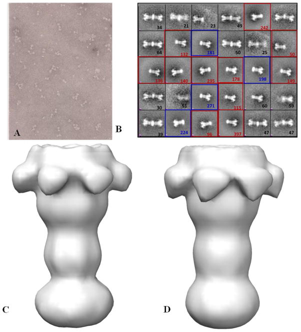
Figure 4.
Scale up of BLI microvolume EM method to generate three dimensional reconstructions of PA pore micelles. Panel A shows a representative negatively stained EM field of the large scale procedure to generate MSP/cholate/POPC micelle (prenanodisc micelle) solublized pore transitioned from thiol bead surfaces. B) 3369 LFN-PA micelle particles were picked to perform reference free 2D class averages and generate 30 separate classes. These classes contain larger ellipsoid prenanodisc micelle solublized pores, highlighted with red square boxes (reconstruction C); single pore inserted smaller rounded prenanodiscs (reconstruction D) highlighted with blue square boxes and a fair number of doubly inserted pore into nanodiscs. C&D) Negative stain 3D structures of first two prominent LFN-PA pore classes at 26 Å. The two prominent classes of prenanodisc micelles that coalse around the PA pore tip appear as expanded ellipsoid (C) or smaller round (D) shaped densities.

