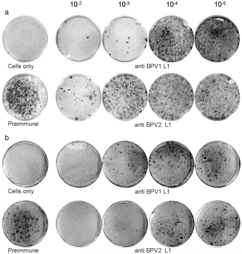
Fig. 3. Comparison of antisera to BPV1 and BPV2 VLPs by focus-forming neutralization assays.
Infectious BPV1 (a) or BPV2 virions (b) purified from cow warts were pre-incubated with immune sera, plated onto C127 fibroblasts, maintained for 3 weeks with regular feeding, and stained. Control plates received either no virus (cells only) or virus incubated with preimmune serum at 10−2 dilution. Antisera raised against BPV1- or BPV2-L1 VLP were serially diluted 10-fold from 10−2 to 10−5 as indicated. Neutralization titers are scored visually as the reciprocal of the highest serum dilution that results in ≥50% reduction in the number of foci obtained with preimmune serum.

