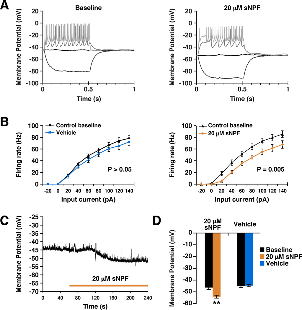
Figure 5. sNPF reduces neuronal resting membrane potential and suppresses action potential generation.
A. Example current clamp recordings made from third instar larval motor neurons expressing sNPFR before (left panel) and after (right panel) treatment with sNPF. Current was injected to depolarize the neuron and elicit action potentials. Only selected sweeps of the current injection protocol are shown for clarity. B. Quantification of current clamp data, plotting firing rate as a function of input current (F/I curve). Vehicle treatment (N = 6) in flies expressing sNPFR did not cause a significant change in F/I curve (left panel). sNPF (N = 11) caused a significant rightward shift in the curve, showing that more current was required to elicit the same spike rate after sNPF treatment (right panel). Data are plotted as mean ± SEM and significance calculated by one-way ANOVA. C. An example trace showing a typical hyperpolarization response to sNPF treatment. Duration of treatment is indicated by bar. D. Quantification of the effects of sNPF and vehicle treatment on resting membrane potential. Numbers in parentheses represent the number of animals in each condition. Data are presented as mean ± SEM. ** represents p <0.001, t-test. See also Figures S4 and S5.

