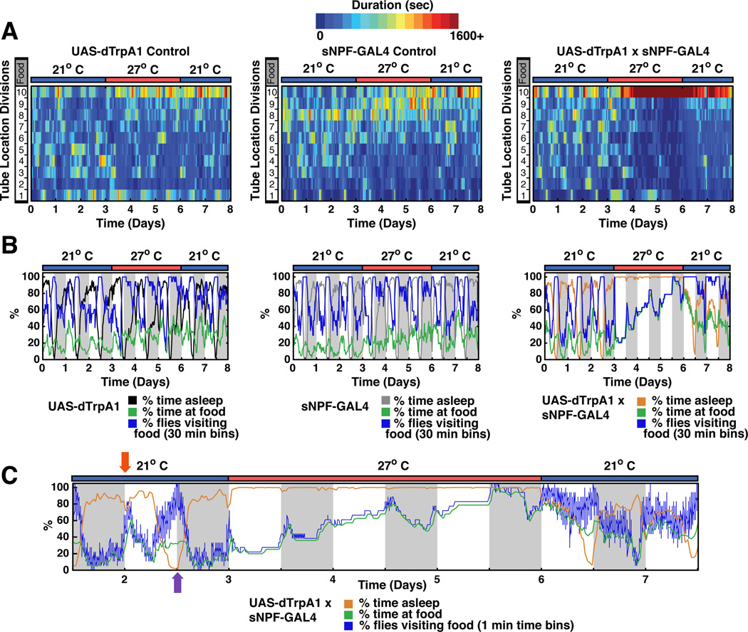
Figure 7. Activation of sNPF neurons alters sleep and food preference on different time scales.
A. Sleep, locomotor activity and position relative to food were monitored by computer video tracking (Donelson et al., 2012). In location heat plots, dark blue indicates flies spent no time at a particular location while dark red indicates flies spent more than 1,600 s at that location. The x-axis time and temperature and the y-axis indicates the location of each genotype within the behavioral tubes relative to food (location 10). Both parental control lines only showed slight increases in the time they spent on food upon heating, while the sNPFGAL4:UAS-dTRPA1 flies spent significantly more time on the food after heat activation. Sleep plots and sleep parameters for this data set are shown in Figure S6. B. Plots of % time asleep, % time at food and % flies visiting food for experiments in panel A. Control lines show a modest change in food dwelling with increased temperature. C. Expanded time scale for experimental fly data in panel B. During the morning peak of activity at 21°C when animals go to food they stay there (red arrow; blue line and green line overlap). During the evening peak of activity flies visit food often but do not stay. This likely reflects “patrolling behavior” (purple arrow; blue line higher than green line). Heat-induced acute firing of sNPF-expressing neurons dramatically and rapidly (within 1 h) increased sleep to maximal levels (orange line). The excessive sleep reversed within 1 h after inactivation by shifting temperature to 21°C. In contrast to the rapid effect on sleep, stimulation of sNPF expressing neurons caused a very slow accumulation of flies at the food which was not rapidly reversible. Flies remained closer to the food for at least 2 days after dTRPA1 N ≥ 17 for each genotype. See also Figure S7.

