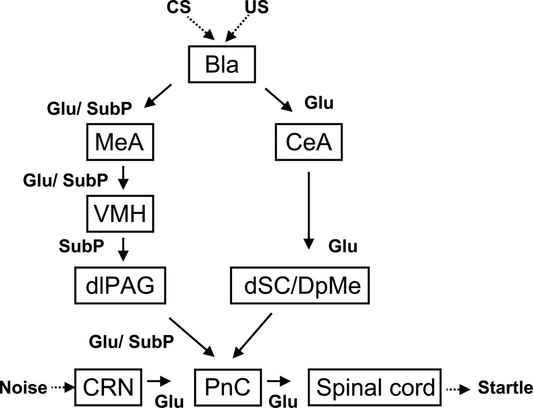
Figure 7.
A schematic diagram that illustrates the hypothesized neural pathways through which the amygdala that may mediate fear potentiated startle. Fear is induced by presence of the conditioning stimulus (CS) that was previously paired with the unconditioned stimulus (US), which is indicated by an increase in startle response in presence of the CS. The fear-induced increase in startle is mediated by the amygdala through two separate pathways, namely, the central-amygdalo-tegmento-pontine pathway and the medial-amygdalo-hypothalamo-periaqueducto-pontine pathway. The central-amygdalo-tegmento-pontine pathway involves the central amygdala (CeA), the deep layers of the superior colliculus/deep mesencephalic nucleus (dSC/DpMe) of the midbrain, and the nucleus reticularis pontis caudalis (PnC) of the brainstem. Glutamatergic neurotransmission is required in this pathway. The medial-amygdalo-hypothalamo-periaqueducto-pontine pathway involves the medial nucleus of the amygdala (MeA), the ventromedial nucleus of the hypothalamus (VMH), the dorsolateral periaqueductal gray (dlPAG) of the midbrain and the PnC. In this circuit substance P neurotransmission is required although we do not know how. We hypothesize that it modulates the release of glutamate in some but not all of these nuclei, including the PnC based on the work of . The startle circuit consists of cochlear root neurons (CN) in the auditory nerve, the PnC and the motor neurons in the spinal cord, where noise impinges onto the CN and subsequently elicit rapid startle reflex. The excitability of this circuit can be elevated by the excitatory inputs from the amygdala to the PnC, thus resulting in increased startle.

