

Sir,
Pap test has been an effective way to screen women for squamous dysplasia and carcinoma of the uterine cervix. Although the Pap test is primarily used for detecting cellular and epithelial abnormalities, certain infections are detected on the Pap test too e.g., donovanosis, tuberculosis, coccidioidomycosis, schistosomiasis, teniasis, molluscum contagiosum.[1] These rare entities pose challenges due to the infrequent occurrence of these entities in the daily practice of cytology. These conditions may also give rise to diagnostic pitfalls to be aware of in Pap tests. We are reporting an incidental finding of Calymmatobacterium granulomatis/ Donovan bodies on Pap test.
A 27-year-old married woman presented with chief complaint of recurrent seropurulent vaginal discharge. She was treated with anti-fungals repeatedly but got only symptomatic relief for some time. Her spouse was also counseled and treated. A complete history and examination was sought in the Indian Council of Medical Research (ICMR) clinic Mandi, Himachal Pradesh. There was a history of seropurulent discharge and itchy ulcerations on the vulva for past one year. There was no promiscuous sexual behavior. Both partners were non reactive for human immunodeficiency virus and Veneral Diseases Research Laboratory test. A Pap test was taken. Pap smears were satisfactory and negative for intraepithelial lesion/malignancy. In addition, smears showed acute inflammatory cell infiltrate with numerous macrophages. The macrophages contained numerous safety pin shaped structures with polar thickening (donovan bodies) and a halo around them [Figure 1]. Pap smears was reported positive for Calymmatobacterium granulomatis/ Donovanosis. It was an incidental finding in a patient who was given erroneous treatment for an intractable infection in an on-going cancer control project in Himachal Pradesh.
Figure 1.
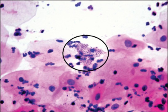
Macrophages show numerous safety pin shaped structures with polar thickening of chromatin (donovan bodies) and a halo around them (circle) (Pap, ×100)
Donovanosis is another name for granuloma inguinale. It is a bacterial sexually transmitted disease (STD) caused by a gram negative, intracellular, non-motile, non-sporing cocco-bacillus Calymmatobacterium granulomatis also known as Donovan body. Pathogenicity is limited to humans only,[2] and is seen more frequently in Africa and Asia.[3] Incubation period ranges from 1 to 12 weeks. It begins as a painless papule on the genitalia, which ulcerates slowly.[2] In early stages donovanosis is difficult to differentiate from chancroid.[4] The chancroid ulcers are painful whereas those of donovanosis are painless. In later stages, it is difficult to differentiate from lymphogranuloma venereum (LGV) and anogenital cutaneous amebiasis.[4] LGV is also a STD caused by Chlamydia trachomatis characterized by painless ulcer and lymphadenopathy (buboes). Anogenital amebiasis is diagnoses by identifying ameba from the ulcers. Microscopically, Donovan body appears as rounded 1-2 μm in diameter, within thin-walled intracytoplasmic vacuoles inside mononuclear cells. They show bipolar condensation of chromatin, giving a closed ‘safety pin’ appearance in stained smears. Neutrophilic infiltrate without giant cells is often seen. Reactive epithelial cells are usually scant and marked reparative change may be found in case of ulceration. The differential diagnosis includes follicular cervicitis, granulation tissue, and other granulomatous diseases, including malakoplakia.[5] In follicular cervicitis, lymphoid cell infiltrate is seen instead of neutrophils along with tingible-body macrophages with cellular debris. In malakoplakia, cytoplasmic inclusions called Michaelis-Gutmann bodies are more dense, laminated and round, compared to Donovan bodies. Ancillary studies that may be helpful include special stains (e.g. Romanowsky and Warthin-Starry stains) to highlight Donovan bodies. In such granulomatous cases it is also important to perform acid-fast stains for mycobacteria and fungal stains (e.g. Gomori methenamine silver).
In conclusion rare entities may be encountered on Pap test. Finding and accurately diagnosing these entities is important, even if their presence does not always signify infection, but rather contamination from vulvar or genital lesions or fecal material. Some of them are sexually transmitted, and their presence should prompt screening for other STDs. Therefore, knowledge of the cytomorphology of unusual entities and their diagnostic pitfalls is important.
References
- 1.Khalbuss WE, Michelow P, Benedict C, Monaco SE, Pantanowitz L. Cytomorphology of unusual infectious entities in the Pap test. Cytojournal. 2012:9–15. doi: 10.4103/1742-6413.97763. [DOI] [PMC free article] [PubMed] [Google Scholar]
- 2.Ananthanarayan R, Panikar CK. Textbook of microbiology. 6th ed. Hyderabad: Orient Longman; 2000. Miscellaneous bacteria-Granuloma inguinale; pp. 373–4. [Google Scholar]
- 3.Chandra M. Cervical smear with intracellular organisms from a case of granuloma venereum (donovanosis) Acta Cytol. 1991;35:143–5. [PubMed] [Google Scholar]
- 4.Ballard RC. Klebsiella granulomatis (Donovanosis, Granuloma Inguinale) In: Mandell GL, Bennett JE, Dolin R, editors. Principles and practice of infectious diseases. 7th ed. Philadelphia: Elsevier Churchill Livingstone; 2009. [Google Scholar]
- 5.Ramdial PK, Sing Y, Chotey NA, Bagratee JS. Concomitant malacoplakia and granuloma inguinale of the cervix in acquired immune deficiency syndrome. Int J Gynecol Pathol. 2008;27:282–7. doi: 10.1097/PGP.0b013e31815788fc. [DOI] [PubMed] [Google Scholar]