Abstract
Homogeneously staining regions (HSR) or double minute chromosomes (dmin) are autonomously replicating extra-chromosomal elements that are frequently associated with gene amplification in a variety of cancers. The diagnosis of leukemia patients was based on characterization of the leukemic cells obtained from bone marrow cytogenetics. This study report two cases, one with Acute Myeloblastic Leukemia without maturation (AML-M1), aged 23-year-old female, and the other with chronic myelogenous leukemia (CML)-blast crisis, a 28-year-old female associated with double minute chromosomes. Most cases of acute myeloid leukemia with dmin in the literature (including our cases) have been diagnosed as having acute myeloid leukemia.
Keywords: AML, cancer, chronic myelogenous leukemia, homogeneously staining regions dmin, HSRs, Iran, incidence, leukemia, new case
INTRODUCTION
Double minute chromosomes (dmin) or Homogeneously Staining Regions (HSRs) was first described in a direct preparation of cells from patient with untreated bronchogenic carcinoma.[1] Sait et al., (2002)[2] reported for the first time that dmin are from chromosome 19. Homogeneously staining of regions or Dmin are the cytogenetic hallmarks of genomic amplification in cancers.[3] Furthermore, dmin/HSRs are from the breakpoint region of translocation event.[4] Although found in a variety of human tumor cells, their presence in hematologic malignancies is rare.[5] Also, their role in leukemogenesis is not clear but they have been reported to be associated with rapid progression and short survival time.[5] Dmin are found in tumor cell proliferations, characteristically varying in number from cell to cell. They are thought to be involved in tumor genesis and in drug resistance.[6] Dmin are small chromatin particles that represent a form of extra chromosomal gene amplification.[7] Gene amplification causes an increase in the gene copy number and, subsequently, elevates the expression of the amplified genes, which modify normal growth control and survival pathway.[8,9,10] In this connection, C-myc was the most frequently amplified gene, but cases with mixed lineage leukemia (MLL) gene amplification have also been reported in the current literature.[2,11] The semi conservative replication of Deoxyribonucleic acid (DNA) in dmin has been demonstrated to occur in both human and mouse cell line.[12] Here, we present two cases with leukemia associated with dmin. The latest data regarding dmin was reported by Mitelman database (http://cgapanci.nih.gov/chromosomes/Mitelman)[9] and own peer reviewed publication.[13]
MATERIALS AND METHODS
We receive bone marrow (BM) and peripheral blood (PB) specimen from AML and CML diagnosed adult patients at initial presentation. The diagnosis of leukemia patients was based on characterization of the leukemic cells, obtained from bone marrow and molecular cytogenetic, when appropriate. In each patient, 0.5 - 1.0 ml BM/PB was obtained and studied using; (a) a 24 h unstimulated culture technique and (b) Methotrexate cell synchronization method,[14] with some modification. For culture, 3-5 × 10⁶ cells were cultured in 4 ml medium (RPMI 1640, Gibco-BRL Grand Island, NY, USA) supplemented with 15% heat inactivated fetal bovine serum (Gibco-BRL Grand Island, NY, USA) at 37°C in an atmosphere containing 5% CO2. For methotrexate (MTX) synchronization, BM/PB cells were synchronized with 10-7 M MTX after 1.0-5.0 h of culture. The S-phase block of synchronized cells was released after 17 h by the adding of 10-5 M Thymidin for 3.0-6.0 h. Standard MTX cell synchronization procedures for cultures, collection of samples, and slide preparation were modified and performed in our laboratory.[14] Briefly, the cultured cells were then treated with Colcemide (Gibco-BRL Grand Island, NY, USA) final concentration, 10 μg/ml and incubated at 37°C for an additional 3 min. The contents of the tube were then centrifuged for 10 min at 1,000 rpm and re-suspend in 10 ml of 75 mM KCl (0.56%) pre-warmed to 37°C for 20 min. At this stage, 1 ml of Carnoys Fixative (3:1 methanol: Acetic acid) was added in to the tube, and this fixation step was repeated four times. Ten slides were prepared for each culture and stained for 3 min with Giemsa G banding.[15] Slides were examined with Yisis/Applied imagine system. Eighty well-spread metaphases were analyzed for each subject. Karyotypes were described according to International System for Chromosome Nomenclature (ISCN).[16]
CASE HISTORY
Two patients with CML and AML were seen between 1994 and 2010, at Shahid Beheshti University of Medical Sciences and various private clinics, in Tehran, Iran.
In patient 1, a 23-year-old female, in previous good health, presented with weight loss, tiredness. Her peripheral blood hemoglobin was 6.2 g/dl with a platelet count of 100 × 109/l (differential: 77% blast, 1% neutrophils, 22% lymphocytes). The marrow aspirate was cellular with reduction in all normal marrow elements and marked increased in Sudan Black-positive myelobalst. G-banding analysis in patient one, AML-M1 showed that all of 20 metaphases cells revealed both numerical and structural abnormalities, including 46, XX, t (9;22)(q34.1;q11) + dmin; 45, XX, t (9;22)(q34.1;q11), -15. Sixteen of the 20 cells (80%) also had 3 − 19 dmin in each cell. Her past medical history noticed without history of prior toxic exposure.
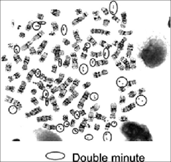
In patient 2, with CML-blast crisis, a 28-year-old female, was the subject of dms. On an admission, splenomegaly was found, count was 15.1 × 10⁴/mm3, hemoglobin was 7.2 g/dl, RBC 224 × 104/mm3 and bone marrow examination revealed hypercellularity with promyelocyte 14.2% and myeloblast 49.7%. G-banding analysis revealed that 14 of the 80 metaphases cells (70%) [Figure 1], had dmin, including [t (9;22)(q34.1;q11)] + dmin. Her past medical history was notable only for occupation for cultivation as Farm Yard Manure. There were no results detected of HSR in chromosomes for both the patients.
Figure 1.

Giemsa staining of bone marrow cell containing dmin in the metaphase of leukemia patients
DISCUSSION
The presence of dmin with two leukemia patients, which were identified in our laboratory, was also observed in other parts of the world. These two new cases of chromosomes will be discussed in the following sections utilizing the largest chromosome aberrations database in cancers (http://cgapanci.nih.gov/chromosomes/Mitelman).[9] Also, Thomas et al., (2004)[5] documented 33 cases with dmin in acute myeloid leukemia.
The relationship of dmin/HSRs and malignancies seems well established and, indeed, dmin/HSRs have not, so far, been observed in non malignant cell.[3] Gene amplification cause an increase in the gene copy number and, subsequently, elevate the expression of the amplified genes, which modify normal growth control and survival pathway.[7,8,9,10] Dmin represent a mechanism for upregulated oncogene expression and are generally associated with a poor prognosis. This type of gene amplification has been found in various solid tumors, such as colon, pancreatic, breast carcinoma, brain tumors, and neuroblastoma (http://cgapanci.nih.gov/chromosomes/Mitelman).[9] However, it appears to be less common in AML.[5] Generally, C-myc amplification results in an over expression of the myc protein, which is known to be a critical nuclear transcription factor.[10,11] Some results suggested that dmin and HSR are alternative manifestations of the same biological phenomenon.[17]
Amplification of the ETS1, FL11, SRPR, NFRKB, and KCNJ4 genes located at 11q23-24 distal to MLL was demonstrated in a patient with AML.[12,18] Many oncogenes have been identified on dmin, for example, MYCN, C-myc, EIFA2, and MDM2.[5,9,19]
Several early reports concluded that dmin, whose number and size vary from cell to cell, were not viral or bacterial contamination. Also, past medical history of both cases presented here, recorded without history of any micro organism contaminations and history of previous malignancies. In case 2, past medical history was notable only for occupation for cultivation as farm yard manure. It is coincident with previous reports indicating that dmin do occur in leukemic patients without previous history of malignancy.[20] On the contrary, a relation between dmin and previous mutagenic exposure has been suggested by other scientists.[21] It is reasonable to suggest that the demonstration of dmin in patients with AML in the present study might be an indication that these patients have a previous history of malignant disease treated with irradiation or chemotherapy or both.
The identification of two new cases of dmin presented here are secondary, apparently non-random associated with leukemia. However, further studies and accumulation of new cases are needed in the hope of defining it as specific abnormalities in the field of leukemia.
ACKNOWLEDGMENT
We thank Dr Fatemeh Isfahani, Dr Abbas Hajfathali, Dr Hamid Attarian, and Dr Hamid Rezvani; Departments Medical Oncology, Internal Medicine. Thanks are also due to Miss Niloofar Safavi for excellent technical assistance and material collection.
Footnotes
Source of Support: Nil
Conflict of Interest: None declared.
REFERENCES
- 1.Spriggs AI, Boddington MM, Clarke CM. Chromosomes of human cancer cells. Br Med J. 1962;2:1431–5. doi: 10.1136/bmj.2.5317.1431. [DOI] [PMC free article] [PubMed] [Google Scholar]
- 2.Sait SN, Qadir MU, Conroy JM, Matsui S, Nowak NJ, Baer MR. Double minute in acute myeloid leukemia and myelodysplastic syndrome: Identification of new amplification regions by fluorescence in situ hybridization and spectral karyotyping. Genes Chromosomes Cancer. 2002;34:42–7. doi: 10.1002/gcc.10038. [DOI] [PubMed] [Google Scholar]
- 3.Storlazzi CT, Lonoce A, Guastadisegni MC, Trombetta D, D’Addabbo P, Daniele G, et al. Gene amplification as double minutes or homogeneously stain regions in solid tumors: Origin and structure. Genome Res. 2010;20:1198–206. doi: 10.1101/gr.106252.110. [DOI] [PMC free article] [PubMed] [Google Scholar]
- 4.Reddy KS. Double minute (dmin) and homogeneously staining region (hsr) in myeloid disordes: A new case suggesting that dmin from hsr in vivo. Cytogenet Genome Res. 2007;119:53–9. doi: 10.1159/000109619. [DOI] [PubMed] [Google Scholar]
- 5.Thomas L, Stamberg J, Gojo I, Ning Y, Rapoport AP. Double minute in monoblastic (M5) and myeloblastic (M2) acute myeloid leukemia: Two case reports and a review of literature. Am J Hematol. 2004;77:55–61. doi: 10.1002/ajh.20151. [DOI] [PubMed] [Google Scholar]
- 6.Von Hoff DD, McGill JR, Forseth BJ, Davidson KK, Bradley TP, Van Devanter DR, et al. Elimination of extrachromosomeally amplified MYC genes from human tumor cells reduceds their tumorigenicity. Pro Natl Acad Sci USA. 1992;89:8165–9. doi: 10.1073/pnas.89.17.8165. [DOI] [PMC free article] [PubMed] [Google Scholar]
- 7.Pappou EP, Ahuja N. The role of oncogenes in gastrointestinal cancer. Gastrointest Cancer Res. 2010;(Suppl 1):S2–15. [PMC free article] [PubMed] [Google Scholar]
- 8.Fichter CD, Herz C, Munch C, Opitz OG, Werner M, Lassmann S. Occurrence of multipolar mitoses and association with Aurora-A/-B kinases and p53 mutations in aneuploid esophageal carcinoma cells. BMC Cell Biol. 2011;12:13. doi: 10.1186/1471-2121-12-13. [DOI] [PMC free article] [PubMed] [Google Scholar]
- 9.Fan Y, Mao R, Lv H, Xu J, Yan L, Liu Y, et al. Frequency of double minute chromosomes and combined cytogenetic abnormalities and their characteristics. J Appl Genet. 2011;52:53–9. doi: 10.1007/s13353-010-0007-z. [DOI] [PubMed] [Google Scholar]
- 10.Yan SY, Hu Y, Fan JG, Tao GQ, Lu YM, Cai X, et al. Clinicopathologic significance of HER-2/neu protein expression and gene amplification in gastric carcinoma. World J Gastroenterol. 2011;17:1501–6. doi: 10.3748/wjg.v17.i11.1501. [DOI] [PMC free article] [PubMed] [Google Scholar]
- 11.Guan XY, Sham JS, Tang TC, Fang Y, Huo KK, Yang JM. Isolation of a novel candidate oncogene within a frequently amplified region at 3q26 in ovarian cancer. Cancer Res. 2001;61:3806–9. [PubMed] [Google Scholar]
- 12.Crossen PE, Morrison MJ, Rodley P, Cochrane J, Morris CM. Identification of amplified genes in a patient with acute myeloid leukemia and double minute chromosomes. Cancer Genet Cytogenet. 1999;113:126–33. doi: 10.1016/s0165-4608(99)00018-7. [DOI] [PubMed] [Google Scholar]
- 13.Movafagh A, Mirfakhraei R, Mousavi-Jarrahi A. Frequent incidence of dmin in cancers, with special up-to-date references to leukemia. Asian Pac J Cancer Prev. 2011;12:3453–6. [PubMed] [Google Scholar]
- 14.Misawa S, Yashige H, Horiike S, Taniwaki M, Nishigaki H, Okuda T, et al. Detection of karyotypic abnormalities in most patients with actue nonlymphocytic leukiemia by adding ethidium bromide to short-term cultures. Leuk Res. 1988;12:719–29. doi: 10.1016/0145-2126(88)90004-5. [DOI] [PubMed] [Google Scholar]
- 15.Seabright M. A rapid banding technique for human chromosomes. Lancet. 1971;2:971–2. doi: 10.1016/s0140-6736(71)90287-x. [DOI] [PubMed] [Google Scholar]
- 16.Brothman AR, Persons DL, Shaffer LG. Nomenclature evolution: Changes in the ISCN from the 2005 to the 2009 edition. Cytogenet Genome Res. 2009;127:1–4. doi: 10.1159/000279442. [DOI] [PubMed] [Google Scholar]
- 17.Uehara M, Kida M, Kamakura M. Ring and double minute in a case with blastic phase of chronic myeloid leukemia. Cancer Genet Cytogenet. 1987;25:253–8. doi: 10.1016/0165-4608(87)90185-3. [DOI] [PubMed] [Google Scholar]
- 18.Kawamata N, Zhang L, Ogawa S, Nannya Y, Dashti A, Lu D, et al. Double minute containing MYB gene and NUP214-ABL1 fusion gene in T-cell leukemia detected by single nucleotide polymorphism DNA microarray and fluorescence in situ hybridization. Leuk Res. 2009;33:569–71. doi: 10.1016/j.leukres.2008.07.030. [DOI] [PMC free article] [PubMed] [Google Scholar]
- 19.Bruckert P, Kappler R, Scherthan H, Link H, Hagmann F, Zankl H. Double minutes and c-MYC amplification in acute myelogenous leukemia: Are they prognostic factors? Cancer Genet Cytogenet. 2000;120:73–9. doi: 10.1016/s0165-4608(99)00235-6. [DOI] [PubMed] [Google Scholar]
- 20.Harteley SE, Toolis F. Dmin in a case of acute myeloblastic leukemia. Cancer Genet Cytogenet. 1980;2:275–80. [Google Scholar]
- 21.Weh HJ, Zschaber R, Hossfeld DK. Double minute chromosome: A frequent marker in leukemic patients with previous history of malignant disease? Cancer Genet Cytogenet. 1982;5:279–80. doi: 10.1016/0165-4608(82)90035-8. [DOI] [PubMed] [Google Scholar]


