Abstract
Notch signaling is frequently hyperactivated in breast cancer, but how the enhanced signaling contributes to the tumor process is less well understood. In this report, we identify the proinflammatory cytokine interleukin-6 (IL-6) as a novel Notch target in breast tumor cells. Enhanced Notch signaling upregulated IL-6 expression, leading to activation of autocrine and paracrine Janus kinase/signal transducers and activators of transcription signaling. IL-6 upregulation was mediated by non-canonical Notch signaling, as it could be effectuated by a cytoplasmically localized Notch intracellular domain and was independent of the DNA-binding protein CSL. Instead, Notch-mediated IL-6 upregulation was controlled by two proteins in the nuclear factor (NF)-κB signaling cascade, IKKα and IKKβ (inhibitor of nuclear factor kappa-B kinase subunit alpha and beta, respectively), as well as by p53. Activation of IL-6 by Notch required IKKα/IKKβ function, but interestingly, did not engage canonical NF-κB signaling, in contrast to IL-6 activation by inflammatory agents such as lipopolysaccharide. With regard to p53 status, IL-6 expression was upregulated by Notch when p53 was mutated or lost, and restoring wild-type p53 into p53-mutated or -deficient cells abrogated the IL-6 upregulation. Furthermore, Notch-induced transcriptomes from p53 wild-type and -mutated breast tumor cell lines differed extensively, and for a subset of genes upregulated by Notch in a p53-mutant cell line, this upregulation was reduced by wild-type p53. In conclusion, we identify IL-6 as a novel non-canonical Notch target gene, and reveal roles for p53 and IKKα/IKKβ in non-canonical Notch signaling in breast cancer and in the generation of cell context-dependent diversity in the Notch signaling output.
Keywords: cytokine, inflammation, tumor stroma, estrogen receptor, breast cancer, BCL
Introduction
Notch signaling is frequently deregulated in breast cancer, and hyperactivation of Notch contributes to the tumor process. Gene rearrangements generating gain-of-function versions of Notch receptors have recently been described in breast tumors,1 and upregulation of Notch signaling is linked to loss or inactivation of Numb, a negative regulator of Notch.2, 3 Similarly, elevated expression of the Notch ligand JAGGED1 correlates with poor prognosis in breast cancer, and overexpression of NOTCH1 and NOTCH4 receptors is observed in triple-negative breast tumors.4, 5 Pharmacological intervention with estrogen receptor (ER) function, by tamoxifen, leads to enhanced Notch signaling,6 which may be an unwanted side effect of the current use of tamoxifen in breast cancer therapy. In addition, deregulation of Notch activity leads to a glycolytic switch in breast tumor cells, and elevated Notch signaling correlates with more invasive growth in breast tumor xenograft experiments.7
The Notch signaling pathway is an evolutionarily highly conserved signaling mechanism for cell–cell communication. Activation of Notch signaling by membrane-bound ligands on juxtaposed cells results in proteolytic processing of the Notch receptor by the γ-secretase complex, ultimately releasing the intracellular domain of the Notch receptor (Notch ICD). Notch ICD is translocated from the cytoplasm to the nucleus, where it binds to the DNA-binding protein CSL (RBP-Jκ) to activate transcription of downstream genes.8 Activation through Notch ICD/CSL is referred to as canonical Notch signaling, and in addition there are non-canonical modes of Notch signaling, including CSL-independent Notch signaling.9 Although we still have a quite limited understanding of how non-canonical Notch signaling works, it appears to be important in breast cancer, as mammary tumors arise in a mouse model overexpressing Notch4 ICD in a CSL-deficient mammary tissue background.10 Furthermore, non-canonical Notch signaling is involved in abrogating Bax-induced apoptosis.11
It is increasingly understood that inflammation and proinflammatory cytokines are important for the tumor process12 and in this report, we explore the effects of hyperactivated Notch signaling in breast cancer, with a particular focus on non-canonical Notch signaling, and the proinflammatory cytokine interleukin-6 (IL-6). IL-6 activates the IL-6 receptor to initiate signaling through the Janus kinase (JAK)/signal transducers and activators of transcription (STAT) signaling pathway,13 and elevated levels of IL-6 correlate with poor prognosis for breast cancer patients.14, 15 We show that Notch upregulates IL-6 expression, leading to activation of JAK/STAT signaling. IL-6 expression was regulated by non-canonical Notch signaling, as a cytoplasmically localized Notch ICD was sufficient to induce IL-6 expression. The non-canonical activation of IL-6 was shown to be dependent on IKKα and IKKβ (inhibitor of nuclear factor kappa-B kinase subunit alpha and beta, respectively), but Notch signaling did not engage canonical NF-κB (nuclear factor kappa-light-chain-enhancer of activated B cells) signaling, in contrast to activation of IL-6 by inflammatory agents, which requires canonical NF-κB signaling. Furthermore, the upregulation of IL-6 was dependent on the p53 status in the cell: IL-6 expression was upregulated by Notch when p53 was mutated or lost. Collectively, these data identify IL-6 as a novel non-canonical Notch target gene, and provide evidence of roles for p53 and IKKα/IKKβ in the regulation of non-canonical Notch signaling in breast cancer and in generating diversity in the Notch downstream response.
Results
Elevated Notch signaling upregulates IL-6 mRNA and protein expression
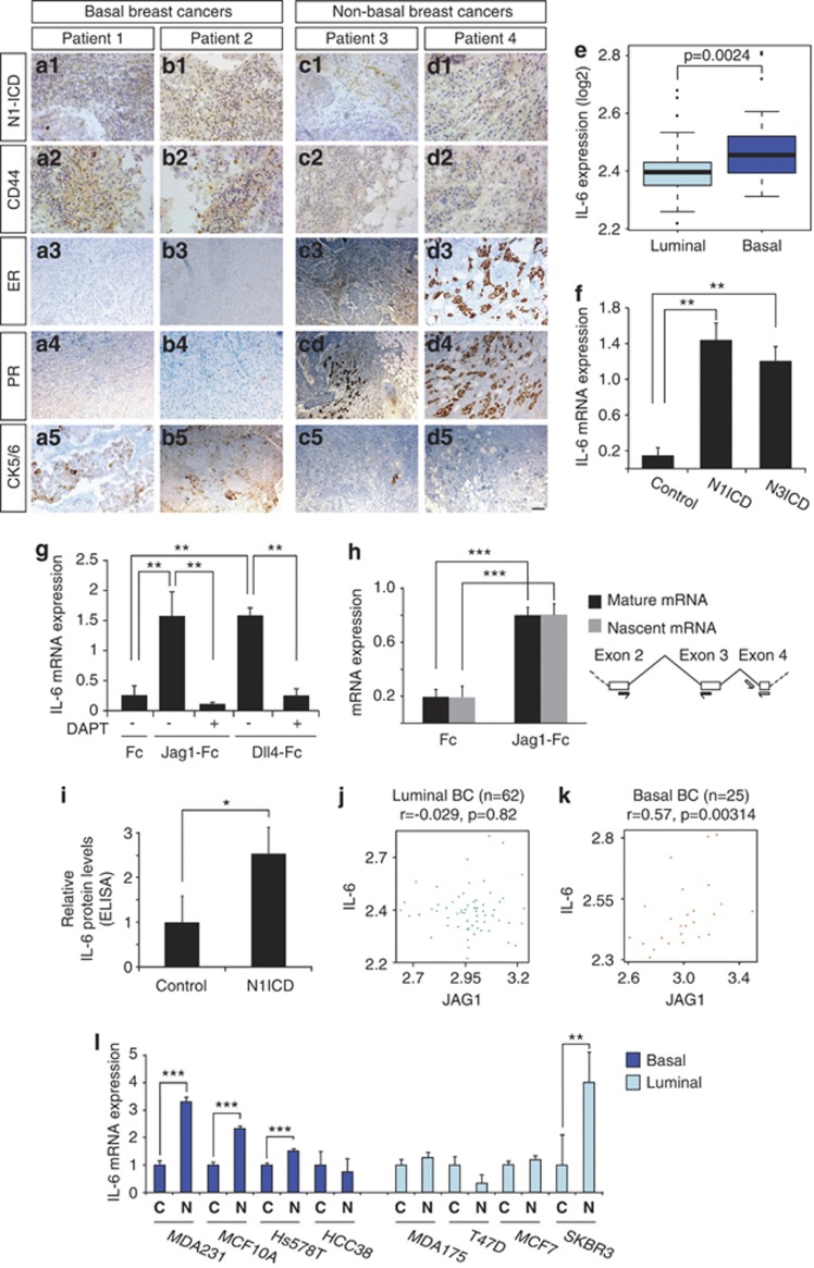
We first analyzed a panel of breast cancers for expression of markers for basal and non-basal breast cancers relative to the status of active Notch signaling. Breast cancers that were of the basal type and thus expressed CD44 and cytokeratin 5/6 but were negative for progesterone and ER-α expression exhibited high expression of nuclear Notch1 ICD immunoreactivity (Figures 1a and b, Supplementary Figure 1). In contrast, breast cancers of the non-basal type, which expressed progesterone and ER-α receptors but only low levels of CD44 and cytokeratin 5/6, showed considerably lower levels of Notch1 ICD immunoreactivity (Figures 1c and d, Supplementary Figure 1). Enhanced IL-6 mRNA expression was also found in basal breast cancers, as compared with luminal breast cancers (Figure 1e; see also Sethi et al.14 and Wang et al.15). This observation, combined with the notion that high IL-6 levels correlate with poor prognosis for breast cancer patients,14, 15 led us to explore a link between Notch signaling and the cytokine response in breast cancer, and to ask whether IL-6 expression was regulated by Notch signaling. Expression of activated forms or Notch1 or Notch3, Notch1 ICD and Notch3 ICD, respectively, led to an increase in IL-6 mRNA expression in the basal type MDA-MB-231 cell line (Figure 1f). Similarly, activation of endogenous Notch signaling by culturing MDA-MB-231 cells on immobilized Jagged1 or Dll4 ligand elevated IL-6 mRNA expression (Figure 1g). The IL-6 mRNA upregulation was dependent on proteolytic processing of the Notch receptors to liberate Notch ICD, as it was abrogated by the γ-secretase inhibitor 5-difluorophenylacetyl-L-alanyl-2-phenylglycine-1,1-di- methylethyl ester (DAPT) (Figure 1g). To learn whether Notch regulated IL-6 expression at the transcriptional or post-transcriptional level, we compared the levels of nascent (pre-splicing) and mature (spliced) IL-6 mRNA in response to Notch activation. Nascent and mature IL-6 mRNA were upregulated to the same extent (Figure 1h), indicating that Notch transcriptionally controls IL-6 expression. The increase in IL-6 mRNA resulted in elevated IL-6 protein levels in the cell culture medium, as determined by an IL-6 enzyme-linked immunosorbent assay (Figure 1i).
Figure 1.
Notch signaling controls IL-6 expression. (a-d) Expression of Notch1 ICD in two basal (a, b) and two non-basal (c, d) breast cancers (data from 12 additional patients are shown in Supplementary Figure 1). Notch1 ICD expression (a1–d1) was analyzed by immunohistochemistry for the N-terminus of Notch1 ICD generated following γ-secretase cleavage. Expression of CD44 (a2–d2), estrogen receptor-α (ER; a3–d3), progesterone receptor (PR; a4–d4) and cytokeratin 5/6 (CK5/6; a5–d5) was analyzed using previously described antibodies. (e) Expression levels of IL-6 in luminal and basal breast tumors16 (f, g) IL-6 mRNA expression measured by quantitative PCR (QPCR) in MDA-MB-231 cells (f) infected with adenoviral vectors expressing GFP, Notch1 ICD (N1ICD) or Notch3 ICD (N3ICD) or (g) cultured on immobilized Jagged1 or Dll4 ligands (Jag1-Fc and Dll4-Fc, respectively) or Fc fragments as control (Fc). In some of the experiments in g, the γ-secretase inhibitor DAPT was used to block Notch receptor proteolytic processing. (h) Analysis of effects of Notch activation on production of nascent versus mature IL-6 mRNA. Primer pairs were designed to capture nascent (pre-splicing) and mature (spliced) IL-6 mRNA (right), and the amount of nascent and mature IL-6 mRNA was analyzed by Q-PCR after culture on Fc-Jag1 or Fc (left). (i) Protein analysis by enzyme-linked immunosorbent assay (ELISA) from MDA-MB-231 cells transfected with Notch1 ICD or enhanced green fluorescent protein (EGFP) (control). (j, k) Correlation between IL-6 and Jagged1 mRNA data from luminal (j) and basal (k) breast cancer transcriptome data.16 (l) Expression of IL-6 mRNA in response to Notch (Notch1 ICD) activation in four basal and four luminal breast cancer cell lines. Values are significant at ***P<0.001, **P<0.01 and *P<0.05. r, correlation coefficient. Graphs represent average of three independent experiments.
To assess whether high levels of IL-6 mRNA correlated with elevated Notch signaling in breast cancers, we examined a previously reported data set containing various types of breast cancer stratified into luminal or basal breast tumors.16 As the Notch transcriptome is quite varied in different tumor types (see Andersson et al.17 and Figure 4 below), we used expression of the Jagged1 ligand as a proxy for Notch signaling, in keeping with a previous report.14 We found that IL-6 and Jagged1 mRNA expression correlated in basal but not in luminal (A+B) breast cancers (Figures 1j and k). We also observed that three out of four basal type of breast cancer cell lines upregulated IL-6 mRNA expression in response to Notch activation, whereas only one out of four luminal cell lines showed enhanced IL-6 expression following Notch stimulation (Figure 1l). In conclusion, the data show that IL-6 is a novel Notch target gene in breast tumor cells, and that both Notch and IL-6 expression is higher in basal breast cancers.
Notch-induced increase in IL-6 expression results in autocrine and paracrine activation of JAK/STAT signaling
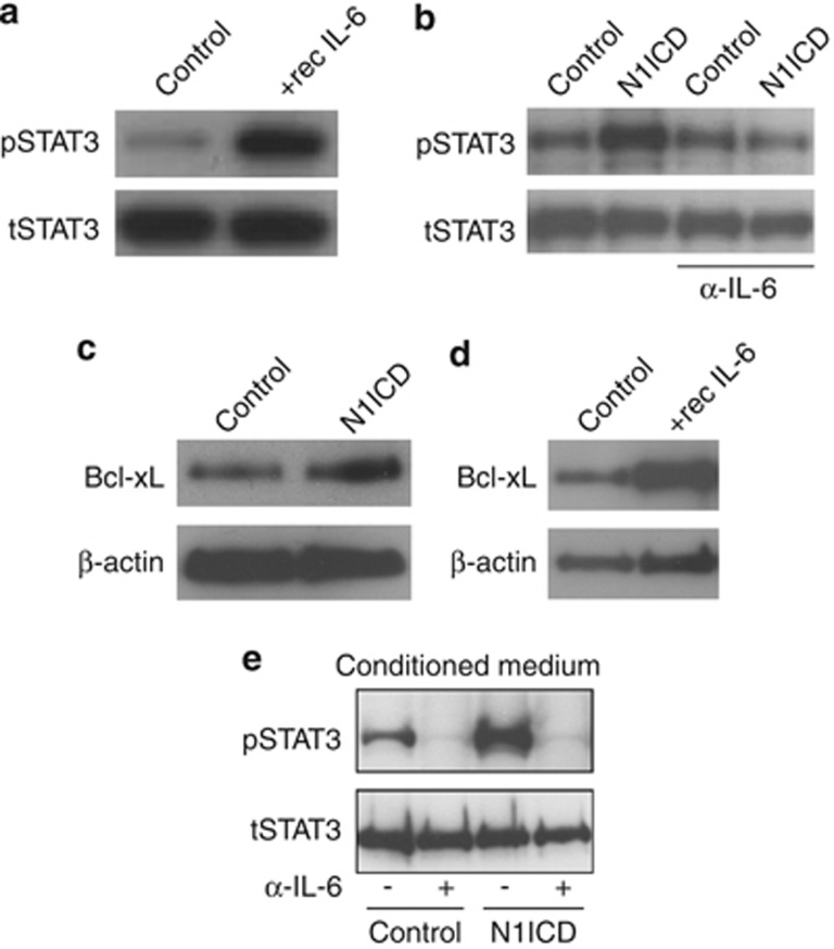
We next assessed whether the observed upregulation of IL-6 expression by Notch led to activated JAK/STAT signaling. Recombinant IL-6 potently enhanced the level of Tyr705 phosphorylated STAT3 in the MDA-MB-231 cells (Figure 2a), and a robust increase in phosphorylated STAT3 was also observed 12 h after expression of Notch1 ICD, which could be reduced by addition of an IL-6 blocking antibody to the cell culture medium (Figure 2b). Furthermore, expression of the JAK/STAT target gene Bcl-xL,18 which encodes an antiapoptotic protein, was elevated by Notch1 ICD (Figure 2c) and as control, by recombinant IL-6 (Figure 2d). To assess paracrine activation, we subjected naive MCF7 cells to conditioned medium from MBA-MB-231 cells, which had been transduced with Notch1 ICD or enhanced green fluorescent protein. Conditioned medium from Notch1 ICD-transduced cells resulted in a robust increase of phosphorylated STAT3 in the MCF7 cells, which could be blocked by an IL-6 blocking antibody (Figure 2e). In sum, these data suggest that IL-6 produced in response to Notch activation can activate JAK/STAT signaling in both an autocrine and paracrine manner.
Figure 2.
Notch activates JAK/STAT signaling via upregulation of IL-6. (a) Addition of recombinant IL-6 to the cell culture medium of MDA-MB-231 cells (+ rec IL-6) led to elevated levels of pTyr705 STAT3 (phosphorylated STAT3 (pSTAT3)) but not total STAT3 (tSTAT3) protein. (b) Analysis of pSTAT3 (upper) and total STAT3 (lower) protein levels in MDA-MB-231 cells infected with adenoviral vectors expressing Notch1 ICD or enhanced green fluorescent protein (EGFP) (control) as indicated. In the two rightmost lanes, an IL-6 blocking antibody (α-IL-6) was added to the cell culture medium before Notch1 ICD or EGFP expression. (c, d) Analysis of (c) Bcl-xL protein levels in MDA-MB-231 cells infected with adenoviral vectors expressing Notch1 ICD or EGFP (control) or (d) in MDA-MB-231 cells supplemented with recombinant IL-6, and with phosphate-buffered saline (PBS) as control. (e) Analysis of paracrine IL-6 activation in MCF7 cells. Conditioned medium from MDA-MB-231 cells transfected with Notch1 ICD or EGFP (control). The conditioned medium was pre-treated with an IL-6 blocking antibody (α-IL-6) before supplemented to the MCF7 cells, and the levels of pSTAT3 and total STAT3 protein were analyzed.
Notch-mediated upregulation of IL-6 depends on the cellular p53 status
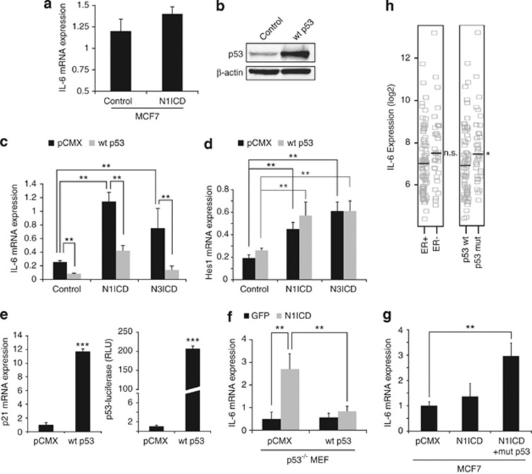
On testing other breast tumor cell lines for induction of IL-6 expression, we noted that IL-6 was not significantly upregulated in MCF7 cells, in contrast to MDA-MB-231 cells. (Figure 3a, see also Figure 1l). MDA-MB-231 and MCF7 cells differ in two important regards: MCF7 cells are ER-positive and carry a wild-type p53 gene, whereas MDA-MB-231 cells are ER-negative and harbor a mutated p53 gene.19
Figure 3.
Notch-mediated IL-6 upregulation is dependent on the cellular p53 status. (a) IL-6 mRNA expression was not significantly upregulated in MCF7 cells following infection with adenoviral vectors expressing Notch1 ICD (N1ICD) or enhanced green fluorescent protein (EGFP) (control). (b) Western blot analysis of p53 protein levels in MDA-MB-231 cells after transfection of empty pCMX vector (control) or wild-type p53 (wt p53). (c, d) IL-6 (c) and Hes1 (d) mRNA expression was analyzed following infection with adenoviral vectors expressing EGFP (control), Notch1 ICD or Notch3 ICD in MDA-MB-231 cells, combined with transfection of wt p53 or empty pCMX vector as control, as indicated. (e) p21 mRNA expression or p53-luciferase activation in MDA-MB-231 cells transfected with wt p53 or control plasmid (pCMX). (f) IL-6 mRNA expression analyzed in p53−/− mouse embryonic fibroblasts (MEFs) transfected with Notch1 ICD or EGFP plasmid and co-transfected with either wt p53 or empty pCMX vector, as indicated. (g) Analysis of IL-6 mRNA expression in MCF7 cells transfected with pCMX, Notch1 ICD or p53R248W (mut p53) co-transfected with Notch1 ICD plasmid, as indicated. (h) Comparison of IL-6 mRNA expression levels in breast tumors stratified according to p53 status (p53 wild-type versus p53 mutated/deficient, right bar) or ER status (ER+ or ER–, left bar). The median is shown as a horizontal line. Values are significant at ***P<0.001, **P<0.01 and *P<0.05, as indicated in the figure. Graphs represent averages of three independent experiments. n.s., not significant. RLU, relative luciferase units.
The difference in the ability to upregulate IL-6 expression did not depend on the ER status, as expression of wild-type ERα in MDA-MB-231 cells did not affect Notch-mediated IL-6 mRNA upregulation, whereas it did activate an estrogen-responsive element-luciferase reporter construct (Supplementary Figure 2). In contrast, the cellular p53 status turned out to be important, as introduction of wild-type p53 into the MDA-MB-231 cells (Figure 3b) led to considerably reduced Notch-mediated IL-6 upregulation (Figure 3c), without affecting the induction of the Notch downstream gene Hes1 (Figure 3d). As a control, introduction of wild-type p53 increased expression of p21 and a p53-responsive reporter (Figure 3e). Introduction of Notch1 ICD into mouse embryonic fibroblasts genetically deficient for p53 led to an upregulation of IL-6 expression, which was abrogated on transduction of wild-type p53 (Figure 3f). To explore the effect of the p53 status in the converse setting, we introduced a mutated form of p53 (p53R248W)20 into MCF7 cells. Introduction of p53R248W was sufficient to induce activation of IL-6 in response to Notch1 ICD (Figure 3g).
Finally, we asked whether a correlation between IL-6 mRNA expression and p53 status was observed in a clinical context. IL-6 expression levels in a group of breast cancer patients with mutations or loss of p5321 were compared with data from a matched control group of breast cancer patients carrying wild-type p53.22, 23, 24 IL-6 was significantly increased in the patients with mutated or lost p53 (Figure 3h) but not when the breast tumor transcriptomes instead were stratified according to ER status (Figure 3h). Taken together, these data show that Notch-induced IL-6 expression depends on the cellular p53 status.
The immediate Notch transcriptomes in basal and luminal type B breast cancer cell lines differ extensively
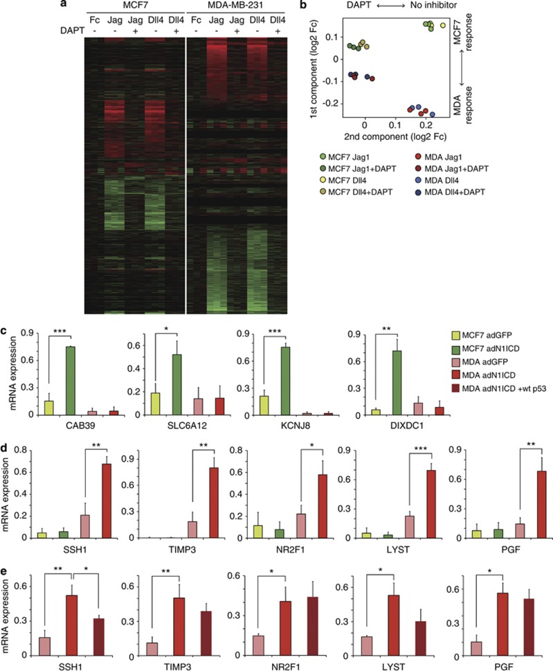
The difference in Notch-induced upregulation of IL-6 between MDA-MB-231 and MCF7 cells prompted us to assess to what extent their Notch-induced transcriptomes differed. To capture genes that are part of the immediate Notch signaling output, the transcriptomes from MDA-MB-231 or MCF7 cells were analyzed following activation by immobilized Jagged1 or Dll4 ligand for 6 h. The Jagged1-induced transcriptome was very similar to the Dll4-induced transcriptome in both cell types, and the ligand-induced expression could largely be abrogated by DAPT treatment, indicating that it indeed reflected Notch-mediated transcriptional upregulation (Figure 4a). However, the Jagged1- or Dll4-induced transcriptomes in MDA-MB-231 and MCF7 turned out to be remarkably different (Figure 4a). In fact, the majority of genes regulated by either Notch ligand in MDA-MB-231 cells were not regulated in MCF7 cells, and vice versa (Figure 4a). Unsupervised principal component analysis of the transcriptome data confirmed that the MDA-MB-231 and MCF7 transcriptomes clustered very differently (Figure 4b). Quantitative PCR analysis following Notch1 ICD activation corroborated the transcriptome analysis data and identified several genes that were upregulated by Notch in MCF7 but not in MDA-MB-231 cells (Figure 4c), or conversely upregulated in MDA-MB-231 but not in MCF7 cells (Figure 4d). We next asked whether transduction of wild-type p53 abrogated the induction of the genes upregulated specifically in MDA-MB-231 cells. Like for IL-6, p53 reduced the Notch-mediated upregulation of SSH1, whereas the Notch-mediated upregulation of TIMP3, NR2F1, LYST and PGF was not significantly affected by p53 (Figure 4e). Analysis of the genes in Figures 4c and e for the presence of functional p53-binding sites, as determined by ChIP-seq data,25 in the vicinity of the gene revealed that four of the genes, IL-6, SSH1, LYST and PGF, contained such sites (Supplementary Figure 3). In conclusion, these data show that Notch signaling induces quite distinct gene sets in two different types of breast cancer cell lines, and that the p53 status affects a subset of genes upregulated by Notch in MDA-MB-231 cells.
Figure 4.
Notch regulates distinct gene sets in MDA-MB-231 and MCF7 cells. (a) A heat map (with triplicates) showing regulation of genes in MDA-MB-231 or MCF7 cells cultured on immobilized Jagged1 or Dll4 ligand or Fc fragment, as indicated. The γ-secretase inhibitor DAPT was added to block Notch signaling as indicated. Red=increased gene expression; green=reduced gene expression. (b) Principal component analysis of the data presented in a, as indicated. (c–e) Quantitative PCR analysis of genes that were (c) upregulated in MCF7 but not in MDA-MB-231 cells; (d) upregulated in MDA-MB-231 but not MCF7 cells; and (e) the genes from d also following transfection of wild-type p53. Values are significant at ***P<0.001, **P<0.01 and *P<0.05, as indicated in the figure. Graphs represent averages of three independent experiments.
IL-6 expression is regulated by non-canonical, CSL-independent, Notch signaling
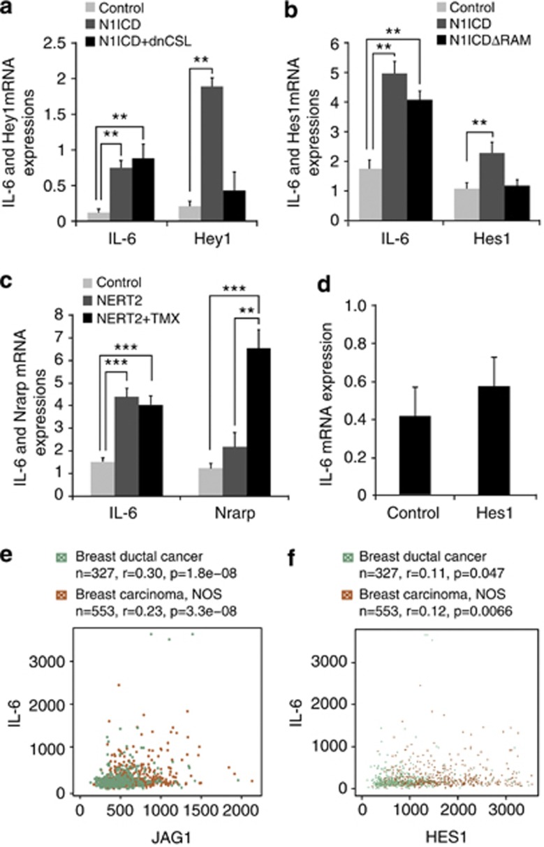
The fact that expression of IL-6, but not the canonical target gene Hes1, was affected by p53 (Figures 3c and d) raised the possibility that IL-6 was regulated by non-canonical Notch signaling. Expression of a dominant-negative version of CSL (CSLR218H), which can bind to Notch ICD but not to DNA,26 did not diminish Notch-mediated increase in IL-6 expression, but as expected, markedly abrogated the Notch-mediated upregulation of the canonical Notch target gene Hey1 (Figure 5a). Furthermore, transfection of Notch1ICDΔRAM, a Notch ICD construct lacking the CSL-binding RAM domain,27 upregulated IL-6 expression, but not expression of Hes1 (Figure 5b). Finally, transfection of a Notch1 ICD–ER fusion protein (NERT2), which is cytoplasmically retained in the absence of tamoxifen,28 upregulated IL-6 expression both in the absence and presence of tamoxifen, whereas the canonical target gene Nrarp was only upregulated when tamoxifen was added (Figure 5c).
Figure 5.
IL-6 expression is activated by non-canonical Notch signaling. (a, b) Analysis of IL-6 and Hey1 (a) or IL-6 and Hes1 (b) mRNA expression following transfection of empty pCMX vector (control), Notch1 ICD or Notch1 ICD combined with dnCSL (a) or pCMX, Notch1 ICD or Notch1 ICDΔRAM (b), as indicated. (c) Analysis of IL-6 and Nrarp mRNA expression following transfection of pCMX vector (control), NERT2 or NERT2 combined with tamoxifen (NERT2+TMX, 50 nM). (d) IL-6 mRNA expression following transfection of Hes1. (e, f) Correlation between IL-6 and Jagged1 (e) or IL-6 and Hes1 mRNA (f) data from the GeneSapiens transcriptome data from breast cancer. Values are significant at ***P<0.001, **P<0.01, as indicated in the figure. r, correlation coefficient. Graphs represent averages of three independent experiments.
To further address the issue of non-canonical Notch signaling, we tested the potential role of Hes1 in regulation of IL-6 mRNA expression. Hes1 is activated by Notch1 ICD via CSL, and has in other cellular contexts been implicated in the regulation of IL-6.14 Expression of Hes1, however, did not activate IL-6 mRNA expression in the MDA-MB-231 cells (Figure 5d). Similarly, we did not observe a high correlation between Hes1 and IL-6 mRNA expression in the breast ductal cancer and breast carcinoma transcriptome data sets from GeneSapiens, whereas a correlation between IL-6 and Jag1 expression was observed from the same data set (Figures 5e and f). In conclusion, these data indicate that Notch controls IL-6 expression by a CSL-independent mode of signaling in breast cancer cells and show that Hes1 is not involved in IL-6 upregulation in the MDA-MB-231 cells.
Notch-mediated IL-6 activation requires IKKα and IKKβ function but not canonical NF-κB signaling
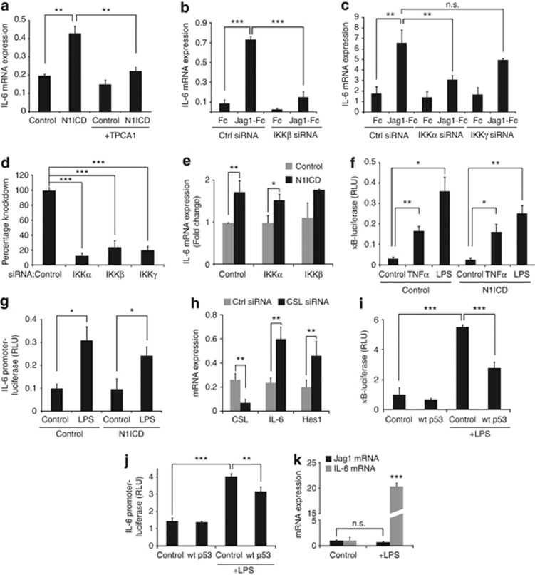
In canonical NF-κB signaling, inflammatory stimuli such as lipopolysaccharide (LPS) or tumor necrosis factor-α cause activation of the IKK complex (IKKα, IKKβ and IKKγ (NEMO)), leading to phosphorylation of IκB and its subsequent ubiquitylation and degradation. The removal of IκB results in translocation of a p50/p65 (RelA) dimer to the nucleus, where it activates transcription of downstream genes containing κB-binding sites, including IL-6.29 As NF-κB has a key role in IL-6 regulation,29 and Notch and NF-κB signaling cross-talk in a number of ways,30 we asked whether NF-κB signaling was involved in the CSL-independent activation of IL-6. Pharmacological blocking of IKKβ by TPCA1 ([5-(p-fluorophenyl)-2-ureido]thiophene-3-carboxamide)31 or small interfering RNA knockdown of IKKβ expression resulted in abrogation of the Notch-induced IL-6 upregulation (Figures 6a and b). Similarly, knockdown of IKKα but not of IKKγ, reduced IL-6 upregulation (Figures 6c and d). IKKα and IKKβ alone could, however, not substitute for Notch in terms of IL-6 induction, as overexpression of IKKα or IKKβ, without activation of Notch, did not increase the IL-6 mRNA levels (Figure 6e). Furthermore, IKKα and IKKβ did not superactivate the Notch-induced IL-6 upregulation (Figure 6e). The observation that Notch signaling did not activate a reporter construct containing κB-binding sites linked to luciferase (κB-luc), which was robustly activated by both tumor necrosis factor-α and LPS, suggests that Notch signaling does not trigger a canonical transcriptional NF-κB response (Figure 6f). Moreover, Notch activation did not alter protein levels of p65 or p105, and knockdown of p50 did not affect the upregulation of IL-6 expression induced by Notch (Supplementary Figure 4). In line with these data, a 2 kb immediate IL-6 proximal promoter reporter construct, which was induced by LPS, was not activated by Notch1 ICD (Figure 6g), despite a CSL-binding site in this region.32 In fact, when CSL expression was knocked down by small interfering RNA, expression of IL-6 and Hes1 were increased in the absence of Notch stimulation (Figure 6h), suggesting that CSL may be positioned at the CSL-binding site in the IL-6 proximal promoter acting as a transcriptional repressor, but is not required for the Notch-mediated activation of IL-6 transcription. As the p53 status was important for the Notch-induced IL-6 upregulation (see Figure 3), we also tested whether the p53 status affected the canonical NF-κB response. Introduction of wild-type p53 into the MDA-MB-231 cells led to a reduction of LPS-induced IL-6-promoter and kB-reporter activation (Figures 6i and j), in keeping with previous reports33 and references therein. However, in contrast to other cell types,34 canonical NF-κB signaling did not upregulate Jagged1 expression in the MDA-MB-231 cells (Figure 6k), suggesting that Notch signaling is not indirectly augmented by NF-κB through regulation of the level of ligand. Taken together, these data show that IKKα and IKKβ are required for the Notch-mediated activation of IL-6, but that canonical NF-κB signaling is not engaged by Notch.
Figure 6.
Notch-mediated upregulation of IL-6 requires IKKα and IKKβ but not canonical NF-κB signaling. (a) Analysis of IL-6 mRNA expression following transfection of Notch1 ICD or enhanced green fluorescent protein (EGFP) (control), in various combinations with the IKKβ inhibitor TPCA1. (b, c) Analysis of IL-6 mRNA expression following small interfering RNA (siRNA) knockdown of IKKβ (b) or IKKα and IKKγ (c) mRNA expression. (d) Analysis of IKKα, β and γ mRNA levels after siRNA knockdown of the respective mRNA. (e) Analysis of IL-6 mRNA expression following transfection of IKKα or IKKβ expression constructs and co-transfection with Notch1 ICD or pCMX (control) plasmid. (f) Analysis of κB-luciferase activity following LPS or tumor necrosis factor alpha (TNFα) stimulation. (g) IL-6 promoter-luciferase activation in response to Notch1 ICD or EGFP (Control), in combination with LPS stimulation, as indicated. (h) Analysis of CSL, IL-6 and Hes1 mRNA expression in response to siRNA for CSL (CSL siRNA) or scrambled siRNA (Ctrl siRNA). (i, j) Analysis of κB-luciferase (i) or IL-6 promoter reporter (j) activity following LPS stimulation and after transfection of wild-type p53 or pCMX plasmid (control). (k) Analysis of Jagged1 and IL-6 mRNA expression following LPS stimulation. Values are significant at ***P<0.001, **P<0.01 and *P<0.05, as indicated in the figure. NS, not significant. RLU, relative luciferase units. Graphs represent averages of three independent experiments.
A role for Notch-mediated, CSL-independent IL-6 activation in the interaction between tumor cells and macrophages
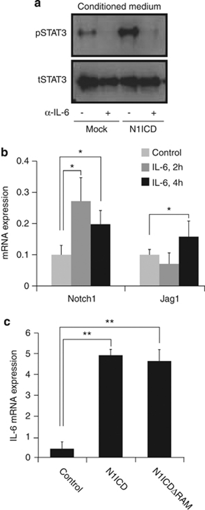
Macrophages constitute an important component of the tumor stroma and tumor cells can influence tumor-associated macrophages to promote tumor growth and metastasis.35, 36 To test whether Notch-mediated IL-6 production in tumor cells could influence tumor-associated macrophages, we exposed RAW264.7 cells, a macrophage-like cell line used as a mimic for tumor-associated macrophages,37 to conditioned medium from Notch-stimulated MDA-MB-231 cells. This resulted in increased JAK/STAT signaling in the RAW264.7 cells, as judged by elevated phosphorylated STAT3 levels, and this increase was abrogated by a IL-6 blocking antibody (Figure 7a). As a previous report has suggested that IL-6 can upregulate expression of components of the Notch pathway,38 we tested whether this was also the case in RAW264.7 cells. Addition of recombinant IL-6 to RAW264.7 cells increased the expression of both Notch 1 and Jagged1 mRNA (Figure 7b). Conversely, introduction of Notch1 ICD or Notch1 ICDΔRAM led to a robust upregulation of mRNA expression of IL-6 (Figure 7c). Collectively, this reveals that IL-6 is activated by CSL-independent Notch signaling in macrophages, and that IL-6 and Notch signaling can reciprocally upregulate each other, indicating the existence of a feed-forward loop, which may lead to sustained Notch and IL-6 expression in macrophages.
Figure 7.
Reciprocal regulation of Notch and IL-6 in RAW264.7 cells. (a) Analysis of paracrine IL-6 activation in RAW264.7 cells. Conditioned medium from MDA-MB-231 cells transfected with Notch1 ICD (N1ICD) or enhanced green fluorescent protein (EGFP) (control) was pre-treated with an IL-6 blocking antibody (α-IL-6) before being supplemented to the RAW264.7 cells, and the levels of pTyr705 STAT3 (upper) and total STAT3 (lower) protein were analyzed. (b) Analysis of Notch1 and Jagged1 mRNA expression in response to IL-6 stimulation for 2 h and 4 h, as indicated. (c) Regulation of IL-6 mRNA expression by transfection of EGFP, Notch1 ICD or Notch1 ICDΔRAM in the RAW264.7 cells, as indicated. Values are significant at **P<0.01 and *P<0.05, as indicated in the figure. Graphs represent averages of three independent experiments.
Discussion
Deregulated Notch signaling is frequently observed in breast cancer1, 2, 3 but how this contributes to tumor progression is less well understood. Here we identify the cytokine IL-6 as a novel Notch target gene in breast tumor cells, and show that the IL-6 upregulation resulted in activation of autocrine and paracrine JAK/STAT signaling. IL-6 was upregulated by non-canonical Notch signaling and was influenced by the cellular p53 status, by two proteins in the NF-κB signaling pathway: IKKα and IKKβ.
A non-canonical CSL-independent mode of activation is supported by the fact that a dominant-negative form of CSL did not affect IL-6 upregulation but reduced Notch-mediated activation of the canonical Notch gene Hey1. Also, a Notch ICD that is not capable of binding CSL upregulated IL-6 without affecting canonical Notch genes. Furthermore, localization of Notch ICD to the cytoplasm was sufficient to upregulate IL-6, whereas nuclear translocation was required for canonical Notch gene activation. This is, to our knowledge, the first example of gene activation from a cytoplasmically localized Notch ICD. The non-canonical, CSL-independent mode of activation also reveals an interesting cell type-specific difference in Notch regulation of IL-6, as it has recently been proposed that Notch activates IL-6 through a Hes1-dependent mechanism in the bone metastatic niche in a mouse mammary tumor model.14
Our data provide evidence that the non-canonical Notch activation of IL-6 is linked to NF-κB signaling, as pharmacological inhibition of IKKβ by TPCA1 or small interfering RNA-mediated knockdown of IKKα or IKKβ mRNA reduced Notch-mediated IL-6 upregulation. There is an emerging view that the Notch and NF-κB pathways interact at different steps in the signaling cascade,30 and interactions between Notch ICD and the IKK signalosome or IKKα have indeed been demonstrated.39, 40 Although we have not been able to demonstrate a direct interaction between Notch and any of the IKKs in the MDA-MB-231 cells, such an interaction is in accordance with a cytoplasmically localized Notch ICD mediating IL-6 upregulation. Interestingly, Notch-mediated IL-6 upregulation was not accompanied by activation of canonical NF-κB signaling, which reveals a pronounced difference between how Notch and inflammatory agents regulate IL-6. Although Notch required IKKα and IKKβ to induce IL-6, it did not induce a κB reporter or the proximal IL-6 promoter, whereas this was the case for LPS and tumor necrosis factor-α. We demonstrate that Notch controls IL-6 expression at the transcriptional level, but how IKKα and IKKβ are involved, not acting via canonical NF-κB signaling, remains to be elucidated. It should be emphasized that NF-κB-independent functions for IKKβ have been described, for example, in the control of FOXO3a.29 Collectively, the data show that IL-6 is subject to complex regulation, and that inflammatory agents and Notch signaling use the NF-κB signaling pathway differently to control IL-6 expression.
The cellular p53 status was important for how Notch controls IL-6 expression. The Notch-mediated induction of IL-6 expression in the p53 mutant MDA-MB-231 cells or in p53−/− mouse embryonic fibroblasts was significantly reduced when wild-type p53 was transduced. Conversely, introduction of a mutant p53 into MCF7 cells was sufficient to induce a Notch-mediated IL-6 upregulation. Given that p53 is frequently lost or mutated not only in mammary tumors but in a large fraction of many different tumors,41 and that p53 is also known to interact with NF-κB signaling at multiple levels in the pathways,42, 43 it will be interesting to learn to what extent the Notch transcriptional response is modified as a result of p53 loss or mutation in different tumor types, and how this contributes to tumor progression. The notion that p53 negatively regulates Notch signaling is corroborated by the finding that the mammosphere-forming potential was increased from p53−/− mice, but only under conditions of intact Notch signaling.44 Similarly, p53 has been shown to suppress Notch4 ICD-mediated growth of mammary epithelial cells.45
The relation between the cellular p53 status and the Notch transcriptional output is also of interest in the quest to better understand why Notch transcriptomes from different cell types differ so extensively.17 The transcriptome data from the MDA-MB-231 and MCF7 cells provide a compelling example of cell context-dependent diversity, and among five genes that were upregulated by Notch in MDA-MB-231 but not in MCF7 cells, two genes, IL-6 and SSH1, showed a Notch-induced upregulation that was reduced by transduction of wild-type p53. This may indicate that a defined subset of Notch-induced genes are sensitive to the cellular p53 status, and that p53 could stratify the Notch transcriptome into a p53-independent and p53-dependent portion, which may contribute, at least in part, to the observed cell context-dependent diversity in signaling output.
In conclusion, the data in this report provide evidence for the involvement of IKKα/IKKβ and p53 in non-canonical Notch signaling. This contributes to our understanding of how diversity in the Notch signaling output is generated, and may provide new ideas for combinatorial treatments for breast cancer in the future. It is also noteworthy that while Notch, NF-κB and p53 in many cases regulate gene activity through transcriptional control in the nucleus, our results point to that for both Notch and NF-κB signaling, and possibly also for p53, the effects appear not to be mediated directly at the transcriptional level, but rather through non-canonical branches of the signaling pathways involving and, in the case of Notch, a cytoplasmic location of the effector molecule.
Materials and methods
Cell culture
MDA-MB-231, MCF7, MCF10A, Hs578T, HCC38, MDA-MB-175-VII, T47D and SKBR3 were purchased from ATCC (American Type Culture Collection, Manassas, VA, USA). RAW267.4 and p53-/- mouse embryonic fibroblast cells were kind gifts from Drs Robert A Harris and Olle Sangfelt (Karolinska Institutet), respectively. Culture conditions are specified in the Supplementary Information.
DNA constructs
The p53 luciferase (#16442), IKKα (#23296) and IKKβ 11103) constructs were purchased from Addgene (Cambridge, MA, USA). N1ICD-GFP, dnCSL, N1ICDΔRAM were generously provided by Dr A Sarin46 and the wild-type and mutated p53 constructs from Drs K Wiman and G Selivanova, respectively. The IL-6 luciferase promoter construct was a kind gift from Drs W Vanden Bergh and G Haegeman.47 The NERT2 plasmid was generated by cloning the Notch-ERT2 from the rNERTneo plasmid48 into the vector pEF1IRESpuro at XhoI and XbaI sites by In-fusion Cloning (Clontech, San Jose, CA, USA).
Western blot analysis
Protein lysates were prepared by directly lysing cells in Laemmli sample buffer containing 5% β-mercaptoethanol. Protein lysates were separated on 4% to 12% Tris–glycine gels (Life Technologies, Carlsbad, CA, USA) and transferred to polyvinylidene difluoride membranes (Millipore, Billerica, MA, USA). After blocking with 10% nonfat dry milk in phosphate-buffered saline, membranes were incubated with primary antibodies (overnight) and peroxidase-conjugated secondary antibody (Amersham). Immunocomplexes were detected with an enhanced chemiluminescence (ECL) kit (Amersham, GE Healthcare, Pittsburgh, PA, USA).
Activation of Notch signaling by immobilized ligand
Activation of Notch signaling by immobilized ligands was performed as previously described.49
Antibodies and reagents
The primary antibodies used were anti-STAT3, anti-phosphoSTAT3, anti-Bcl-xL, -IKKα, -IKKβ, -NF-κB -p65 and -p105 Cell signaling (Boston, MA, USA), mouse anti-β-actin (Sigma-Aldrich, St Louis, MO, USA), rabbit anti-Notch1 C-20 (Santa Cruz, CA, USA) anti-IL6 (Sigma-Aldrich). The secondary antibodies used were anti-rabbit horseradish peroxidase and anti-mouse horseradish peroxidase (Dako, Glostrup, Denmark). Recombinant human IL-6, TPCA-1, LPS, tumor necrosis factor-α, 4-hydroxytamoxifen and actinomycin D were purchased from Sigma-Aldrich.
Immunohistochemistry
Eight cases of basal and eight cases of non-basal breast cancer with associated clinical and pathological data were analyzed for Notch1 ICD expression (using anti activated Notch 1 antibody ab8925 (Abcam, Cambridge, UK), which recognizes the newly formed N-terminus of Notch ICD following γ-secretase cleavage). Tissue sections were also stained for expression of ER, progesterone receptor, CD44 and cytokeratin 5/6. Tissue section preparation is described in the Supplementary Information.
Real-time quantitative reverse transcriptase–PCR analysis
RNA was extracted using the RNeasy mini kit (Qiagen, Hilden, Germany) and complementary DNA was synthesized from 1 μg total RNA using Superscript III Reverse Transcriptase, oligo(dT)12–18 primers or random primers according to the manufacturer's protocol (Invitrogen). Reverse transcriptase–PCR was carried out on a 7500 Fast Real-Time PCR system with Fast SYBR Green Master Mix (Applied Biosystems, Carlsbad, CA, USA) according to the manufacturer's recommendations. Primers used for quantitative PCR are listed in Supplementary Figure 5. Primers used for analysis of mouse and human Hes1, Hey1 have been published previously.49, 50 Expression levels were normalized to human β-actin mRNA expression.
Statistical analysis
The unpaired Student's t-test was applied to evaluate differences between experimental groups. P-values ⩽0.05 were considered statistically significant.
Transcriptome analysis, bioinformatics and GeneSapiens analysis
For each array experiment, 2 million cells each were plated. In experiments where Notch signaling was pharmacologically blocked, DAPT was applied before ligand stimulation. After stimulation with immobilized ligand (Jagged1-Fc or Dll4-Fc) for 6 h, cells were washed with phosphate-buffered saline, trypsinized, resuspended in RNA later and stored at −20 °C. the method for RNA preparation is described in the Supplementary Information. The micro-array data were subjected to background subtraction on the BeadStudio Data Analysis software (Illumina, San Diego, CA, USA) and normalized using the cross-correlation method.51 Differentially expressed transcripts were identified and sorted based on the mean Log2 fold change in expression values compared with the controls. The micro-array data were deposited into NCBI GEO with the accession number GSE36051. For the gene expression analysis in a clinical breast cancer material, the database is available from the GeneSapiens website (www.genesapiens.org), and the analysis of data from GeneSapiens has been previously described.24, 52
Transfections, adenoviral infections, enzyme-linked immunosorbent assay and analysis of IL-6 signaling
The procedures for transfections, adenoviral infections, enzyme-linked immunosorbent assay and analysis of IL-6 signaling are described in the Supplementary Information. All cellular transfections were carried out with Lipofectamine 2000 (Invitrogen) according to the manufacturer's protocol. Luciferase assays were performed as described previously.53
Acknowledgments
This work was financially supported by the Swedish Cancer Society, the Swedish Research Council (DBRM and Project Grant), the EC projects EuroSyStem and NotchIT, Karolinska Institutet (BRECT, Theme Center in Regenerative Medicine and a Distinguished Professor Award). We are grateful to Susanne Bergstedt, Rasmus Niemi and Sinikka Kollanus for technical assistance.
The authors declare no conflict of interest.
Footnotes
Supplementary Information accompanies the paper on the Oncogene website (http://www.nature.com/onc)
Supplementary Material
References
- Robinson DR, Kalyana-Sundaram S, Wu Y-M, Shankar S, Cao X, Ateeq B, et al. Functionally recurrent rearrangements of the MAST kinase and Notch gene families in breast cancer. Nat Med. 2011;17:1646–1651. doi: 10.1038/nm.2580. [DOI] [PMC free article] [PubMed] [Google Scholar]
- Pece S, Serresi M, Santolini E, Capra M, Hulleman E, Galimberti V, et al. Loss of negative regulation by Numb over Notch is relevant to human breast carcinogenesis. J Cell Biol. 2004;167:215–221. doi: 10.1083/jcb.200406140. [DOI] [PMC free article] [PubMed] [Google Scholar]
- Stylianou S, Clarke RB, Brennan K. Aberrant activation of notch signaling in human breast cancer. Cancer Res. 2006;66:1517–1525. doi: 10.1158/0008-5472.CAN-05-3054. [DOI] [PubMed] [Google Scholar]
- Reedijk M, Odorcic S, Chang L, Zhang H, Miller N, McCready DR, et al. High-level coexpression of JAG1 and NOTCH1 is observed in human breast cancer and is associated with poor overall survival. Cancer Res. 2005;65:8530–8537. doi: 10.1158/0008-5472.CAN-05-1069. [DOI] [PubMed] [Google Scholar]
- Speiser J, Foreman K, Drinka E, Godellas C, Perez C, Salhadar A, et al. Notch-1 and Notch-4 biomarker expression in triple-negative breast cancer. Int J Surg Pathol. 2012;20:139–145. doi: 10.1177/1066896911427035. [DOI] [PubMed] [Google Scholar]
- Rizzo P, Miao H, D'Souza G, Osipo C, Yun J, Zhao H, et al. Cross-talk between Notch and the estrogen receptor in breast cancer suggests novel therapeutic approaches. Cancer Res. 2008;68:5226–5235. doi: 10.1158/0008-5472.CAN-07-5744. [DOI] [PMC free article] [PubMed] [Google Scholar]
- Landor SK-J, Mutvei AP, Mamaeva V, Jin S, Busk M, Borra R, et al. Hypo- and hyperactivated Notch signaling induce a glycolytic switch through distinct mechanisms. Proc Natl Acad Sci USA. 2011;108:18814–18819. doi: 10.1073/pnas.1104943108. [DOI] [PMC free article] [PubMed] [Google Scholar]
- Kopan R, Ilagan MXG. The canonical Notch signaling pathway: unfolding the activation mechanism. Cell. 2009;137:216–233. doi: 10.1016/j.cell.2009.03.045. [DOI] [PMC free article] [PubMed] [Google Scholar]
- Heitzler P. Biodiversity and noncanonical Notch signaling. Curr Top Dev Biol. 2010;92:457–481. doi: 10.1016/S0070-2153(10)92014-0. [DOI] [PubMed] [Google Scholar]
- Raafat A, Lawson S, Bargo S, Klauzinska M, Strizzi L, Goldhar AS, et al. Rbpj conditional knockout reveals distinct functions of Notch4/Int3 in mammary gland development and tumorigenesis. Oncogene. 2008;28:219–230. doi: 10.1038/onc.2008.379. [DOI] [PMC free article] [PubMed] [Google Scholar]
- Perumalsamy LR, Nagala M, Sarin A. Notch-activated signaling cascade interacts with mitochondrial remodeling proteins to regulate cell survival. Proc Natl Acad Sci USA. 2010;107:6882–6887. doi: 10.1073/pnas.0910060107. [DOI] [PMC free article] [PubMed] [Google Scholar]
- Hanahan D, Weinberg RA. Hallmarks of cancer: the next generation. Cell. 2011;144:646–674. doi: 10.1016/j.cell.2011.02.013. [DOI] [PubMed] [Google Scholar]
- Yu H, Pardoll D, Jove R. STATs in cancer inflammation and immunity: a leading role for STAT3. Nat Rev Cancer. 2009;9:798–809. doi: 10.1038/nrc2734. [DOI] [PMC free article] [PubMed] [Google Scholar]
- Sethi N, Dai X, Winter CG, Kang Y. Tumor-derived JAGGED1 promotes osteolytic bone metastasis of breast cancer by engaging notch signaling in bone cells. Cancer Cell. 2011;19:192–205. doi: 10.1016/j.ccr.2010.12.022. [DOI] [PMC free article] [PubMed] [Google Scholar]
- Wang Y, Klijn JGM, Zhang Y, Sieuwerts AM, Look MP, Yang F, et al. Gene-expression profiles to predict distant metastasis of lymph-node-negative primary breast cancer. Lancet. 2005;365:671–679. doi: 10.1016/S0140-6736(05)17947-1. [DOI] [PubMed] [Google Scholar]
- Pawitan Y, Bjöhle J, Amler L, Borg A-L, Egyhazi S, Hall P, et al. Gene expression profiling spares early breast cancer patients from adjuvant therapy: derived and validated in two population-based cohorts. Breast Cancer Res. 2005;7:R953–R964. doi: 10.1186/bcr1325. [DOI] [PMC free article] [PubMed] [Google Scholar]
- Andersson ER, Sandberg R, Lendahl U. Notch signaling: simplicity in design, versatility in function. Development. 2011;138:3593–3612. doi: 10.1242/dev.063610. [DOI] [PubMed] [Google Scholar]
- Turkson J. STAT proteins as novel targets for cancer drug discovery. Expert Opin Ther Targets. 2004;8:409–422. doi: 10.1517/14728222.8.5.409. [DOI] [PubMed] [Google Scholar]
- Wang YA, Johnson SK, Brown BL, McCarragher LM, Al-Sakkaf K, Royds JA, et al. Enhanced anti-cancer effect of a phosphatidylinositol-3 kinase inhibitor and doxorubicin on human breast epithelial cell lines with different p53 and oestrogen receptor status. Int J Cancer. 2008;123:1536–1544. doi: 10.1002/ijc.23671. [DOI] [PubMed] [Google Scholar]
- Selivanova G, Ryabchenko L, Jansson E, Iotsova V, Wiman KG. Reactivation of mutant p53 through interaction of a C-terminal peptide with the core domain. Mol Cell Biol. 1999;19:3395–3402. doi: 10.1128/mcb.19.5.3395. [DOI] [PMC free article] [PubMed] [Google Scholar]
- Miller LD, Smeds J, George J, Vega VB, Vergara L, Ploner A, et al. An expression signature for p53 status in human breast cancer predicts mutation status, transcriptional effects, and patient survival. Proc Natl Acad Sci USA. 2005;102:13550–13555. doi: 10.1073/pnas.0506230102. [DOI] [PMC free article] [PubMed] [Google Scholar]
- Enerly E, Steinfeld I, Kleivi K, Leivonen S-K, Aure MR, Russnes HG, et al. miRNA-mRNA integrated analysis reveals roles for miRNAs in primary breast tumors. PLoS ONE. 2011;6:e16915. doi: 10.1371/journal.pone.0016915. [DOI] [PMC free article] [PubMed] [Google Scholar]
- Muggerud AA, Edgren H, Wolf M, Kleivi K, Dejeux E, Tost J, et al. Data integration from two microarray platforms identifies bi-allelic genetic inactivation of RIC8A in a breast cancer cell line. BMC Med Genomics. 2009;2:26. doi: 10.1186/1755-8794-2-26. [DOI] [PMC free article] [PubMed] [Google Scholar]
- Kilpinen S, Autio R, Ojala K, Iljin K, Bucher E, Sara H, et al. Systematic bioinformatic analysis of expression levels of 17 330 human genes across 9783 samples from 175 types of healthy and pathological tissues. Genome Biol. 2008;9:R139. doi: 10.1186/gb-2008-9-9-r139. [DOI] [PMC free article] [PubMed] [Google Scholar]
- Botcheva K, McCorkle SR, McCombie WR, Dunn JJ, Anderson CW. Distinct p53 genomic binding patterns in normal and cancer-derived human cells. Cell Cycle. 2011;10:4237–4249. doi: 10.4161/cc.10.24.18383. [DOI] [PMC free article] [PubMed] [Google Scholar]
- Chung CN, Hamaguchi Y, Honjo T, Kawaichi M. Site-directed mutagenesis study on DNA binding regions of the mouse homologue of suppressor of hairless, RBP-J kappa. Nucleic Acids Res. 1994;22:2938–2944. doi: 10.1093/nar/22.15.2938. [DOI] [PMC free article] [PubMed] [Google Scholar]
- Kovall RA, Blacklow SC. Mechanistic insights into Notch receptor signaling from structural and biochemical studies. Curr Top Dev Biol. 2010;92:31–71. doi: 10.1016/S0070-2153(10)92002-4. [DOI] [PubMed] [Google Scholar]
- Meier-Stiegen F, Schwanbeck R, Bernoth K, Martini S, Hieronymus T, Ruau D, et al. Activated Notch1 target genes during embryonic cell differentiation depend on the cellular context and include lineage determinants and inhibitors. PLoS ONE. 2010;5:e11481. doi: 10.1371/journal.pone.0011481. [DOI] [PMC free article] [PubMed] [Google Scholar]
- Oeckinghaus A, Hayden MS, Ghosh S. Crosstalk in NF-κB signaling pathways. Nat Immunol. 2011;12:695–708. doi: 10.1038/ni.2065. [DOI] [PubMed] [Google Scholar]
- Osipo C, Golde TE, Osborne BA, Miele LA. Off the beaten pathway: the complex cross talk between Notch and NF-kappaB. Lab Invest. 2008;88:11–17. doi: 10.1038/labinvest.3700700. [DOI] [PubMed] [Google Scholar]
- Podolin PL, Callahan JF, Bolognese BJ, Li YH, Carlson K, Davis TG, et al. Attenuation of murine collagen-induced arthritis by a novel, potent, selective small molecule inhibitor of IkappaB kinase 2, TPCA-1 (2-[(aminocarbonyl)amino]-5-(4-fluorophenyl)-3-thiophenecarboxamide), occurs via reduction of proinflammatory cytokines and antigen-induced T cell Proliferation. J Pharmacol Exp Ther. 2005;312:373–381. doi: 10.1124/jpet.104.074484. [DOI] [PubMed] [Google Scholar]
- Wongchana W, Palaga T. Direct regulation of interleukin-6 expression by Notch signaling in macrophages. Cell Mol Immunol. 2012;9:155–162. doi: 10.1038/cmi.2011.36. [DOI] [PMC free article] [PubMed] [Google Scholar]
- Murphy SH, Suzuki K, Downes M, Welch GL, De Jesus P, Miraglia LJ, et al. Tumor suppressor protein (p)53, is a regulator of NF-kappaB repression by the glucocorticoid receptor. Proc Natl Acad Sci USA. 2011;108:17117–17122. doi: 10.1073/pnas.1114420108. [DOI] [PMC free article] [PubMed] [Google Scholar]
- Johnston DA, Dong B, Hughes CCW. TNF induction of jagged-1 in endothelial cells is NFkappaB-dependent. Gene. 2009;435:36–44. doi: 10.1016/j.gene.2009.01.003. [DOI] [PMC free article] [PubMed] [Google Scholar]
- Biswas SK, Mantovani A. Macrophage plasticity and interaction with lymphocyte subsets: cancer as a paradigm. Nat Immunol. 2010;11:889–896. doi: 10.1038/ni.1937. [DOI] [PubMed] [Google Scholar]
- Mantovani A, Sozzani S, Locati M, Allavena P, Sica A. Macrophage polarization: tumor-associated macrophages as a paradigm for polarized M2 mononuclear phagocytes. Trends Immunol. 2002;23:549–555. doi: 10.1016/s1471-4906(02)02302-5. [DOI] [PubMed] [Google Scholar]
- Zeng B, Li H, Liu Y, Zhang Z, Zhang Y, Yang R. Tumor-induced suppressor of cytokine signaling 3 inhibits Toll-like receptor 3 signaling in dendritic cells via binding to tyrosine kinase 2. Cancer Res. 2008;68:5397–5404. doi: 10.1158/0008-5472.CAN-07-6792. [DOI] [PubMed] [Google Scholar]
- Sansone P, Storci G, Tavolari S, Guarnieri T, Giovannini C, Taffurelli M, et al. IL-6 triggers malignant features in mammospheres from human ductal breast carcinoma and normal mammary gland. J Clin Invest. 2007;117:3988–4002. doi: 10.1172/JCI32533. [DOI] [PMC free article] [PubMed] [Google Scholar]
- Song LL, Peng Y, Yun J, Rizzo P, Chaturvedi V, Weijzen S, et al. Notch-1 associates with IKKα and regulates IKK activity in cervical cancer cells. Oncogene. 2008;27:5833–5844. doi: 10.1038/onc.2008.190. [DOI] [PubMed] [Google Scholar]
- Vilimas T, Mascarenhas J, Palomero T, Mandal M, Buonamici S, Meng F, et al. Targeting the NF-kappaB signaling pathway in Notch1-induced T-cell leukemia. Nat Med. 2007;13:70–77. doi: 10.1038/nm1524. [DOI] [PubMed] [Google Scholar]
- Vousden KH, Lane DP. p53 in health and disease. Nat Rev Mol Cell Biol. 2007;8:275–283. doi: 10.1038/nrm2147. [DOI] [PubMed] [Google Scholar]
- Ak P, Levine AJ. p53 and NF-κB: different strategies for responding to stress lead to a functional antagonism. FASEB J. 2010;24:3643–3652. doi: 10.1096/fj.10-160549. [DOI] [PubMed] [Google Scholar]
- Schneider G, Krämer OH. NF κB/p53 crosstalk-a promising new therapeutic target. Biochim Biophys Acta. 2011;1815:90–103. doi: 10.1016/j.bbcan.2010.10.003. [DOI] [PubMed] [Google Scholar]
- Tao L, Roberts AL, Dunphy KA, Bigelow C, Yan H, Jerry DJ. Repression of mammary stem/progenitor cells by p53 is mediated by Notch and separable from apoptotic activity. Stem Cells. 2011;29:119–127. doi: 10.1002/stem.552. [DOI] [PMC free article] [PubMed] [Google Scholar]
- Sun Y, Klauzinska M, Lake RJ, Lee JM, Santopietro S, Raafat A, et al. Trp53 regulates Notch 4 signaling through Mdm2. J Cell Sci. 2011;124 (Part 7:1067–1076. doi: 10.1242/jcs.068965. [DOI] [PMC free article] [PubMed] [Google Scholar]
- Perumalsamy LR, Nagala M, Banerjee P, Sarin A. A hierarchical cascade activated by non-canonical Notch signaling and the mTOR-Rictor complex regulates neglect-induced death in mammalian cells. Cell Death Differ. 2009;16:879–889. doi: 10.1038/cdd.2009.20. [DOI] [PubMed] [Google Scholar]
- Vanden Berghe W, Plaisance S, Boone E, De Bosscher K, Schmitz ML, Fiers W, et al. p38 and extracellular signal-regulated kinase mitogen-activated protein kinase pathways are required for nuclear factor-kappaB p65 transactivation mediated by tumor necrosis factor. J Biol Chem. 1998;273:3285–3290. doi: 10.1074/jbc.273.6.3285. [DOI] [PubMed] [Google Scholar]
- Schroeder T, Just U. Notch signalling via RBP-J promotes myeloid differentiation. EMBO J. 2000;19:2558–2568. doi: 10.1093/emboj/19.11.2558. [DOI] [PMC free article] [PubMed] [Google Scholar]
- Sahlgren C, Gustafsson MV, Jin S, Poellinger L, Lendahl U. Notch signaling mediates hypoxia-induced tumor cell migration and invasion. Proc Natl Acad Sci USA. 2008;105:6392–6397. doi: 10.1073/pnas.0802047105. [DOI] [PMC free article] [PubMed] [Google Scholar]
- Gustafsson MV, Zheng X, Pereira T, Gradin K, Jin S, Lundkvist J, et al. Hypoxia requires Notch signaling to maintain the undifferentiated cell state. Dev Cell. 2005;9:617–628. doi: 10.1016/j.devcel.2005.09.010. [DOI] [PubMed] [Google Scholar]
- Chua S-W, Vijayakumar P, Nissom PM, Yam C-Y, Wong VVT, Yang H. A novel normalization method for effective removal of systematic variation in microarray data. Nucleic Acids Res. 2006;34:e38. doi: 10.1093/nar/gkl024. [DOI] [PMC free article] [PubMed] [Google Scholar]
- Main H, Lee KL, Yang H, Haapa-Paananen S, Edgren H, Jin S, et al. Interactions between Notch- and hypoxia-induced transcriptomes in embryonic stem cells. Exp Cell Res. 2010;27:1610–1624. doi: 10.1016/j.yexcr.2009.12.012. [DOI] [PubMed] [Google Scholar]
- Chapman G, Liu L, Sahlgren C, Dahlqvist C, Lendahl U. High levels of Notch signaling down-regulate Numb and Numblike. J Cell Biol. 2006;175:535–540. doi: 10.1083/jcb.200602009. [DOI] [PMC free article] [PubMed] [Google Scholar]
Associated Data
This section collects any data citations, data availability statements, or supplementary materials included in this article.


