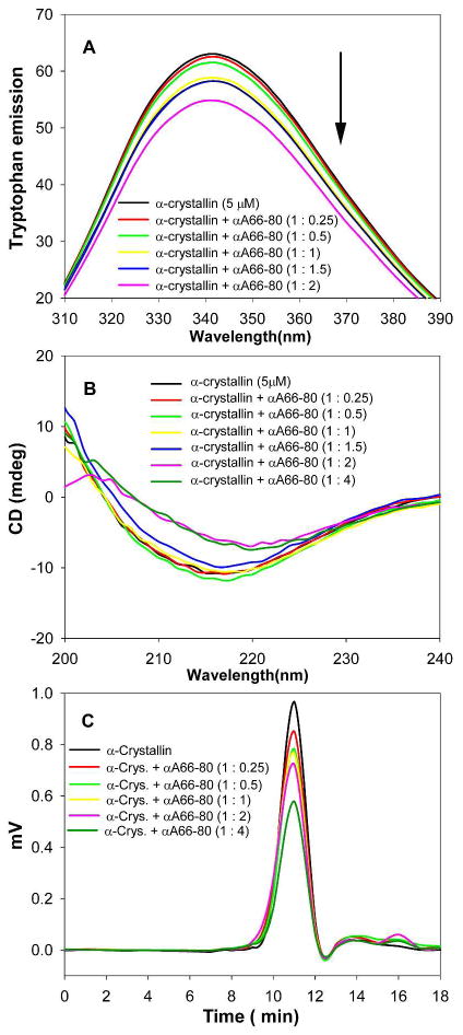
Figure 4.
The interaction between α-crystallin and the αA66-80 peptide analyzed by (A) tryptophan fluorescence measurements. The arrow points to the decrease in intrinsic tryptophan fluorescence of α-crystallin in the presence of increasing concentrations of peptide. (B) Far-UV CD spectra. The signal intensity decreases, in concomitance with the formation of insoluble precipitate. (C) Size exclusion chromatography. Chromatograms represent the decrease in the soluble α-crystallin fraction in the presence of αA66-80 peptide.

