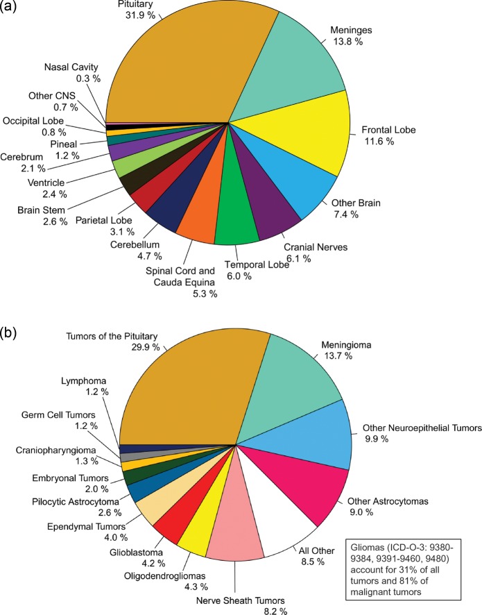
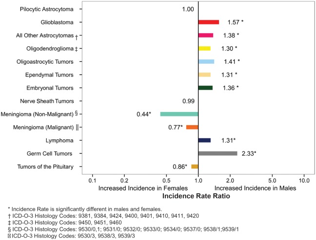
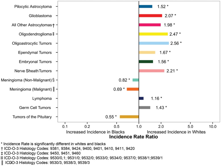
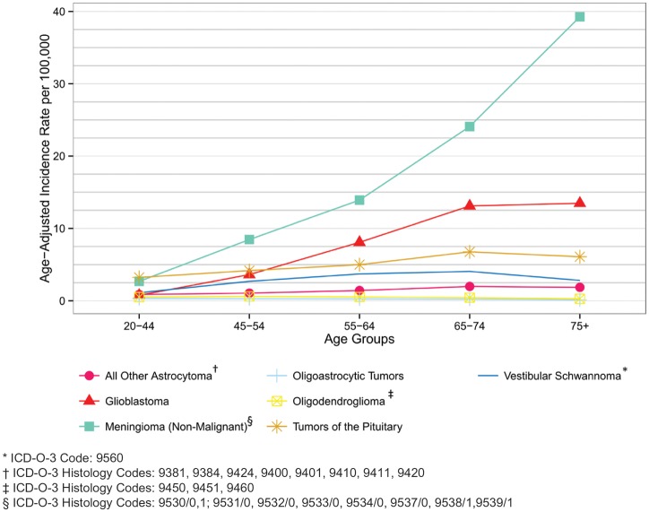
Introduction
The objective of CBTRUS Statistical Report: Primary Brain and Central Nervous System Tumors Diagnosed in the United States in 2006-2010 is to provide a current comprehensive review of the descriptive epidemiology of primary brain and central nervous system (CNS) tumors in the United States population. The Central Brain Tumor Registry of the United States (CBTRUS) obtained data on all newly diagnosed primary brain and CNS tumors from the Centers for Disease Control and Prevention (CDC), National Program of Cancer Registries (NPCR) and the National Cancer Institute (NCI), Surveillance, Epidemiology and End Results (SEER) program for diagnosis years 2006-2010. Incidence counts and rates of primary malignant and non-malignant brain and CNS tumors are documented by histology, gender, age, race, and Hispanic ethnicity. Mortality and relative survival rates for selected malignant histologies calculated using SEER data for the period 1995-2010 are also presented.
Background
The Central Brain Tumor Registry of the United States (CBTRUS) is a unique professional research organization that focuses exclusively on providing quality statistical data on population-based primary brain and CNS incident tumors in the United States. CBTRUS was incorporated as a nonprofit 501 (c) 3 with a founding and sustaining grant from the Pediatric Brain Tumor Foundation in 1992 following a two-year study conducted by the American Brain Tumor Association to determine the feasibility of a central registry for all primary brain and CNS tumor cases in the United States. Until that time, standard data reporting in the United States had been limited to malignant cases only.
CBTRUS is currently the only population-based site-specific registry in the United States that works in partnership with a public cancer surveillance organization, the NPCR (www.cdc.gov/cancer/npcr) of the CDC and from which data are directly received under a special agreement. This agreement permits transfer of data through the National Program of Cancer Registries Cancer Surveillance System (NPCR-CSS) Submission Specifications mechanism1, the system utilized for collection of central (state) cancer data as mandated in 1992 by Public Law 102-515, the Cancer Registries Amendment Act.2 Passed in 2002, the Benign Brain Tumor Cancer Registries Amendment Act (Public Law 107-260)3 expanded the collection of cancer by NPCR to include primary non-malignant brain and CNS tumors having International Classification of Diseases for Oncology Third Edition (ICD-O-3)4 codes beginning with the 2004 diagnosis year. All central cancer registries now include these data in their cancer collection practices and send them to NPCR. The passage of Public Law 107-260 paved the way for CBTRUS to access brain and CNS data through NPCR. CBTRUS researchers combine the NPCR data with data from the SEER (www.seer.cancer.gov) program5 of the NCI which was established for national cancer surveillance in the early 1970s. All data from NPCR and from SEER originate from tumor registrars who adhere to the Uniform Data Standards (UDS) for malignant and non-malignant brain and CNS tumors as directed by the North American Association of Cancer Registries (NAACCR) (www.naaccr.org). Along with the UDS, there are quality control checks and a system for rating each central registry to further insure that these data are reported as accurately and completely as possible. As a surveillance partner, CBTRUS can, therefore, report high quality data on brain and CNS tumors with histological specificity useful to the communities it serves. Its database contains the largest aggregation of population-based data on the incidence of all primary brain and CNS tumors in the United States.
This sixteenth statistical report, and second one published as a supplement to Neuro-Oncology, the official journal of the Society for Neuro-Oncology (www.soc-neuro-onc.org), continues the past efforts that CBTRUS has made to provide population-based incidence rates for all primary brain and CNS tumors by histology, age, gender, race, and Hispanic ethnicity. These data have been organized by clinically relevant histology groupings which were revised in 2012 in collaboration with three neuropathologists, Drs. Janet Bruner (University of Texas M.D. Anderson Medical Center), Roger McLendon (Duke University), and Tarik Tihan (University of California at San Francisco) to reflect the 2007 WHO Classification of Tumours of the Central Nervous System.6 These data provide information important for allocation and planning of specialty healthcare services, in the planning of disease prevention and control programs, in research activities and may lead to clues that will stimulate research into the causes of this terrible disease.
Technical Notes
Data Collection
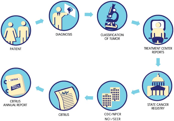
CBTRUS does not collect data directly from patients' medical records. As noted, data for CBTRUS analyses come from the NPCR and SEER programs. By law, cancer and benign brain tumors are reportable diseases and tumor registrars in treatment centers send these data and other pertinent information to central cancer registries in their states where they are collated and sent to NPCR and SEER. On an annual basis, NPCR secures permission from the central cancer registries to release their data on brain and CNS tumors to CBTRUS. Central cancer registries play an essential role in the collection process, diagrammatically presented in Figure 1. These data are population-based and, therefore, represent by definition a comprehensive documentation of all cancers diagnosed within a region over a period of time.
Fig. 1.
Schematic of Cancer Registration Process
CBTRUS obtained incidence data from 50 cancer registries (45 NPCR and 5 SEER) that include cases of malignant and non-malignant (benign and uncertain) primary brain and CNS tumors. The 50 population-based cancer registries include 49 state registries (excluding Minnesota, as this registry did not provide data to NPCR in 2013) and the District of Columbia. It should be noted that metastatic tumors including those found in the brain and CNS are not collected by surveillance organization in the United States. Data were requested for all newly-diagnosed primary malignant and non-malignant tumors from 2006 to 2010 at any of the following anatomic sites (ICD-O-3 topography codes in parentheses): brain (C71.0-C71.9), meninges (C70.0-C70.9), spinal cord, cranial nerves, and other parts of the central nervous system (C72.0-C72.9), pituitary and pineal glands (C75.1-C75.3), and olfactory tumors of the nasal cavity [C30.0 (ICD-O-3 histology codes 9522 and 9523)].4
NPCR provided data on 313,780 primary brain and CNS tumors diagnosed from 2006 to 2010. NPCR cancer registries agree to participate in the CBTRUS Statistical Report and to pass certain data quality standards required by NPCR, thus allowing CBTRUS to receive and analyze the data. An additional 12,991 primary brain and CNS tumor case records for the time period were obtained from SEER. These data were combined into a single data set for analyses. A total of 8,175 records (2.4%) were deleted from the final analytic data set because of one or more of the following reasons: invalid site/histology combinations, duplicate records that included a less accurate reporting source than microscopic confirmation (e.g. radiographic versus microscopic confirmation), duplicate records for bilateral vestibular schwannoma or meningioma, duplicate record for recurrent disease, and errors in time sequence of diagnosis. The final analytic data set included 326,711 records from 50 population-based central cancer registries, including 49 state registries and the District of Columbia.
Survival data for malignant brain and CNS tumors were obtained from 18 SEER registries for years 1995 to 2010. Survival information derived from active patient follow-up is not available in the data that CBTRUS receives from NPCR registries, so this alternate data source is used for the generation of these tables. This dataset includes information from 9 state registries, 5 regional registries and the Alaska Native Tumor Registry. Together, this provides population-based information for about 26% of the United States Population.7 Survival was not calculated for non-malignant tumors as collection of these cases has only been mandated since 2004, and, therefore, not enough time has elapsed to accurately calculate relative survival.
Mortality data used in this report are from the National Center for Health Statistics (NCHS) and includes deaths where primary brain or CNS tumor was listed as cause of death on the death certificate for all 50 states and the District of Columbia.
Definitions
Measures in Surveillance Epidemiology
The Incidence Rate is a basic measure of disease occurrence that measures the occurrence count of newly-diagnosed cases of disease per 100,000 population over a time period. Mortality Rates quantify the number of people who have died from the disease per 100,000 population in a specific time period. Prevalence Rates measure the number of people with a disease per 100,000 population at a particular point in time or during a particular period of time. Survival Rates represent the percentage of individuals who survive their disease to a specified time period. Relative Survival Rates are defined as the observed probability of survival adjusted for the expected survival rate of the United States population for that age, gender, and calendar year.
Incidence and mortality rates in this report are expressed per unit population in which the events are observed. For cancer, rates are usually expressed per 100,000 population. The unadjusted rate of disease in an entire population is the Crude Rate. Crude rates are generally adjusted by age because as a population ages the crude rate would increase, and this may only reflect the aging of the population rather than an actual rate increase. Age-Adjusted Rates to a common standard population allow for comparisons of rates in populations across regions with different age structures. Incidence and mortality rates in this report are age-adjusted to the Year 2000 U.S. Standard Population. Rates for a subset of a population are termed specific rates. Specific rates by gender, race, and Hispanic ethnicity are also reported. The variability around the estimates of rates is reflected in the Standard Error, which is incorporated into the formula for computing the confidence interval associated with a certain rate. A Confidence Interval is the computed interval with a given probability (e.g., 95 percent) that the true value of a variable such as a mean, proportion, or rate occurs within the interval. For example, the age-adjusted primary brain and CNS tumor incidence rate is 21.03 cases per 100,000. We can assume with 95 percent certainty that the actual incidence rate is within the range of 20.96 and 21.11 cases per 100,000. Incidence Rate Ratio is calculated to compare incidence rates between selected populations, and is generated by dividing the incidence rate of one group by the incidence rate of the comparison group. For example, for the age-adjusted incidence rate of glioblastoma is 3.97 case per 100,000 for males and 2.53 cases per 100,000 for females. This results in an incidence rate ratio of 1.57, meaning that men have an incidence rate of glioblastoma that is 1.57 times the rate of women.
Comparing incidence rates between statistical reports produced by multiple reporting organizations is not recommended. To compare incidence rates for brain and CNS tumors among statistical reports, agencies, or registries, a number of factors should be considered. These factors include whether the case definition, data collection, and rate calculation are similar between comparison sources. The following questions may be helpful in determining the comparability of reporting sources:
How is an incident case determined and defined?
Does the data set include both malignant and non-malignant tumors?
What anatomic locations (primary sites) are included?
Are lymphomas and hematopoietic neoplasms included in the incidence rates?
Are the populations comparable by time period, geography, and age distribution?
Are the incidence rates age-adjusted? If so, to which standard population are they age-adjusted?
Classification by Behavior and Histology
Brain and CNS tumor classifications according to behavior ICD-O-3 standards for benign, uncertain, and malignant behaviors are coded 0, 1, and 3, respectively. The histology groupings in CBTRUS statistical reports were initially developed in collaboration with the CBTRUS consulting neuropathologist, Dr. Janet Bruner of the University of Texas M.D. Anderson Cancer Center. In 2012, Drs. Roger McLendon and Tarik Tihan joined Dr. Bruner and CBTRUS staff to synchronize CBTRUS histology grouping scheme with the 2007 World Health Organization (WHO) classification of tumors of the central nervous system.6,8 This report uses the most recent 2012 CBTRUS Histology Grouping Scheme (Table 1). The classification scheme utilizes ICD-O-3 codes4 and may include morphology codes that were not previously reported to CBTRUS.9 Tables 1a and 1b list malignant only and non-malignant only histologies, respectively. In this report, incidence rates are provided by major histology grouping and detailed histology.
Table 1.
Central Brain Tumor Registry of the United States (CBTRUS), Brain and Central Nervous System Tumor Histology Groupings∥
| Histology | ICD-O-3† Histology Code |
|---|---|
| Tumors of Neuroepithelial Tissue | |
| Pilocytic astrocytoma | 9421 |
| Diffuse astrocytoma | 9400, 9410, 9411, 9420 |
| Anaplastic astrocytoma | 9401 |
| Unique astrocytoma variants | 9381, 9384, 9424 |
| Glioblastoma | 9440, 9441, 9442/3‡ |
| Oligodendroglioma | 9450 |
| Anaplastic oligodendroglioma | 9451, 9460 |
| Oligoastrocytic tumors | 9382 |
| Ependymal tumors | 9383, 9391, 9392, 9393, 9394 |
| Glioma malignant, NOS | 9380 |
| Choroid plexus tumors | 9390 |
| Other neuroepithelial tumors | 9363, 9423, 9430, 9444 |
| Neuronal and mixed neuronal-glial tumors | 8680, 8681, 8690, 8693, 9412, 9413, 9442/1§, |
| 9492 (excluding site C75.1), 9493, 9505, 9506, 9522, 9523 | |
| Tumors of the pineal region | 9360, 9361, 9362 |
| Embryonal tumors | 8963, 9364, 9470, 9471, 9472, 9473, 9474, 9480, |
| 9490, 9500, 9501, 9502, 9508 | |
| Tumors of Cranial and Spinal Nerves | |
| Nerve sheath tumors | 9540, 9541, 9550, 9560, 9561, 9570, 9571 |
| Other tumors of cranial and spinal nerves | 9562 |
| Tumors of Meninges | |
| Meningioma | 9530, 9531, 9532, 9533, 9534, 9537, 9538, 9539 |
| Mesenchymal tumors | 8324, 8800, 8801, 8802, 8803, 8804, 8805, 8806, 8810, 8815, 8824, 8830, |
| 8831, 8835, 8836, 8850, 8851, 8852, 8853, 8854, 8857, 8861, 8870, 8880, | |
| 8890, 8897, 8900, 8901, 8902, 8910, 8912, 8920, 8921, 8935, 8990, 9040, 9136, | |
| 9150, 9170, 9180, 9210, 9241, 9260, 9373, | |
| Primary melanocytic lesions | 8720, 8728, 8770, 8771 |
| Other neoplasms related to the meninges | 9161, 9220, 9231, 9240, 9243, 9370, 9371, 9372, 9535 |
| Lymphomas and Hemopoietic Neoplasms | |
| Lymphoma | 9590, 9591, 9596, 9650, 9651, 9652, 9653, 9654, 9655, 9659, 9661, |
| 9662, 9663, 9664, 9665, 9667, 9670, 9671, 9673, 9675, 9680, 9684, | |
| 9687, 9690, 9691, 9695, 9698, 9699, 9701, 9702, 9705, 9714, 9719, | |
| 9728, 9729 | |
| Other hemopoietic neoplasms | 9727, 9731, 9733, 9734, 9740, 9741, 9750, 9751, 9752, 9753, 9754, 9755, |
| 9756, 9757, 9758, 9760, 9766, 9823, 9826, 9827, 9832, 9837, 9860, 9861, 9866, 9930, 9970 | |
| Germ Cell Tumors and Cysts | |
| Germ cell tumors, cysts and heterotopias | 8020, 8440, 9060, 9061, 9064, 9065, 9070, 9071, 9072, 9080, 9081, 9082, |
| 9083, 9084, 9085, 9100, 9101 | |
| Tumors of Sellar Region | |
| Tumors of the pituitary | 8040, 8140, 8146, 8246, 8260, 8270, 8271, 8272, |
| 8280, 8281, 8290, 8300, 8310, 8323, 9492 (Site C75.1 only), 9582 | |
| Craniopharyngioma | 9350, 9351, 9352 |
| Unclassified Tumors | |
| Hemangioma | 9120, 9121, 9122, 9123, 9125, 9130, 9131, 9133, 9140 |
| Neoplasm, unspecified | 8000, 8001, 8002, 8003, 8004, 8005, 8010, 8021 |
| All other | 8320, 8452, 8710, 8711, 8713, 8811, 8840, 8896, 8980, 9173, 9503, 9580 |
∥Based on 2007 WHO Classification of Tumours of the Central Nervous System6
†International Classification of Diseases for Oncology, 3rd Edition, 2000. World Health Organization, Geneva, Switzerland.
‡Morphology 9442/3 only.
§Morphology 9442/1 only.
CBTRUS defines the broad category of gliomas to include ICD-O-3 histology codes 9380-9384,9391-9460,9480.
Abbreviations: CBTRUS, Central Brain Tumor Registry of the United States; NOS, not otherwise specified.
Table 1a.
Central Brain Tumor Registry of the United States (CBTRUS), Brain and Central Nervous System Tumor Malignant Histologies†
| Histology | ICD-O-3‡ Histology Code |
|---|---|
| Tumors of Neuroepithelial Tissue | |
| Pilocytic astrocytoma | 9421/1 [Included with malignant tumors as explained in Brain Tumor Definition Differences] |
| Diffuse astrocytoma | 9400/3, 9410/3, 9411/3, 9420/3 |
| Anaplastic astrocytoma | 9401/3 |
| Unique astrocytoma variants | 9381/3, 9424/3 |
| Glioblastoma | 9440/3, 9441/3, 9442/3 |
| Oligodendroglioma | 9450/3 |
| Anaplastic oligodendroglioma | 9451/3, 9460/3 |
| Oligoastrocytic tumors | 9382/3 |
| Ependymal tumors | 9391/3, 9392/3, 9393/3 |
| Glioma malignant, NOS | 9380/3 |
| Choroid plexus | 9390/3 |
| Other neuroepithelial tumors | 9423/3, 9430/3 |
| Neuronal and mixed neuronal- glial tumors | 8680/3, 8693/3, 9505/3, 9522/3, 9523/3 |
| Tumors of the pineal region | 9362/3 |
| Embryonal tumors | 8963/3, 9364/3, 9470/3, 9471/3, 9472/3,9473/3, 9474/3, 9480/3, |
| 9490/3, 9500/3, 9501/3, 9502/3, 9508/3 | |
| Tumors of Cranial and Spinal Nerves | |
| Nerve sheath tumors | 9540/3, 9560/3, 9561/3, 9571/3 |
| Tumors of Meninges | |
| Meningioma | 9530/3, 9538/3, 9539/3 |
| Mesenchymal tumors | 8800/3, 8801/3, 8802/3, 8803/3, 8804/3, 8805/3, 8806/3, 8810/3, 8815/3, 8830/3, |
| 8850/3, 8851/3, 8852/3, 8853/3, 8854/3, 8857/3, 8890/3, 8900/3, 8901/3, 8902/3, | |
| 8910/3, 8912/3, 8920/3, 8921/3, 8990/3, 9040/3, 9150/3, 9170/3, 9180/3, 9260/3 | |
| Primary melanocytic lesions | 8720/3, 8728/3, 8770/3, 8771/3 |
| Other neoplasms related to the meninges | 9220/3, 9231/3, 9240/3, 9243/3, 9370/3, 9371/3, 9372/3 |
| Lymphomas and Hemopoietic Neoplasms | |
| Lymphoma | 9590/3, 9591/3, 9596/3, 9650/3, 9651/3, 9652/3, 9653/3, 9654/3, 9655/3, 9659/3, |
| 9661/3, 9662/3, 9663/3, 9664/3, 9665/3, 9667/3, 9670/3, 9671/3, 9673/3, 9675/3, | |
| 9680/3, 9684/3, 9687/3, 9690/3, 9691/3, 9695/3, 9698/3, 9699/3, 9701/3, 9702/3, | |
| 9705/3, 9714/3, 9719/3, 9728/3, 9729/3 | |
| Other hemopoietic neoplasms | 9727/3, 9731/3, 9733/3, 9734/3, 9740/3, 9741/3, 9750/3, 9754/3, 9755/3, 9756/3, 9757/3, 9758/3, |
| 9760/3, 9823/3, 9826/3, 9827/3, 9832/3, 9837/3, 9860/3, 9861/3, 9866/3, 9930/3 | |
| Germ Cell Tumors and Cysts | |
| Germ cell tumors, cysts and | 8020/3, 8440/3, 9060/3, 9061/3, 9064/3, 9065/3, 9070/3, 9071/3, 9072/3, 9080/3, |
| heterotopias | 9081/3, 9082/3, 9083/3, 9084/3, 9085/3, 9100/3, 9101/3 |
| Tumors of Sellar Region | |
| Tumors of the pituitary | 8140/3, 8246/3, 8260/3, 8270/3, 8272/3, 8280/3, 8281/3, 8290/3, 8300/3, 8310/3, 8323/3 |
| Unclassified Tumors | |
| Hemangioma | 9120/3, 9130/3, 9133/3, 9140/3 |
| Neoplasm, unspecified | 8000/3, 8001/3, 8002/3, 8003/3, 8004/3, 8005/3, 8010/3, 8021/3 |
| All other | 8320/3, 8710/3, 8711/3, 8811/3, 8840/3, 8896/3, 8980/3, 9503/3, 9580/3 |
∥Based on 2007 WHO Classification of Tumours of the Central Nervous System6
†Includes all the histologies listed in the standard definition of reportable brain tumors from the Consensus Conference on Brain Tumor Definitions.
‡International Classification of Diseases for Oncology, 3rd Edition, 2000. World Health Organization, Geneva, Switzerland.
Abbreviations: CBTRUS, Central Brain Tumor Registry of the United States; NOS, not otherwise specified.
Table 1b.
Central Brain Tumor Registry of the United States (CBTRUS), Brain and Central Nervous System Tumor Non-Malignant Histologies†
| Histology | ICD-O-3‡ Histology Code |
|---|---|
| Tumors of Neuroepithelial Tissue | |
| Pilocytic astrocytoma | 9421/1 [Included with malignant tumors as explained in Brain Tumor Definition Differences] |
| Unique astrocytoma variants | 9384/1 |
| Ependymal tumors | 9383/1; 9394/1 |
| Choroid plexus | 9390/0,1 |
| Other neuroepithelial tumors | 9363/0; 9444/1 |
| Neuronal and mixed neuronal- | 8680/0,1; 8681/1; 8690/1; 8693/1; 9412/1; 9413/0; 9442/1; |
| glial tumors | 9492/0 (excluding site C75.1); 9493/0; 9505/1; 9506/1 |
| Tumors of the pineal region | 9360/1; 9361/1 |
| Embryonal tumors | 9490/0 |
| Tumors of Cranial and Spinal Nerves | |
| Nerve sheath tumors | 9540/0,1; 9541/0, 9550/0; 9560/0,1; 9570/0; 9571/0 |
| Other tumors of cranial and spinal nerves | 9562/0 |
| Tumors of Meninges | |
| Meningioma | 9530/0,1; 9531/0; 9532/0; 9533/0; 9534/0; 9537/0; 9538/1; 9539/1 |
| Mesenchymal tumors | 8324/0; 8800/0; 8810/0; 8815/0; 8824/0,1; 8830/0,1; 8831/0; 8835/1; 8836/1; |
| 8850/0,1; 8851/0; 8852/0, 8854/0; 8857/0; 8861/0; 8870/0; 8880/0, 8890/0,1; 8897/1; | |
| 8900/0; 8920/1; 8935/0,1; 8990/0,1; 9040/0; 9136/1, 9150/0,1; 9170/0; 9180/0; 9210/0; 9241/0; 9373/0 | |
| Primary melanocytic lesions | 8728/0,1; 8770/0; 8771/0 |
| Other neoplasms related to the meninges | 9161/1; 9220/0,1; 9535/0 |
| Lymphomas and Hemopoietic Neoplasms | |
| Other hemopoietic neoplasms | 9740/1; 9751/1; 9752/1; 9753/1; 9766/1; 9970/1 |
| Germ Cell Tumors and Cysts | |
| Germ cell tumors, cysts and heterotopias | 8440/0; 9080/0,1; 9084/0 |
| Tumors of Sellar Region | |
| Tumors of the pituitary | 8040/0,1; 8140/0,1; 8146/0; 8260/0; 8270/0; 8271/0; 8272/0; |
| 8280/0; 8281/0; 8290/0; 8300/0; 8310/0; 8323/0; 9492/0 (site C75.1 only); 9582/0 | |
| Craniopharyngioma | 9350/1; 9351/1; 9352/1 |
| Unclassified Tumors | |
| Hemangioma | 9120/0; 9121/0; 9122/0; 9123/0; 9125/0; 9130/0,1; 9131/0; 9133/1 |
| Neoplasm, unspecified | 8000/0,1; 8001/0,1; 8005/0; 8010/0 |
| All other | 8452/1; 8711/0; 8713/0; 8811/0; 8840/0; 9173/0; 9580/0 |
∥Based on 2007 WHO Classification of Tumours of the Central Nervous System6
†Includes all the histologies listed in the standard definition of reportable brain tumors from the Consensus Conference on Brain Tumor Definition.
‡International Classification of Diseases for Oncology, 3rd Edition, 2000. World Health Organization, Geneva, Switzerland.
Abbreviations: CBTRUS, Central Brain Tumor Registry of the United States; NOS, not otherwise specified.
Gliomas are tumors that arise from glial or precursor cells, and include astrocytoma (ICD-O-3 Histology codes: 9381, 9384, 9400, 9401, 9410, 9411, 9420, 9421, 9424), glioblastoma (ICD-O-3 Histology codes: 9440-9442), oligodendroglioma (ICD-O-3 Histology codes: 9450, 9451, 9460), ependymoma (ICD-O-3 Histology codes: 9383, 9391, 9392, 9393, 9394), mixed glioma (ICD-O-3 Histology code: 9382), malignant glioma, not otherwise specified (NOS) (ICD-O-3 Histology codes: 9380), and a few more rare histologies (ICD-O-3 Histology codes: 9363, 9423, 9430, 9444). Because there is no standard definition for gliomas, CBTRUS defines glioma as ICD-O-3 histology codes 9380-9384, 9391-9460, and 9480. It has been noted by Dr. Janet Bruner, CBTRUS Consulting Neuropathologist, that 9480/3 Cerebellar sarcoma, NOS is no longer in use. CBTRUS continues to list this tumor as it is included in ICD-O-3 as 9480/3 Cerebellar sarcoma, NOS (C71.6) [obs]. It is also important to note that the statistics for lymphomas and hematopoietic neoplasms contained in this report refer only to those lymphomas and hematopoietic neoplasms that arise in the brain and CNS.
This report also utilizes the International Classification of Childhood Cancer (ICCC) grouping system for pediatric cancers. ICCC categories for this report were generated using the SEER Site/Histology ICCC-3 Recode10 based on the ICCC, Third edition11 and 2008 WHO Classification of Tumours of Haematopoietic and Lymphoid Tissues.
Anatomic Location of Tumor Sites
Various terms are used to describe the regions of the brain and central nervous system. The specific sites used in this report are broadly based on the categories and site codes defined in the SEER Site/Histology Validation List.12 Tumors include olfactory tumors of the nasal cavity in addition to brain tumors located in sites included in the standard definition from the Consensus Conference on Brain Tumor Definition for Registration.9 According to the standard definition from the Consensus Conference, reportable primary brain-related tumors (intracranial and central nervous system tumors) are all primary tumors, irrespective of histology and behavior, occurring in the following sites: brain; meninges; pineal gland; pituitary gland and craniopharyngeal duct; and spinal cord, cranial nerves, and other parts of the central nervous system. As per the site definition outlined by the Consensus Conference, brain lymphomas coded to any of the brain or CNS site codes listed above are included in the CBTRUS report. The group of tumors known as spinal cord tumors is coded to the following sites: spinal meninges, spinal cord, and cauda equina.
Statistics by ICD-O-3 primary sites are grouped in the following manner for Brain, C71: the frontal lobe (C71.1); temporal lobe (C71.2); parietal lobe (C71.3); and occipital lobe (C71.4) are grouped together. Cerebrum (C71.0), ventricle (C71.5), cerebellum (C71.6), and brain stem (C71.7) are each grouped independently. For purposes of analysis, the category other brain is used to refer to ICD-O codes C71.8-C71.9, which are defined as overlapping regions of the brain and brain, not otherwise specified respectively as listed under C71. The Spinal Cord, Cranial Nerves, and Other Parts of the Central Nervous System are listed under C72. The cranial nerve category (C72.2-C72.5) includes the olfactory nerve, optic nerve, acoustic nerve, and other cranial nerves. The spinal cord (C72.0) and cauda equina (C72.1) are grouped together. The category other central nervous system is used to refer to ICD-O codes C72.8-C72.9, which includes overlapping regions of the brain and central nervous system, and central nervous system, not otherwise specified in C72. The meninges C70 include the cerebral meninges and spinal meninges (C70.0-C70.9). Pituitary tumors (C75.1-C75.2) include tumors located in the pituitary gland and craniopharyngeal duct. Pineal tumors (C75.3) include tumors located in the pineal gland. In this report, tumors located in the nasal cavity (C30.0) are olfactory tumors (defined by ICD-O-3 histology codes 9522 and 9523).
Measurement Methods
Counts, means, rates, ratios, proportions, and other relevant statistics were calculated using R and/or SEER*Stat 8.04 statistical software.13,14 Statistics are suppressed when counts are fewer than 16 within a cell. However, the data in the suppressed cells are included in the counts and rates for the totals.
Population data for each geographic region were obtained from the SEER program website15 for the purpose of rate calculation.
Age-adjusted incidence rates and 95% confidence intervals16 for malignant and non-malignant tumors and for selected histology groupings by gender, race, Hispanic ethnicity, and pediatric, young adult, and adult age groups were estimated. Age-adjustment was based on one-year age groupings and standardized to the 2000 U.S. standard population. The age distribution of the 2000 U.S. standard population is shown in Appendix A. Combined populations for the regions included in this report are shown in Appendix B and Appendix C.
CBTRUS presents statistics on the pediatric age group 0-19 years in order to include and describe specific brain and CNS tumor patterns in age groups 0-4, 5-9, 10-14, and 15-19 years. However, the 0-14 year age group is a standard age category for childhood cancer used by other cancer surveillance organizations and has been included in this report for consistency and comparison purposes. Race categories in this report are all races, white, black, American Indian/Alaskan Native (AIAN), and Asian Pacific Islander (API). State cancer registries utilize codes that allow documentation of cancer occurrence in Asian/Pacific Islander subpopulations, but these have been grouped into a single API race category for the purpose of this analysis, in accordance with NAACCR data standards.17 Other race, unspecified, and unknown race are included in all race statistics. Hispanic ethnicity was defined using the NAACCR Hispanic Identification Algorithm, version 2, data element, which utilizes a combination of cancer registry data fields (Spanish/Hispanic Origin data element, birthplace, race, and surnames) to directly and indirectly classify cases as Hispanic or non-Hispanic.18
Trends across annual incidence rates were not estimated because a timeframe of five years is not sufficient to detect a real change in the rate pattern with any degree of confidence.
Brain Tumor Definition Differences
It should be noted that NPCR, SEER, and NAACCR report brain tumors differently than CBTRUS. The definition of brain and CNS tumors used by these organizations (in their published incidence and mortality statistics) includes tumors located in the following sites with their ICD-O-3 site codes in parentheses: brain, meninges, and other central nervous system tumors (C70.0-9, C71.0-9, and C72.0-9), but excludes lymphoma and leukemia histologies (9590-9989) from all brain and CNS sites. NPCR and SEER include separate tables for malignant and non-malignant brain and CNS tumors reflecting the 2002 Consensus Conference9 definition in their respective publications. With the inclusion of non-malignant brain tumors, an increase in incidence rates may result, especially for the following histology groups and subgroups: (groups) tumors of meninges; tumors of cranial and spinal nerves; tumors of the sellar region; and (subgroups) unique astrocytoma variants; ependymal tumors; choroid plexus; neuronal and mixed neuronal-glial tumors; tumors of the pineal region; nerve sheath tumors; meningioma; mesenchymal tumors; other neoplasms related to the meninges; germ cell tumors; tumors of the pituitary; craniopharyngioma; hemangioma; neoplasm, unspecified; and all other.
In contrast, the CBTRUS reports data on all tumor morphologies located within the Consensus Conference site definition including lymphoma and other hematopoietic histologies (9590-9989) as well as olfactory tumors of the nasal cavity [C30.0 (9522-9523)].9 NPCR, SEER, and NAACCR include Pilocytic astrocytoma in their malignant (ICD-0-3 behavior code of 3) brain tumor data and statistics, though it is listed in the WHO Classification of Tumours of the Central Nervous System6 as having uncertain behavior (ICD-0-3 behavior code of 1). Since the CBTRUS Histology Grouping Scheme is reflective of WHO 2007, CBTRUS also includes Pilocytic astrocytoma in Table 1a. In support of consistency within cancer surveillance reporting, the CBTRUS categorizes Pilocytic astrocytomas in the malignant tumor category (Table 1b) to enhance comparability of rates to those reported by NPCR, SEER, and NAACCR, especially for comparison of childhood brain and CNS tumor rates. It is important to understand these differences in definition, as they influence the direct comparison of published rates.
Estimation of Expected Numbers of Brain and CNS Tumors in 2013 and 2014
Estimated numbers of expected malignant and non-malignant brain and CNS tumors were calculated for 2013 and 2014. To project 2013 and 2014 estimates of all primary brain and CNS tumors, age-adjusted brain tumor incidence rates for a state were multiplied by the projected population for that state. Projected population estimates for 2013 and 2014 were derived using the US Census Bureau 2000-2009 population data (www.seer.cancer.gov/popdata/index.html).15
Estimation of Mortality Rates for Underlying Cause from Brain and CNS Tumors
Age-adjusted mortality rates for deaths resulting from all brain and CNS tumors were calculated using SEER Stat 8.0.4.14,19 The underlying mortality data were provided to the SEER program by NCHS (www.cdc.gov/nchs). In addition to total age-adjusted rate for the United States, age-adjusted rates are presented by gender and state.
Estimation of Survival Rates
SEER*Stat 8.0.4 statistical software was used to estimate one, two, three, four, five, and ten-year relative survival rates for primary malignant brain tumor cases diagnosed between 1995-2010 in eighteen SEER areas.14,20 This software utilizes life-table (actuarial) methods to compute survival estimates and accounts for current follow-up. Survival was estimated for brain (C71.0-C71.9), meninges (C70.0-C70.9), spinal cord, cranial nerves, and other parts of the central nervous system (C72.0-C72.9), pituitary and pineal glands (C75.1-C75.3), and olfactory tumors of the nasal cavity [C30.0 (9522-9523)]. Lymphomas and leukemias (morphology codes 9590-9989) and meningiomas (9530-9539) occurring at any brain or CNS site are included. Second or later primary tumors, cases diagnosed at autopsy, cases in which race or sex is coded as other or unknown, and cases known to be alive but for whom follow-up time could not be calculated were excluded from the SEER survival data analyses.
Data Interpretation
The CBTRUS works diligently to support the broader surveillance efforts aimed at improving the collection and reporting of primary brain and CNS tumors. The central cancer registry data provided to NPCR and SEER and, subsequently, to CBTRUS vary from year-to-year due to ongoing updates in collection and data refinement aimed to improve completeness and accuracy. The data presented in this report must be interpreted within this surveillance framework as well as taking into account the information provided in the technical notes. Therefore, it is important to note that data from previous CBTRUS Reports cannot be compared to data in CBTRUS Statistical Report: Primary Brain and Central Nervous System Tumors Diagnosed in the United States in 2006-2010.
Random fluctuations in average annual rates are usual especially for rates based on small incidence counts. The CBTRUS policy to suppress data presentation for cells with counts of less than 16 is consistent with the NPCR policy. The suppression of data with counts of fewer than 16 is more than adequate to protect confidentiality given the CBTRUS data set is an aggregate of 49 states (excluding Minnesota) and the District of Columbia for 2006-2010.
Delays in reporting and late ascertainment are a reality and a known issue influencing registry completeness and, consequently, rate underestimations - especially for more recent data collection years.21 CBTRUS also recognizes that the problem may be even more likely to occur in the reporting of non-malignant brain and CNS tumors, where reporting often comes from non-hospital based sources and mandated collection is relatively recent (2004).
Reporting from Veteran's Health Administration (VHA) hospitals, the sole source of data for cancer cases diagnosed among Veterans served by those institutions, affects completeness of data. Cancer cases from VHA facilities account for at least three percent and possibly as much as eight percent of all cancer cases diagnosed among men. VHA policy that went into effect in 2007 restricting veterans' health data sharing has resulted in the underreporting of cancer incidence data for diagnosis years 2005 through 2007. Since late 2008, VHA facilities and states with central cancer registries have been working to establish data transfer agreements that correct the problem to assure more complete ascertainment of national cancer incidence including brain and CNS tumor incidence data used in CBTRUS statistical reports.22 This continues to be an ongoing process.
CBTRUS editing practices conducted yearly aim to refine the data for accuracy and clinical relevance should also be recognized in interpreting these report data. Exclusion of site and histology combinations considered to be invalid by the consulting neuropathologists, who revised the CBTRUS site/histology validation list in 2012, may have the impact of conservatively underestimating the incidence of brain and CNS tumors. Editing changes also incorporate updates to the cancer registration coding rules that influence case ascertainment and data collection. For example, beginning in 2004 some brain and CNS site codes were reconsidered as paired sites, meaning that having a tumor in both the left and right hemisphere would result in multiple tumors being reported rather than one bilateral tumor. This has likely caused some increase in reported incidence. Another relevant coding tool affecting reporting was the 2007 Multiple Primary and Histology Coding Rules. These rules revised the way malignant and non-malignant brain tumors are reported and may affect incident rate changes for certain histologies.
Population estimates used for denominators affect incidence rates. CBTRUS has utilized population data estimates for years 2006-2010 available on the SEER website for rate calculations in this report. These population estimates are provided to SEER on an annual basis from the United States Bureau of the Census. It should be noted that these estimates do not reflect the 2010 decennial census which may or may not impact the rates published.
Results
Primary Brain and CNS Tumors: Distributions and Incidence by Gender, Race, Hispanic Ethnicity, Age Group, Cancer Registry, and Behavior
Counts of the 326,711 incident tumors (112,458 malignant; 214,253 non-malignant) reported during 2006-2010 by histology and demographic characteristics for all ages and for children ages 0-19 are presented in Tables 2–8. Approximately 7% of the cases were in individuals younger than 20 years of age at the time of diagnosis, and 93% were in individuals 20 years of age or older. Approximately 42% of all brain and CNS tumors overall occurred in males and 58% in females; malignant: 55% in males and 45% in females and non-malignant: 36% in males and 64% in females. The overall number of all reported tumors is listed by central cancer registry in Table 9. The average annual combined 2006-2010 population of 298,719,439 represents approximately 98% of the U.S. population for those years. The overall percent of non-malignant tumors varied considerably by cancer registry (range: 53.2-73.0%). About 62.9% of all tumors had a histologically confirmed diagnosis, with substantial regional variation (see range: 53.3-75.2% in Table 9).
Table 2.
Average Annual Age-Adjusted Incidence Rates† for Brain and Central Nervous System Tumors by Major Histology Groupings, Histology and Gender, CBTRUS Statistical Report: NPCR and SEER, 2006-2010
| Histology | Total |
Male |
Female |
||||||
|---|---|---|---|---|---|---|---|---|---|
| N | Rate | 95% CI | N | Rate | 95% CI | N | Rate | 95% CI | |
| Tumors of Neuroepithelial Tissue | 101,825 | 6.60 | (6.56-6.64) | 56,695 | 7.76 | (7.70-7.83) | 45,130 | 5.60 | (5.55-5.65) |
| Pilocytic astrocytoma | 4,741 | 0.33 | (0.32-0.34) | 2,425 | 0.33 | (0.32-0.35) | 2,316 | 0.33 | (0.31-0.34) |
| Diffuse astrocytoma | 8,535 | 0.56 | (0.55-0.58) | 4,785 | 0.66 | (0.64-0.68) | 3,750 | 0.48 | (0.47-0.50) |
| Anaplastic astrocytoma | 5,621 | 0.37 | (0.36-0.38) | 3,169 | 0.43 | (0.42-0.45) | 2,452 | 0.31 | (0.29-0.32) |
| Unique astrocytoma variants | 943 | 0.06 | (0.06-0.07) | 515 | 0.07 | (0.06-0.08) | 428 | 0.06 | (0.05-0.06) |
| Glioblastoma | 50,872 | 3.19 | (3.16-3.21) | 29,019 | 3.97 | (3.92-4.01) | 21,853 | 2.53 | (2.50-2.57) |
| Oligodendroglioma | 4,020 | 0.27 | (0.26-0.28) | 2,218 | 0.30 | (0.29-0.32) | 1,802 | 0.24 | (0.23-0.25) |
| Anaplastic oligodendroglioma | 1,658 | 0.11 | (0.10-0.11) | 931 | 0.13 | (0.12-0.13) | 727 | 0.09 | (0.09-0.10) |
| Oligoastrocytic tumors | 3,045 | 0.20 | (0.20-0.21) | 1,734 | 0.24 | (0.23-0.25) | 1,311 | 0.17 | (0.16-0.18) |
| Ependymal tumors | 6,304 | 0.42 | (0.41-0.43) | 3,508 | 0.47 | (0.46-0.49) | 2,796 | 0.36 | (0.35-0.38) |
| Glioma malignant, NOS | 6,765 | 0.46 | (0.44-0.47) | 3,397 | 0.48 | (0.46-0.50) | 3,368 | 0.44 | (0.42-0.45) |
| Choroid plexus tumors | 797 | 0.05 | (0.05-0.06) | 374 | 0.05 | (0.05-0.06) | 423 | 0.06 | (0.05-0.06) |
| Other neuroepithelial tumors | 93 | 0.01 | (0.01-0.01) | 33 | 0.00 | (0.00-0.01) | 60 | 0.01 | (0.01-0.01) |
| Neuronal and mixed neuronal-glial tumors | 4,036 | 0.27 | (0.26-0.28) | 2,143 | 0.29 | (0.28-0.30) | 1,893 | 0.26 | (0.24-0.27) |
| Tumors of the pineal region | 605 | 0.04 | (0.04-0.04) | 237 | 0.03 | (0.03-0.04) | 368 | 0.05 | (0.04-0.05) |
| Embryonal tumors | 3,790 | 0.26 | (0.26-0.27) | 2,207 | 0.30 | (0.29-0.32) | 1,583 | 0.22 | (0.21-0.23) |
| Tumors of Cranial and Spinal Nerves | 26,564 | 1.69 | (1.67-1.71) | 12,671 | 1.69 | (1.66-1.72) | 13,893 | 1.70 | (1.67-1.73) |
| Nerve sheath tumors | 26,548 | 1.69 | (1.67-1.71) | 12,664 | 1.69 | (1.66-1.72) | 13,884 | 1.70 | (1.67-1.72) |
| Other tumors of cranial and spinal nerves | 16 | 0.00 | (0.00-0.00) | – | – | – | – | – | – |
| Tumors of Meninges | 121,110 | 7.71 | (7.67-7.75) | 33,090 | 4.73 | (4.68-4.79) | 88,020 | 10.27 | (10.20-10.34) |
| Meningioma | 116,986 | 7.44 | (7.40-7.48) | 30,888 | 4.44 | (4.39-4.49) | 86,098 | 10.02 | (9.96-10.09) |
| Mesenchymal tumors | 1,229 | 0.08 | (0.08-0.09) | 584 | 0.08 | (0.07-0.09) | 645 | 0.08 | (0.08-0.09) |
| Primary melanocytic lesions | 116 | 0.01 | (0.01-0.01) | 70 | 0.01 | (0.01-0.01) | 46 | 0.01 | (0.00-0.01) |
| Other neoplasms related to the meninges | 2,779 | 0.18 | (0.17-0.19) | 1,548 | 0.21 | (0.20-0.22) | 1,231 | 0.16 | (0.15-0.17) |
| Lymphomas and Hemopoietic Neoplasms | 7,122 | 0.46 | (0.45-0.47) | 3,751 | 0.52 | (0.51-0.54) | 3,371 | 0.40 | (0.39-0.41) |
| Lymphoma | 6,919 | 0.44 | (0.43-0.46) | 3,648 | 0.51 | (0.49-0.53) | 3,271 | 0.39 | (0.37-0.40) |
| Other hemopoietic neoplasms | 203 | 0.01 | (0.01-0.02) | 103 | 0.01 | (0.01-0.02) | 100 | 0.01 | (0.01-0.02) |
| Germ Cell Tumors and Cysts | 1,464 | 0.10 | (0.10-0.11) | 1,005 | 0.14 | (0.13-0.14) | 459 | 0.06 | (0.06-0.07) |
| Germ cell tumors, cysts and heterotopias | 1,464 | 0.10 | (0.10-0.11) | 1,005 | 0.14 | (0.13-0.14) | 459 | 0.06 | (0.06-0.07) |
| Tumors of Sellar Region | 50,709 | 3.32 | (3.29-3.34) | 22,795 | 3.11 | (3.07-3.15) | 27,914 | 3.60 | (3.55-3.64) |
| Tumors of the pituitary | 47,958 | 3.13 | (3.10-3.16) | 21,482 | 2.93 | (2.89-2.97) | 26,476 | 3.41 | (3.37-3.45) |
| Craniopharyngioma | 2,751 | 0.18 | (0.18-0.19) | 1,313 | 0.18 | (0.17-0.19) | 1,438 | 0.19 | (0.18-0.20) |
| Unclassified Tumors | 17,917 | 1.16 | (1.14-1.18) | 7,954 | 1.16 | (1.13-1.18) | 9,963 | 1.17 | (1.14-1.19) |
| Hemangioma | 3,934 | 0.26 | (0.25-0.27) | 1,724 | 0.24 | (0.22-0.25) | 2,210 | 0.28 | (0.27-0.29) |
| Neoplasm, unspecified | 13,895 | 0.90 | (0.88-0.91) | 6,188 | 0.92 | (0.89-0.94) | 7,707 | 0.88 | (0.86-0.90) |
| All other | 88 | 0.01 | (0.00-0.01) | 42 | 0.01 | (0.00-0.01) | 46 | 0.01 | (0.00-0.01) |
| TOTAL‡ | 326,711 | 21.03 | (20.96-21.11) | 137,961 | 19.11 | (19.00-19.21) | 188,750 | 22.79 | (22.68-22.89) |
†Rates are per 100,000 and are age-adjusted to the 2000 US standard population.
‡Refers to all brain tumors including histologies not presented in this table.
–Counts are not presented when fewer than 16 cases were reported for the specific histology category. Suppressed cases are included in the total count.
Abbreviations: CBTRUS, Central Brain Tumor Registry of the United States; NPCR, CDC's National Program of Cancer Registries; SEER, NCI's Surveillance, Epidemiology and End Results program; CI, confidence interval; NOS, not otherwise specified.
Table 3.
Average Annual Age-Adjusted Incidence Rates† for Brain and Central Nervous System Tumors by Major Histology Groupings, Histology and Race, CBTRUS Statistical Report: NPCR and SEER, 2006-2010
| Histology | White |
Black |
AIAN |
API |
||||||||
|---|---|---|---|---|---|---|---|---|---|---|---|---|
| N | Rate | 95% CI | N | Rate | 95% CI | N | Rate | 95% CI | N | Rate | 95% CI | |
| Tumors of Neuroepithelial Tissue | 90,357 | 7.12 | (7.07-7.17) | 7,158 | 3.79 | (3.70-3.88) | 528 | 3.24 | (2.95-3.56) | 3,205 | 4.35 | (4.20-4.51) |
| Pilocytic astrocytoma | 3,946 | 0.35 | (0.34-0.37) | 523 | 0.23 | (0.21-0.25) | 30 | 0.13 | (0.09-0.20) | 214 | 0.27 | (0.24-0.31) |
| Diffuse astrocytoma | 7,476 | 0.61 | (0.59-0.62) | 625 | 0.32 | (0.30-0.35) | 71 | 0.41 | (0.32-0.53) | 302 | 0.40 | (0.35-0.45) |
| Anaplastic astrocytoma | 5,035 | 0.40 | (0.39-0.41) | 342 | 0.19 | (0.17-0.21) | 35 | 0.20 | (0.13-0.28) | 167 | 0.24 | (0.20-0.28) |
| Unique astrocytoma variants | 751 | 0.06 | (0.06-0.07) | 130 | 0.06 | (0.05-0.07) | – | – | – | 45 | 0.06 | (0.04-0.08) |
| Glioblastoma | 46,480 | 3.45 | (3.41-3.48) | 2,847 | 1.67 | (1.60-1.73) | 192 | 1.48 | (1.26-1.72) | 1,124 | 1.67 | (1.57-1.78) |
| Oligodendroglioma | 3,591 | 0.30 | (0.29-0.31) | 233 | 0.12 | (0.11-0.14) | 24 | 0.12 | (0.08-0.19) | 150 | 0.19 | (0.16-0.22) |
| Anaplastic oligodendroglioma | 1,474 | 0.12 | (0.11-0.13) | 88 | 0.05 | (0.04-0.06) | – | – | – | 72 | 0.09 | (0.07-0.11) |
| Oligoastrocytic tumors | 2,708 | 0.23 | (0.22-0.24) | 176 | 0.09 | (0.08-0.11) | 20 | 0.10 | (0.06-0.16) | 113 | 0.14 | (0.11-0.17) |
| Ependymal tumors | 5,431 | 0.45 | (0.43-0.46) | 538 | 0.27 | (0.25-0.29) | 36 | 0.19 | (0.13-0.27) | 266 | 0.33 | (0.29-0.37) |
| Glioma malignant, NOS | 5,729 | 0.48 | (0.47-0.49) | 652 | 0.33 | (0.31-0.36) | 35 | 0.20 | (0.14-0.29) | 306 | 0.42 | (0.37-0.47) |
| Choroid plexus tumors | 688 | 0.06 | (0.06-0.06) | 55 | 0.03 | (0.02-0.03) | – | – | – | 42 | 0.05 | (0.04-0.07) |
| Other neuroepithelial tumors | 79 | 0.01 | (0.01-0.01) | – | – | – | – | – | – | – | – | – |
| Neuronal and mixed neuronal-glial tumors | 3,356 | 0.29 | (0.28-0.30) | 419 | 0.20 | (0.18-0.22) | 30 | 0.14 | (0.09-0.21) | 211 | 0.26 | (0.22-0.30) |
| Tumors of the pineal region | 462 | 0.04 | (0.04-0.04) | 101 | 0.05 | (0.04-0.06) | – | – | – | 25 | 0.03 | (0.02-0.04) |
| Embryonal tumors | 3,151 | 0.28 | (0.27-0.30) | 419 | 0.18 | (0.17-0.20) | 25 | 0.11 | (0.07-0.18) | 165 | 0.21 | (0.18-0.24) |
| Tumors of Cranial and Spinal Nerves | 22,985 | 1.78 | (1.75-1.80) | 1,499 | 0.80 | (0.76-0.85) | 136 | 0.86 | (0.71-1.03) | 1,764 | 2.34 | (2.23-2.46) |
| Nerve sheath tumors | 22,974 | 1.77 | (1.75-1.80) | 1,498 | 0.80 | (0.76-0.85) | 136 | 0.86 | (0.71-1.03) | 1,761 | 2.34 | (2.23-2.45) |
| Other tumors of cranial and spinal nerves | – | – | – | – | – | – | – | – | – | – | – | – |
| Tumors of Meninges | 99,668 | 7.51 | (7.46-7.55) | 14,650 | 9.01 | (8.86-9.17) | 652 | 5.29 | (4.85-5.75) | 5,581 | 8.44 | (8.21-8.68) |
| Meningioma | 96,197 | 7.23 | (7.18-7.27) | 14,270 | 8.81 | (8.66-8.96) | 617 | 5.10 | (4.67-5.56) | 5,366 | 8.17 | (7.94-8.40) |
| Mesenchymal tumors | 1,014 | 0.08 | (0.08-0.09) | 125 | 0.07 | (0.06-0.08) | – | – | – | 63 | 0.08 | (0.06-0.10) |
| Primary melanocytic lesions | 106 | 0.01 | (0.01-0.01) | – | – | – | – | – | – | – | – | – |
| Other neoplasms related to the meninges | 2,351 | 0.19 | (0.18-0.20) | 249 | 0.13 | (0.12-0.15) | 19 | 0.10 | (0.06-0.16) | 150 | 0.19 | (0.16-0.23) |
| Lymphomas and Hemopoietic Neoplasms | 5,990 | 0.45 | (0.44-0.47) | 708 | 0.39 | (0.36-0.42) | 43 | 0.31 | (0.22-0.43) | 334 | 0.50 | (0.45-0.56) |
| Lymphoma | 5,827 | 0.44 | (0.43-0.45) | 682 | 0.38 | (0.35-0.41) | 40 | 0.30 | (0.20-0.41) | 325 | 0.49 | (0.44-0.55) |
| Other hemopoietic neoplasms | 163 | 0.01 | (0.01-0.02) | 26 | 0.01 | (0.01-0.02) | – | – | – | – | – | – |
| Germ Cell Tumors and Cysts | 1,163 | 0.10 | (0.10-0.11) | 151 | 0.07 | (0.06-0.08) | – | – | – | 137 | 0.18 | (0.15-0.21) |
| Germ cell tumors, cysts and heterotopias | 1,163 | 0.10 | (0.10-0.11) | 151 | 0.07 | (0.06-0.08) | – | – | – | 137 | 0.18 | (0.15-0.21) |
| Tumors of Sellar Region | 37,412 | 3.00 | (2.97-3.03) | 9,631 | 5.38 | (5.27-5.49) | 420 | 2.53 | (2.27-2.80) | 2,935 | 3.85 | (3.70-3.99) |
| Tumors of the pituitary | 35,344 | 2.83 | (2.80-2.86) | 9,129 | 5.12 | (5.01-5.23) | 402 | 2.42 | (2.17-2.69) | 2,786 | 3.65 | (3.51-3.79) |
| Craniopharyngioma | 2,068 | 0.17 | (0.16-0.18) | 502 | 0.26 | (0.23-0.28) | 18 | 0.10 | (0.06-0.17) | 149 | 0.20 | (0.17-0.23) |
| Unclassified Tumors | 15,182 | 1.17 | (1.15-1.19) | 1,806 | 1.10 | (1.05-1.15) | 119 | 0.93 | (0.75-1.13) | 748 | 1.12 | (1.04-1.21) |
| Hemangioma | 3,378 | 0.27 | (0.26-0.28) | 297 | 0.16 | (0.14-0.18) | 29 | 0.17 | (0.11-0.25) | 220 | 0.29 | (0.25-0.33) |
| Neoplasm, unspecified | 11,730 | 0.89 | (0.87-0.91) | 1,501 | 0.94 | (0.89-0.99) | 90 | 0.76 | (0.59-0.95) | 522 | 0.82 | (0.75-0.90) |
| All other | 74 | 0.01 | (0.00-0.01) | – | – | – | – | – | – | – | – | – |
| TOTAL‡ | 272,757 | 21.13 | (21.05-21.21) | 35,603 | 20.54 | (20.32-20.77) | 1,904 | 13.2 | (12.54-13.85) | 14,704 | 20.78 | (20.44-21.13) |
†Rates are per 100,000 and are age-adjusted to the 2000 US standard population.
‡Refers to all brain tumors including histologies not presented in this table. Does not include persons with unknown race in totals.
–Counts and rates are not presented when fewer than 16 cases were reported for the specific histology category. The suppressed cases are included in the counts and rates for totals.
Abbreviations: CBTRUS, Central Brain Tumor Registry of the United States; NPCR, CDC's National Program of Cancer Registries; SEER, NCI's Surveillance, Epidemiology and End Results program; CI, confidence interval; NOS, not otherwise specified; AIAN, American Indian/Alaskan Native; API, Asian Pacific Islander.
Table 4.
Average Annual Age-Adjusted Incidence Rates† for Brain and Central Nervous System Tumor by Major Histology Groupings, Histology and Hispanic Ethnicity‡, CBTRUS Statistical Report: NPCR and SEER, 2006-2010
| Histology | Hispanic |
Non-Hispanic |
||||
|---|---|---|---|---|---|---|
| N | Rate | 95% CI | N | Rate | 95% CI | |
| Tumors of Neuroepithelial Tissue | 9,556 | 5.13 | (5.02-5.24) | 92,269 | 6.83 | (6.79-6.88) |
| Pilocytic astrocytoma | 669 | 0.22 | (0.21-0.24) | 4,072 | 0.36 | (0.35-0.37) |
| Diffuse astrocytoma | 863 | 0.43 | (0.40-0.47) | 7,672 | 0.59 | (0.58-0.60) |
| Anaplastic astrocytoma | 489 | 0.26 | (0.24-0.29) | 5,132 | 0.38 | (0.37-0.39) |
| Unique astrocytoma variants | 134 | 0.05 | (0.04-0.06) | 809 | 0.07 | (0.06-0.07) |
| Glioblastoma | 3,445 | 2.45 | (2.36-2.53) | 47,427 | 3.26 | (3.23-3.29) |
| Oligodendroglioma | 428 | 0.21 | (0.19-0.23) | 3,592 | 0.28 | (0.27-0.29) |
| Anaplastic oligodendroglioma | 169 | 0.09 | (0.08-0.11) | 1,489 | 0.11 | (0.11-0.12) |
| Oligoastrocytic tumors | 311 | 0.15 | (0.13-0.16) | 2,734 | 0.22 | (0.21-0.22) |
| Ependymal tumors | 763 | 0.34 | (0.32-0.37) | 5,541 | 0.43 | (0.42-0.44) |
| Glioma malignant, NOS | 811 | 0.38 | (0.35-0.41) | 5,954 | 0.48 | (0.46-0.49) |
| Choroid plexus tumors | 138 | 0.05 | (0.04-0.06) | 659 | 0.06 | (0.05-0.06) |
| Other neuroepithelial tumors | – | – | – | 82 | 0.01 | (0.01-0.01) |
| Neuronal and mixed neuronal-glial tumors | 472 | 0.19 | (0.17-0.20) | 3,564 | 0.29 | (0.28-0.30) |
| Tumors of the pineal region | 92 | 0.04 | (0.03-0.05) | 513 | 0.04 | (0.04-0.05) |
| Embryonal tumors | 761 | 0.26 | (0.24-0.28) | 3,029 | 0.27 | (0.26-0.28) |
| Tumors of Cranial and Spinal Nerves | 2,177 | 1.24 | (1.19-1.30) | 24,387 | 1.76 | (1.73-1.78) |
| Nerve sheath tumors | 2,176 | 1.24 | (1.19-1.30) | 24,372 | 1.76 | (1.73-1.78) |
| Other tumors of cranial and spinal nerves | – | – | – | – | – | – |
| Tumors of Meninges | 10,381 | 7.59 | (7.43-7.74) | 110,729 | 7.76 | (7.72-7.81) |
| Meningioma | 9,881 | 7.34 | (7.19-7.50) | 107,105 | 7.49 | (7.44-7.53) |
| Mesenchymal tumors | 127 | 0.06 | (0.05-0.07) | 1,102 | 0.08 | (0.08-0.09) |
| Primary melanocytic lesions | – | – | – | 101 | 0.01 | (0.01-0.01) |
| Other neoplasms related to the meninges | 358 | 0.18 | (0.16-0.20) | 2,421 | 0.18 | (0.17-0.19) |
| Lymphomas and Hemopoietic Neoplasms | 761 | 0.50 | (0.46-0.54) | 6,361 | 0.45 | (0.44-0.46) |
| Lymphoma | 722 | 0.48 | (0.44-0.52) | 6,197 | 0.44 | (0.43-0.45) |
| Other hemopoietic neoplasms | 39 | 0.02 | (0.01-0.02) | 164 | 0.01 | (0.01-0.01) |
| Germ Cell Tumors and Cysts | 284 | 0.10 | (0.09-0.12) | 1,180 | 0.10 | (0.10-0.11) |
| Germ cell tumors, cysts and heterotopias | 284 | 0.10 | (0.09-0.12) | 1,180 | 0.10 | (0.10-0.11) |
| Tumors of Sellar Region | 7,342 | 3.96 | (3.86-4.06) | 43,367 | 3.25 | (3.22-3.28) |
| Tumors of the pituitary | 6,912 | 3.78 | (3.68-3.87) | 41,046 | 3.07 | (3.04-3.10) |
| Craniopharyngioma | 430 | 0.19 | (0.17-0.21) | 2,321 | 0.18 | (0.17-0.19) |
| Unclassified Tumors | 1,917 | 1.25 | (1.18-1.31) | 16,000 | 1.15 | (1.14-1.17) |
| Hemangioma | 470 | 0.24 | (0.22-0.26) | 3,464 | 0.26 | (0.25-0.27) |
| Neoplasm, unspecified | 1,430 | 0.99 | (0.94-1.05) | 12,465 | 0.89 | (0.87-0.90) |
| All other | 17 | 0.01 | (0.01-0.02) | 71 | 0.01 | (0.00-0.01) |
| TOTAL§ | 32,418 | 19.77 | (19.53-20.01) | 294,293 | 21.30 | (21.23-21.38) |
†Rates are per 100,000 and age-adjusted to the 2000 US standard population.
‡Hispanic ethnicity is not mutually exclusive of race; Classified using the North American Association of Central Cancer Registries Hispanic Identification Algorithm, version 2 (NHIA v2).
§Refers to all brain tumors including histologies not presented in this table.
–Counts and rates are not presented when fewer than 16 cases were reported for the specific histology category. The suppressed cases are included in the counts and rates for totals.
Abbreviations: CBTRUS, Central Brain Tumor Registry of the United States; NPCR, CDC's National Program of Cancer Registries; SEER, NCI's Surveillance, Epidemiology and End Results program; CI, confidence interval; NOS, not otherwise specified.
Table 5.
Average Annual Age-Adjusted Incidence Rates† for Childhood (Ages 0-19) Brain and Central Nervous System Tumors by Major Histology Groupings, Histologies and Gender, CBTRUS Statistical Report: NPCR and SEER, 2006-2010
| Histology | Total |
Male |
Female |
||||||
|---|---|---|---|---|---|---|---|---|---|
| N | Rate | 95% CI | N | Rate | 95% CI | N | Rate | 95% CI | |
| Tumors of Neuroepithelial Tissue | 14,596 | 3.59 | (3.53-3.65) | 7,872 | 3.78 | (3.7-3.87) | 6,724 | 3.39 | (3.31-3.47) |
| Pilocytic astrocytoma | 3,328 | 0.82 | (0.79-0.85) | 1,730 | 0.83 | (0.8-0.87) | 1,598 | 0.81 | (0.77-0.85) |
| Diffuse astrocytoma | 1,115 | 0.27 | (0.26-0.29) | 594 | 0.28 | (0.26-0.31) | 521 | 0.26 | (0.24-0.28) |
| Anaplastic astrocytoma | 353 | 0.09 | (0.08-0.1) | 199 | 0.1 | (0.08-0.11) | 154 | 0.08 | (0.07-0.09) |
| Unique astrocytoma variants | 415 | 0.1 | (0.09-0.11) | 223 | 0.11 | (0.09-0.12) | 192 | 0.1 | (0.08-0.11) |
| Glioblastoma | 588 | 0.14 | (0.13-0.16) | 337 | 0.16 | (0.14-0.18) | 251 | 0.13 | (0.11-0.14) |
| Oligodendroglioma | 231 | 0.06 | (0.05-0.06) | 124 | 0.06 | (0.05-0.07) | 107 | 0.05 | (0.04-0.06) |
| Anaplastic oligodendroglioma | 44 | 0.01 | (0.01-0.01) | 22 | 0.01 | (0.01-0.02) | 22 | 0.01 | (0.01-0.02) |
| Oligoastrocytic tumors | 144 | 0.04 | (0.03-0.04) | 67 | 0.03 | (0.02-0.04) | 77 | 0.04 | (0.03-0.05) |
| Ependymal tumors | 1,132 | 0.28 | (0.26-0.29) | 660 | 0.31 | (0.29-0.34) | 472 | 0.24 | (0.21-0.26) |
| Glioma malignant, NOS | 2,477 | 0.61 | (0.59-0.64) | 1,215 | 0.59 | (0.56-0.62) | 1,262 | 0.64 | (0.61-0.68) |
| Choroid plexus tumors | 407 | 0.1 | (0.09-0.11) | 224 | 0.11 | (0.09-0.12) | 183 | 0.09 | (0.08-0.11) |
| Other neuroepithelial tumors | 31 | 0.01 | (0.01-0.01) | – | – | – | 25 | 0.01 | (0.01-0.02) |
| Neuronal and mixed neuronal-glial tumors | 1,478 | 0.36 | (0.34-0.38) | 808 | 0.38 | (0.36-0.41) | 670 | 0.34 | (0.31-0.36) |
| Tumors of the pineal region | 172 | 0.04 | (0.04-0.05) | 80 | 0.04 | (0.03-0.05) | 92 | 0.05 | (0.04-0.06) |
| Embryonal tumors | 2,681 | 0.66 | (0.64-0.69) | 1,583 | 0.77 | (0.73-0.8) | 1,098 | 0.55 | (0.52-0.59) |
| Tumors of Cranial and Spinal Nerves | 1,094 | 0.27 | (0.25-0.28) | 570 | 0.27 | (0.25-0.29) | 524 | 0.26 | (0.24-0.29) |
| Nerve sheath tumors | 1,094 | 0.27 | (0.25-0.28) | 570 | 0.27 | (0.25-0.29) | 524 | 0.26 | (0.24-0.29) |
| Other tumors of cranial and spinal nerves | – | – | – | – | – | – | – | – | – |
| Tumors of Meninges | 837 | 0.2 | (0.19-0.22) | 413 | 0.19 | (0.18-0.21) | 424 | 0.21 | (0.19-0.23) |
| Meningioma | 530 | 0.13 | (0.12-0.14) | 264 | 0.12 | (0.11-0.14) | 266 | 0.13 | (0.12-0.15) |
| Mesenchymal tumors | 156 | 0.04 | (0.03-0.04) | 62 | 0.03 | (0.02-0.04) | 94 | 0.05 | (0.04-0.06) |
| Primary melanocytic lesions | 16 | 0.00 | (0.00-0.01) | – | – | – | – | – | – |
| Other neoplasms related to the meninges | 135 | 0.03 | (0.03-0.04) | 79 | 0.04 | (0.03-0.05) | 56 | 0.03 | (0.02-0.04) |
| Lymphomas and Hemopoietic Neoplasms | 101 | 0.02 | (0.02-0.03) | 65 | 0.03 | (0.02-0.04) | 36 | 0.02 | (0.01-0.02) |
| Lymphoma | 58 | 0.01 | (0.01-0.02) | 41 | 0.02 | (0.01-0.03) | 17 | 0.01 | (0.00-0.01) |
| Other hemopoietic neoplasms | 43 | 0.01 | (0.01-0.01) | 24 | 0.01 | (0.01-0.02) | 19 | 0.01 | (0.01-0.01) |
| Germ Cell Tumors and Cysts | 866 | 0.21 | (0.2-0.23) | 611 | 0.29 | (0.27-0.31) | 255 | 0.13 | (0.11-0.15) |
| Germ cell tumors, cysts and heterotopias | 866 | 0.21 | (0.2-0.23) | 611 | 0.29 | (0.27-0.31) | 255 | 0.13 | (0.11-0.15) |
| Tumors of Sellar Region | 2,874 | 0.69 | (0.66-0.71) | 958 | 0.45 | (0.43-0.48) | 1,916 | 0.94 | (0.90-0.98) |
| Tumors of the pituitary | 2,088 | 0.49 | (0.47-0.52) | 565 | 0.26 | (0.24-0.29) | 1,523 | 0.74 | (0.70-0.78) |
| Craniopharyngioma | 786 | 0.20 | (0.18-0.21) | 393 | 0.19 | (0.17-0.21) | 393 | 0.20 | (0.18-0.22) |
| Unclassified Tumors | 1,144 | 0.28 | (0.26-0.29) | 603 | 0.29 | (0.26-0.31) | 541 | 0.27 | (0.25-0.29) |
| Hemangioma | 358 | 0.09 | (0.08-0.1) | 195 | 0.09 | (0.08-0.11) | 163 | 0.08 | (0.07-0.09) |
| Neoplasm, unspecified | 776 | 0.19 | (0.18-0.2) | 403 | 0.19 | (0.17-0.21) | 373 | 0.19 | (0.17-0.21) |
| All other | – | – | – | – | – | – | – | – | – |
| TOTAL‡ | 21,512 | 5.26 | (5.19-5.33) | 11,092 | 5.31 | (5.21-5.41) | 10,420 | 5.21 | (5.11-5.31) |
†Rates are per 100,000 and are age-adjusted to the 2000 US standard population.
‡Refers to all brain tumors including histologies not presented in this table.
–Counts and rates are not presented when fewer than 16 cases were reported for the specific histology category. Suppressed cases are included in the total counts and rates.
Abbreviations: CBTRUS, Central Brain Tumor Registry of the United States; NPCR, CDC's National Program of Cancer Registries; SEER, NCI's Surveillance, Epidemiology and End Results program; CI, confidence interval; NOS, not otherwise specified.
Table 6.
Average Annual Age-Adjusted Incidence Rates† for Childhood (Ages 0-19) Brain and Central Nervous System Tumors by Major Histology Groupings and Race, CBTRUS Statistical Report: NPCR and SEER, 2006-2010
| White |
Black |
AIAN |
API |
|||||||||
|---|---|---|---|---|---|---|---|---|---|---|---|---|
| N | Rate | 95% CI | N | Rate | 95% CI | N | Rate | 95% CI | N | Rate | 95% CI | |
| Tumors of Neuroepithelial Tissue | 11,794 | 3.80 | (3.73-3.87) | 1,827 | 2.70 | (2.58-2.83) | 113 | 1.60 | (1.32-1.93) | 755 | 3.46 | (3.22-3.72) |
| Pilocytic astrocytoma | 2,732 | 0.88 | (0.85-0.92) | 396 | 0.59 | (0.53-0.65) | 22 | 0.31 | (0.19-0.47) | 157 | 0.72 | (0.61-0.84) |
| Diffuse astrocytoma | 888 | 0.29 | (0.27-0.30) | 150 | 0.22 | (0.19-0.26) | – | – | – | 60 | 0.28 | (0.21-0.36) |
| Anaplastic astrocytoma | 285 | 0.09 | (0.08-0.10) | 48 | 0.07 | (0.05-0.09) | – | – | – | – | – | – |
| Unique astrocytoma variants | 312 | 0.10 | (0.09-0.11) | 71 | 0.10 | (0.08-0.13) | – | – | – | 25 | 0.12 | (0.08-0.17) |
| Glioblastoma | 445 | 0.14 | (0.13-0.16) | 101 | 0.15 | (0.12-0.18) | – | – | – | 32 | 0.15 | (0.10-0.21) |
| Oligodendroglioma | 185 | 0.06 | (0.05-0.07) | 32 | 0.05 | (0.03-0.07) | – | – | – | – | – | – |
| Anaplastic oligodendroglioma | 35 | 0.01 | (0.01-0.02) | – | – | – | – | – | – | – | – | – |
| Oligoastrocytic tumors | 124 | 0.04 | (0.03-0.05) | – | – | – | – | – | – | – | – | – |
| Ependymal tumors | 905 | 0.29 | (0.27-0.31) | 143 | 0.21 | (0.18-0.25) | – | – | – | 64 | 0.29 | (0.22-0.37) |
| Glioma malignant, NOS | 2,006 | 0.65 | (0.62-0.68) | 290 | 0.44 | (0.39-0.49) | – | – | – | 147 | 0.68 | (0.57-0.80) |
| Choroid plexus tumors | 349 | 0.11 | (0.1-0.12) | 26 | 0.04 | (0.02-0.06) | – | – | – | 25 | 0.11 | (0.07-0.16) |
| Other neuroepithelial tumors | 23 | 0.01 | (0.00-0.01) | – | – | – | – | – | – | – | – | – |
| Neuronal and mixed neuronal-glial tumors | 1,218 | 0.39 | (0.37-0.41) | 165 | 0.24 | (0.2-0.28) | 16 | 0.23 | (0.13-0.37) | 74 | 0.34 | (0.27-0.43) |
| Tumors of the pineal region | 104 | 0.03 | (0.03-0.04) | 55 | 0.08 | (0.06-0.11) | – | – | – | – | – | – |
| Embryonal tumors | 2,183 | 0.71 | (0.68-0.74) | 321 | 0.48 | (0.42-0.53) | 21 | 0.29 | (0.18-0.45) | 132 | 0.60 | (0.50-0.71) |
| Tumors of Cranial and Spinal Nerves | 846 | 0.27 | (0.25-0.29) | 147 | 0.22 | (0.18-0.25) | – | – | – | 81 | 0.37 | (0.30-0.46) |
| Nerve sheath tumors | 846 | 0.27 | (0.25-0.29) | 147 | 0.22 | (0.18-0.25) | – | – | – | 81 | 0.37 | (0.30-0.46) |
| Other tumors of cranial and spinal nerves | – | – | – | – | – | – | – | – | – | – | – | – |
| Tumors of Meninges | 661 | 0.21 | (0.19-0.23) | 108 | 0.15 | (0.13-0.19) | – | – | – | 52 | 0.24 | (0.18-0.31) |
| Meningioma | 406 | 0.13 | (0.12-0.14) | 77 | 0.11 | (0.09-0.14) | – | – | – | 34 | 0.16 | (0.11-0.22) |
| Mesenchymal tumors | 133 | 0.04 | (0.04-0.05) | – | – | – | – | – | – | – | – | – |
| Primary melanocytic lesions | 13 | 0.00 | (0.00-0.01) | – | – | – | – | – | – | – | – | – |
| Other neoplasms related to the meninges | 109 | 0.03 | (0.03-0.04) | 17 | 0.02 | (0.01-0.04) | – | – | – | – | – | – |
| Lymphomas and Hemopoietic Neoplasms | 77 | 0.02 | (0.02-0.03) | – | – | – | – | – | – | – | – | – |
| Lymphoma | 40 | 0.01 | (0.01-0.02) | – | – | – | – | – | – | – | – | – |
| Other hemopoietic neoplasms | 37 | 0.01 | (0.01-0.02) | – | – | – | – | – | – | – | – | – |
| Germ Cell Tumors and Cysts | 665 | 0.21 | (0.20-0.23) | 98 | 0.14 | (0.11-0.17) | – | – | – | 95 | 0.45 | (0.36-0.55) |
| Germ cell tumors, cysts and heterotopias | 665 | 0.21 | (0.20-0.23) | 98 | 0.14 | (0.11-0.17) | – | – | – | 95 | 0.45 | (0.36-0.55) |
| Tumors of Sellar Region | 2,173 | 0.68 | (0.66-0.71) | 437 | 0.62 | (0.56-0.68) | 45 | 0.62 | (0.45-0.83) | 201 | 0.93 | (0.81-1.07) |
| Tumors of the pituitary | 1,572 | 0.49 | (0.46-0.51) | 316 | 0.44 | (0.39-0.49) | 36 | 0.49 | (0.34-0.68) | 151 | 0.70 | (0.60-0.82) |
| Craniopharyngioma | 601 | 0.20 | (0.18-0.21) | 121 | 0.18 | (0.15-0.22) | – | – | – | 50 | 0.23 | (0.17-0.30) |
| Unclassified Tumors | 929 | 0.30 | (0.28-0.32) | 114 | 0.17 | (0.14-0.20) | – | – | – | 85 | 0.39 | (0.31-0.48) |
| Hemangioma | 310 | 0.10 | (0.09-0.11) | 23 | 0.03 | (0.02-0.05) | – | – | – | 22 | 0.10 | (0.06-0.15) |
| Neoplasm, unspecified | 610 | 0.19 | (0.18-0.21) | 91 | 0.13 | (0.11-0.16) | – | – | – | 62 | 0.29 | (0.22-0.37) |
| All other | – | – | – | – | – | – | – | – | – | – | – | – |
| TOTAL‡ | 17,145 | 5.50 | (5.42-5.58) | 2,746 | 4.03 | (3.88-4.18) | 196 | 2.75 | (2.38-3.17) | 1,277 | 5.89 | (5.57-6.22) |
†Rates are per 100,000 and are age-adjusted to the 2000 US standard population.
‡Refers to all brain tumors including histologies not presented in this table. Does not include persons with unknown race in totals.
–Counts and rates are not presented when fewer than 16 cases were reported for the specific histology category. Suppressed cases are included in the total counts and rates.
Abbreviations: CBTRUS, Central Brain Tumor Registry of the United States; NPCR, CDC's National Program of Cancer Registries; SEER, NCI's Surveillance, Epidemiology and End Results program; CI, confidence interval; NOS, not otherwise specified; AIAN, American Indian/Alaskan Native; API, Asian Pacific Islander.
Table 7.
Average Annual Age-Adjusted Incidence Rates† for Childhood (Ages 0-19) Brain and Central Nervous System Tumors by Major Histology Groupings, Histology and Hispanic Ethnicity‡, CBTRUS Statistical Report: NPCR and SEER, 2006-2010
| Histology | Hispanic |
Non-Hispanic |
||||
|---|---|---|---|---|---|---|
| N | Rate | 95% CI | N | Rate | 95% CI | |
| Tumors of Neuroepithelial Tissue | 2,573 | 2.81 | (2.70-2.92) | 12,023 | 3.81 | (3.74-3.88) |
| Pilocytic astrocytoma | 523 | 0.57 | (0.53-0.63) | 2,805 | 0.89 | (0.86-0.92) |
| Diffuse astrocytoma | 190 | 0.21 | (0.18-0.24) | 925 | 0.29 | (0.27-0.31) |
| Anaplastic astrocytoma | 68 | 0.08 | (0.06-0.10) | 285 | 0.09 | (0.08-0.10) |
| Unique astrocytoma variants | 76 | 0.09 | (0.07-0.11) | 339 | 0.11 | (0.10-0.12) |
| Glioblastoma | 117 | 0.13 | (0.11-0.16) | 471 | 0.15 | (0.13-0.16) |
| Oligodendroglioma | 34 | 0.04 | (0.03-0.05) | 197 | 0.06 | (0.05-0.07) |
| Anaplastic oligodendroglioma | – | – | – | 37 | 0.01 | (0.01-0.02) |
| Oligoastrocytic tumors | 21 | 0.02 | (0.01-0.04) | 123 | 0.04 | (0.03-0.05) |
| Ependymal tumors | 247 | 0.27 | (0.24-0.30) | 885 | 0.28 | (0.26-0.30) |
| Glioma malignant, NOS | 419 | 0.45 | (0.41-0.50) | 2,058 | 0.66 | (0.63-0.69) |
| Choroid plexus tumors | 87 | 0.09 | (0.07-0.11) | 320 | 0.10 | (0.09-0.11) |
| Other neuroepithelial tumors | – | – | – | 29 | 0.01 | (0.01-0.01) |
| Neuronal and mixed neuronal-glial tumors | 216 | 0.24 | (0.21-0.28) | 1,262 | 0.39 | (0.37-0.42) |
| Tumors of the pineal region | 34 | 0.04 | (0.03-0.05) | 138 | 0.04 | (0.04-0.05) |
| Embryonal tumors | 532 | 0.56 | (0.52-0.61) | 2,149 | 0.69 | (0.66-0.72) |
| Tumors of Cranial and Spinal Nerves | 172 | 0.19 | (0.16-0.22) | 922 | 0.29 | (0.27-0.31) |
| Nerve sheath tumors | 172 | 0.19 | (0.16-0.22) | 922 | 0.29 | (0.27-0.31) |
| Other tumors of cranial and spinal nerves | – | – | – | – | – | – |
| Tumors of Meninges | 141 | 0.16 | (0.14-0.19) | 696 | 0.21 | (0.20-0.23) |
| Meningioma | 72 | 0.08 | (0.07-0.11) | 458 | 0.14 | (0.13-0.15) |
| Mesenchymal tumors | 27 | 0.03 | (0.02-0.04) | 129 | 0.04 | (0.03-0.05) |
| Primary melanocytic lesions | – | – | – | – | – | – |
| Other neoplasms related to the meninges | 37 | 0.04 | (0.03-0.06) | 98 | 0.03 | (0.02-0.04) |
| Lymphomas and Hemopoietic Neoplasms | 25 | 0.03 | (0.02-0.04) | 76 | 0.02 | (0.02-0.03) |
| Lymphoma | – | – | – | 46 | 0.01 | (0.01-0.02) |
| Other hemopoietic neoplasms | – | – | – | 30 | 0.01 | (0.01-0.01) |
| Germ Cell Tumors and Cysts | 185 | 0.21 | (0.18-0.24) | 681 | 0.21 | (0.20-0.23) |
| Germ cell tumors, cysts and heterotopias | 185 | 0.21 | (0.18-0.24) | 681 | 0.21 | (0.20-0.23) |
| Tumors of Sellar Region | 657 | 0.77 | (0.71-0.83) | 2,217 | 0.67 | (0.64-0.70) |
| Tumors of the pituitary | 472 | 0.56 | (0.51-0.61) | 1,616 | 0.48 | (0.46-0.50) |
| Craniopharyngioma | 185 | 0.21 | (0.18-0.24) | 601 | 0.19 | (0.18-0.21) |
| Unclassified Tumors | 261 | 0.29 | (0.26-0.33) | 883 | 0.27 | (0.26-0.29) |
| Hemangioma | 80 | 0.09 | (0.07-0.11) | 278 | 0.09 | (0.08-0.10) |
| Neoplasm, unspecified | 179 | 0.20 | (0.17-0.23) | 597 | 0.19 | (0.17-0.20) |
| All other | – | – | – | – | – | – |
| TOTAL§ | 4,014 | 4.46 | (4.32-4.60) | 17,498 | 5.49 | (5.41-5.57) |
†Rates are per 100,000 and are age-adjusted to the 2000 US standard population.
‡Hispanic ethnicity is not mutually exclusive of race; Classified using the North American Association of Central Cancer Registries Hispanic Identification Algorithm, version 2 (NHIA v2).
§Refers to all brain tumors including histologies not presented in this table.
–Counts and rates are not presented when fewer than 16 cases were reported for the specific histology category. The suppressed cases are included in the counts and rates for totals.
Abbreviations: CBTRUS, Central Brain Tumor Registry of the United States; NPCR, CDC's National Program of Cancer Registries; SEER, NCI's Surveillance, Epidemiology and End Results program; CI, confidence interval; NOS, not otherwise specified.
Table 8.
Average Annual Age-Adjusted Incidence Rates† for Childhood Brain and Central Nervous System Tumors by Major Histology Groupings, Histologies and Age at Diagnosis, CBTRUS Statistical Report: NPCR and SEER, 2006-2010
| Age At Diagnosis |
||||||||||||||||||
|---|---|---|---|---|---|---|---|---|---|---|---|---|---|---|---|---|---|---|
| 0-4 |
5-9 |
10-14 |
15-19 |
0-19‡ |
0-14‡ |
|||||||||||||
| N | Rate | 95% CI | N | Rate | 95% CI | N | Rate | 95% CI | N | Rate | 95% CI | N | Rate | 95% CI | N | Rate | 95% CI | |
| Tumors of Neuroepithelial Tissue | 4,750 | 4.8 | (4.66-4.93) | 3,723 | 3.79 | (3.67-3.91) | 3,215 | 3.15 | (3.04-3.26) | 2,908 | 2.68 | (2.58-2.78) | 14,596 | 3.59 | (3.53-3.65) | 11,688 | 3.9 | (3.82-3.97) |
| Pilocytic astrocytoma | 951 | 0.96 | (0.90-1.03) | 898 | 0.91 | (0.85-0.97) | 846 | 0.83 | (0.77-0.89) | 633 | 0.58 | (0.54-0.63) | 3,328 | 0.82 | (0.79-0.85) | 2,695 | 0.90 | (0.87-0.93) |
| Diffuse astrocytoma | 338 | 0.34 | (0.31-0.38) | 233 | 0.24 | (0.21-0.27) | 266 | 0.26 | (0.23-0.29) | 278 | 0.26 | (0.23-0.29) | 1,115 | 0.27 | (0.26-0.29) | 837 | 0.28 | (0.26-0.30) |
| Anaplastic astrocytoma | 66 | 0.07 | (0.05-0.09) | 90 | 0.09 | (0.07-0.11) | 92 | 0.09 | (0.07-0.11) | 105 | 0.10 | (0.08-0.12) | 353 | 0.09 | (0.08-0.10) | 248 | 0.08 | (0.07-0.09) |
| Unique astrocytoma variants | 69 | 0.07 | (0.05-0.09) | 89 | 0.09 | (0.07-0.11) | 130 | 0.13 | (0.11-0.15) | 127 | 0.12 | (0.10-0.14) | 415 | 0.10 | (0.09-0.11) | 288 | 0.10 | (0.09-0.11) |
| Glioblastoma | 93 | 0.09 | (0.08-0.12) | 152 | 0.16 | (0.13-0.18) | 163 | 0.16 | (0.14-0.19) | 180 | 0.17 | (0.14-0.19) | 588 | 0.14 | (0.13-0.16) | 408 | 0.14 | (0.12-0.15) |
| Oligodendroglioma | 27 | 0.03 | (0.02-0.04) | 42 | 0.04 | (0.03-0.06) | 55 | 0.05 | (0.04-0.07) | 107 | 0.10 | (0.08-0.12) | 231 | 0.06 | (0.05-0.06) | 124 | 0.04 | (0.03-0.05) |
| Anaplastic oligodendroglioma | – | – | – | – | – | – | – | – | – | 25 | 0.02 | (0.01-0.03) | 44 | 0.01 | (0.01-0.01) | 19 | 0.01 | (0.00-0.01) |
| Oligoastrocytic tumors | 31 | 0.03 | (0.02-0.04) | 27 | 0.03 | (0.02-0.04) | 36 | 0.04 | (0.02-0.05) | 50 | 0.05 | (0.03-0.06) | 144 | 0.04 | (0.03-0.04) | 94 | 0.03 | (0.03-0.04) |
| Ependymal tumors | 430 | 0.43 | (0.39-0.48) | 215 | 0.22 | (0.19-0.25) | 212 | 0.21 | (0.18-0.24) | 275 | 0.25 | (0.22-0.29) | 1,132 | 0.28 | (0.26-0.29) | 857 | 0.28 | (0.26-0.30) |
| Glioma malignant, NOS | 887 | 0.90 | (0.84-0.96) | 848 | 0.86 | (0.80-0.92) | 455 | 0.45 | (0.41-0.49) | 287 | 0.26 | (0.23-0.30) | 2,477 | 0.61 | (0.59-0.64) | 2,190 | 0.73 | (0.70-0.76) |
| Choroid plexus tumors | 275 | 0.28 | (0.24-0.31) | 43 | 0.04 | (0.03-0.06) | 39 | 0.04 | (0.03-0.05) | 50 | 0.05 | (0.03-0.06) | 407 | 0.10 | (0.09-0.11) | 357 | 0.12 | (0.10-0.13) |
| Other neuroepithelial tumors | – | – | – | – | – | – | – | – | – | – | – | – | 31 | 0.01 | (0.01-0.01) | 26 | 0.01 | (0.01-0.01) |
| Neuronal and mixed neuronal-glial tumors | 256 | 0.26 | (0.23-0.29) | 305 | 0.31 | (0.28-0.35) | 435 | 0.42 | (0.39-0.47) | 482 | 0.44 | (0.41-0.49) | 1,478 | 0.36 | (0.34-0.38) | 996 | 0.33 | (0.31-0.35) |
| Tumors of the pineal region | 57 | 0.06 | (0.04-0.07) | 36 | 0.04 | (0.03-0.05) | 35 | 0.03 | (0.02-0.05) | 44 | 0.04 | (0.03-0.05) | 172 | 0.04 | (0.04-0.05) | 128 | 0.04 | (0.04-0.05) |
| Embryonal tumors | 1,263 | 1.27 | (1.20-1.35) | 736 | 0.75 | (0.70-0.80) | 422 | 0.41 | (0.38-0.46) | 260 | 0.24 | (0.21-0.27) | 2,681 | 0.66 | (0.64-0.69) | 2,421 | 0.8 | (0.77-0.84) |
| Tumors of Cranial and Spinal Nerves | 287 | 0.29 | (0.26-0.32) | 226 | 0.23 | (0.20-0.26) | 224 | 0.22 | (0.19-0.25) | 357 | 0.33 | (0.30-0.37) | 1,094 | 0.27 | (0.25-0.28) | 737 | 0.25 | (0.23-0.26) |
| Nerve sheath tumors | 287 | 0.29 | (0.26-0.32) | 226 | 0.23 | (0.20-0.26) | 224 | 0.22 | (0.19-0.25) | 357 | 0.33 | (0.3-0.37) | 1,094 | 0.27 | (0.25-0.28) | 737 | 0.25 | (0.23-0.26) |
| Other tumors of cranial and spinal nerves | – | – | – | – | – | – | – | – | – | – | – | – | – | – | – | – | – | – |
| Tumors of Meninges | 124 | 0.12 | (0.10-0.15) | 94 | 0.10 | (0.08-0.12) | 209 | 0.2 | (0.18-0.23) | 410 | 0.38 | (0.34-0.42) | 837 | 0.20 | (0.19-0.22) | 427 | 0.14 | (0.13-0.16) |
| Meningioma | 56 | 0.06 | (0.04-0.07) | 53 | 0.05 | (0.04-0.07) | 149 | 0.14 | (0.12-0.17) | 272 | 0.25 | (0.22-0.28) | 530 | 0.13 | (0.12-0.14) | 258 | 0.09 | (0.08-0.10) |
| Mesenchymal tumors | 56 | 0.06 | (0.04-0.07) | 32 | 0.03 | (0.02-0.05) | 30 | 0.03 | (0.02-0.04) | 38 | 0.03 | (0.02-0.05) | 156 | 0.04 | (0.03-0.04) | 118 | 0.04 | (0.03-0.05) |
| Primary melanocytic lesions | – | – | – | – | – | – | – | – | – | – | – | – | 16 | 0.00 | (0.00-0.01) | – | – | – |
| Other neoplasms related to the meninges | – | – | – | – | – | – | 28 | 0.03 | (0.02-0.04) | 95 | 0.09 | (0.07-0.11) | 135 | 0.03 | (0.03-0.04) | 40 | 0.01 | (0.01-0.02) |
| Lymphomas and Hemopoietic Neoplasms | 21 | 0.02 | (0.01-0.03) | 27 | 0.03 | (0.02-0.04) | 19 | 0.02 | (0.01-0.03) | 34 | 0.03 | (0.02-0.04) | 101 | 0.02 | (0.02-0.03) | 67 | 0.02 | (0.02-0.03) |
| Lymphoma | – | – | – | 16 | 0.02 | (0.01-0.03) | – | – | – | 22 | 0.02 | (0.01-0.03) | 58 | 0.01 | (0.01-0.02) | 36 | 0.01 | (0.01-0.02) |
| Other hemopoietic neoplasms | – | – | – | – | – | – | – | – | – | – | – | – | 43 | 0.01 | (0.01-0.01) | 31 | 0.01 | (0.01-0.01) |
| Germ Cell Tumors and Cysts | 122 | 0.12 | (0.10-0.15) | 148 | 0.15 | (0.13-0.18) | 295 | 0.29 | (0.26-0.33) | 301 | 0.28 | (0.25-0.31) | 866 | 0.21 | (0.2-0.23) | 565 | 0.19 | (0.17-0.21) |
| Germ cell tumors, cysts and heterotopias | 122 | 0.12 | (0.10-0.15) | 148 | 0.15 | (0.13-0.18) | 295 | 0.29 | (0.26-0.33) | 301 | 0.28 | (0.25-0.31) | 866 | 0.21 | (0.20-0.23) | 565 | 0.19 | (0.17-0.21) |
| Tumors of Sellar Region | 178 | 0.18 | (0.15-0.21) | 385 | 0.39 | (0.36-0.44) | 638 | 0.62 | (0.57-0.67) | 1,673 | 1.54 | (1.47-1.62) | 2,874 | 0.69 | (0.66-0.71) | 1,201 | 0.4 | (0.38-0.43) |
| Tumors of the pituitary | 36 | 0.04 | (0.03-0.05) | 116 | 0.12 | (0.1-0.14) | 427 | 0.41 | (0.37-0.45) | 1,509 | 1.39 | (1.32-1.46) | 2,088 | 0.49 | (0.47-0.52) | 579 | 0.19 | (0.18-0.21) |
| Craniopharyngioma | 142 | 0.14 | (0.12-0.17) | 269 | 0.27 | (0.24-0.31) | 211 | 0.21 | (0.18-0.24) | 164 | 0.15 | (0.13-0.18) | 786 | 0.20 | (0.18-0.21) | 622 | 0.21 | (0.19-0.23) |
| Unclassified Tumors | 238 | 0.24 | (0.21-0.27) | 192 | 0.20 | (0.17-0.23) | 283 | 0.28 | (0.25-0.31) | 431 | 0.4 | (0.36-0.44) | 1,144 | 0.28 | (0.26-0.29) | 713 | 0.24 | (0.22-0.26) |
| Hemangioma | 52 | 0.05 | (0.04-0.07) | 52 | 0.05 | (0.04-0.07) | 96 | 0.09 | (0.08-0.11) | 158 | 0.15 | (0.12-0.17) | 358 | 0.09 | (0.08-0.10) | 200 | 0.07 | (0.06-0.08) |
| Neoplasm, unspecified | 183 | 0.18 | (0.16-0.21) | 139 | 0.14 | (0.12-0.17) | 184 | 0.18 | (0.16-0.21) | 270 | 0.25 | (0.22-0.28) | 776 | 0.19 | (0.18-0.20) | 506 | 0.17 | (0.15-0.18) |
| All other | – | – | – | – | – | – | – | – | – | – | – | – | – | – | – | – | – | – |
| TOTAL§ | 5,720 | 5.77 | (5.63-5.93) | 4,795 | 4.89 | (4.75-5.03) | 4,883 | 4.78 | (4.64-4.91) | 6,114 | 5.64 | (5.5-5.78) | 21,512 | 5.26 | (5.19-5.33) | 15,398 | 5.14 | (5.05-5.22) |
†Rates are per 100,000 and are age-adjusted to the 2000 US standard population.
§Refers to all brain tumors including histologies not presented in this table.
–Counts and rates are not presented when fewer than 16 cases were reported for the specific histology category. The suppressed cases are included in the counts and rates for totals.
Abbreviations: CBTRUS, Central Brain Tumor Registry of the United States; NPCR, CDC's National Program of Cancer Registries; SEER, NCI's Surveillance, Epidemiology and End Results program; CI, confidence interval; NOS, not otherwise specified.
Table 9.
Characteristics of Brain and Central Nervous System Tumors by Central Cancer Registry, CBTRUS Statistical Report: NPCR and SEER, 2006-2010
| State | No. Of Newly Diagnosed Tumors | Percent Non-Malignant Tumors | Histologically Confirmed Percent | Radiographically Confirmed Percent | Average Annual 2006-2010 Population† |
|---|---|---|---|---|---|
| Alabama | 4,187 | 56.8 | 72.4 | 23.8 | 4,712,673 |
| Alaska | 697 | 65.1 | 61.5 | 35.6 | 691,220 |
| Arizona | 6,647 | 64.7 | 63.2 | 30.9 | 6,246,699 |
| Arkansas | 3,001 | 63.7 | 59.5 | 35.4 | 2,872,679 |
| California | 35,034 | 65.4 | 67.9 | 28.8 | 36,635,055 |
| Colorado | 6,285 | 72.1 | 53.3 | 43.7 | 4,886,782 |
| Connecticut | 3,663 | 60.2 | 70.4 | 27.5 | 3,545,523 |
| Delaware | 913 | 61.8 | 68.8 | 27.7 | 881,283 |
| District of Columbia | 543 | 63.5 | 66.3 | 28.4 | 584,492 |
| Florida | 24,121 | 68.6 | 60.2 | 36.7 | 18,510,679 |
| Georgia | 9,437 | 66.9 | 61.7 | 35.1 | 9,468,729 |
| Hawaii | 1,317 | 73.0 | 59.4 | 37.1 | 1,333,539 |
| Idaho | 1,466 | 61.3 | 67.9 | 27.6 | 1,526,727 |
| Illinois | 14,205 | 66.9 | 59.9 | 37.8 | 12,745,123 |
| Indiana | 6,848 | 63.2 | 57.7 | 39.8 | 6,417,404 |
| Iowa | 3,486 | 63.2 | 62.6 | 35.1 | 3,016,332 |
| Kansas | 2,756 | 62.2 | 62.9 | 32.9 | 2,809,328 |
| Kentucky | 5,769 | 68.9 | 53.3 | 43.1 | 4,286,017 |
| Louisiana | 4,450 | 67.2 | 65.3 | 31.3 | 4,430,165 |
| Maine | 1,344 | 53.2 | 75.2 | 21.2 | 1,327,627 |
| Maryland | 5,558 | 63.9 | 67.7 | 26.7 | 5,696,362 |
| Massachusetts | 6,580 | 58.8 | 73.9 | 22.5 | 6,476,738 |
| Michigan | 11,306 | 64.7 | 62.7 | 33.2 | 9,952,598 |
| Mississippi | 2,875 | 65.3 | 63.1 | 32.7 | 2,941,996 |
| Missouri | 6,934 | 66.3 | 59.9 | 36.1 | 5,922,207 |
| Montana | 1,150 | 66.2 | 63.0 | 33.3 | 973,751 |
| Nebraska | 1,783 | 59.7 | 68.5 | 28.1 | 1,799,067 |
| Nevada | 2,097 | 60.6 | 72.7 | 21.9 | 2,633,262 |
| New Hampshire | 1,397 | 58.8 | 72.8 | 24.4 | 1,313,949 |
| New Jersey | 9,263 | 61.5 | 67.9 | 27.8 | 8,721,170 |
| New Mexico | 1,758 | 63.6 | 71.8 | 22.2 | 2,013,117 |
| New York | 24,074 | 69.1 | 60.6 | 36.7 | 19,230,335 |
| North Carolina | 10,096 | 66.0 | 68.4 | 28.5 | 9,270,911 |
| North Dakota | 543 | 58.9 | 61.1 | 33.9 | 659,882 |
| Ohio | 10,861 | 57.7 | 68.9 | 25.4 | 11,512,787 |
| Oklahoma | 3,382 | 57.6 | 63.1 | 33.5 | 3,675,034 |
| Oregon | 4,041 | 60.3 | 72.5 | 25.4 | 3,761,796 |
| Pennsylvania | 16,288 | 66.1 | 60.8 | 34.0 | 12,614,322 |
| Rhode Island | 1,138 | 66.2 | 70.0 | 27.8 | 1,056,318 |
| South Carolina | 4,766 | 64.4 | 61.6 | 32.2 | 4,511,586 |
| South Dakota | 735 | 61.6 | 63.1 | 32.2 | 799,489 |
| Tennessee | 7,622 | 68.0 | 58.1 | 38.8 | 6,235,072 |
| Texas | 26,859 | 69.6 | 55.4 | 38.6 | 24,311,166 |
| Utah | 2,707 | 67.6 | 70.2 | 29.0 | 2,657,036 |
| Vermont | 922 | 66.2 | 60.6 | 37.5 | 624,250 |
| Virginia | 7,509 | 64.1 | 68.0 | 28.9 | 7,841,622 |
| Washington | 8,837 | 70.1 | 56.6 | 40.1 | 6,560,989 |
| West Virginia | 2,097 | 61.8 | 61.4 | 35.2 | 1,840,883 |
| Wisconsin | 6,801 | 63.1 | 58.9 | 36.8 | 5,638,070 |
| Wyoming | 563 | 60.9 | 71.2 | 28.2 | 545,598 |
| TOTAL | 326,711 | 65.6 | 62.9 | 33.3 | 298,719,439 |
†Population estimates were obtained from the United States Bureau of the Census available on the SEER program website.
Abbreviations: CBTRUS, Central Brain Tumor Registry of the United States; CNS, central nervous system; NPCR, CDC's National Program of Cancer Registries; SEER, NCI's Surveillance, Epidemiology and End Results program.
Overall Incidence Rates
The overall average annual age-adjusted incidence rate for 2006-2010 for primary brain and CNS tumors was 21.03 per 100,000. The overall incidence rate was 5.26 per 100,000 for children 0-19 years of age (5.14 per 100,000 for children less than 15 years), and 27.38 per 100,000 for adults (20+ years). The overall incidence rates of tumors by behavior and age group (0-19 years and 20+ years) are shown in Figure 2.
Fig. 2.
Average Annual Age-Adjusted Incidence Rates† of Primary Brain and CNS Tumors by Age and Behavior, CBTRUS Statistical Report: NPCR and SEER data from 2006-2010
Overall Incidence Rates by Year
Figure 3 presents annual age-adjusted incidence rates of all primary brain and CNS tumors by behavior from 2006 through 2010. The incidence rates of all primary brain and CNS tumors during diagnostic years 2006-2010 did not differ statistically significantly from each other.
Fig. 3.
Annual Age-Adjusted Incidence Rates† of Primary Brain and CNS Tumors by Year and Behavior, CBTRUS Statistical Report: NPCR and SEER data from 2006-2010
Incidence Rates by Central Cancer Registry, Age, and Behavior
The overall average annual age-adjusted incidence rates by central cancer registry, age group, and behavior are presented in Table 10. The overall average annual age-adjusted incidence rates of all primary brain and CNS tumors (malignant and non-malignant) for each individual central cancer registry ranged from 15.19 to 26.46 per 100,000 (Figure 4a). In addition, the average annual age-adjusted incidence rates of all primary malignant brain and CNS tumors ranged from 5.03 to 8.84 per 100,000 (Figure 4b), and average annual age-adjusted incidence rates of all primary non-malignant brain and CNS tumors ranged from 8.99 to 19.13 per 100,000 (Figure 4c). Among adults 20 years of age and older, the central cancer registry-specific incidence rates ranged from 5.96 to 11.51 per 100,000 for malignant tumors and from 12.29 to 26.04 per 100,000 for non-malignant tumors. For several central cancer registries, the numbers of non-malignant tumors in those less than 20 years of age were too small to report; the highest reported incidence rate was 4.20 per 100,000 for malignant tumors and 3.43 per 100,000 for non-malignant tumors among the age group.
Table 10.
Average Annual Age-Adjusted Incidence Rates† for Brain and Central Nervous System Tumors by Age, Behavior and Central Cancer Registry, CBTRUS Statistical Report: NPCR and SEER, 2006-2010
| State | 0-19 Years |
20+ Years |
All Ages |
|||||||||||
|---|---|---|---|---|---|---|---|---|---|---|---|---|---|---|
| Malignant |
Non-Malignant |
Malignant |
Non-Malignant |
Malignant |
Non-Malignant |
All Tumors |
||||||||
| Rate | 95% CI | Rate | 95% CI | Rate | 95% CI | Rate | 95% CI | Rate | 95% CI | Rate | 95% CI | Rate | 95% CI | |
| Alabama | 3.15 | (2.73-3.62) | 0.93 | (0.71-1.20) | 8.87 | (8.43-9.32) | 12.87 | (12.35-13.42) | 7.23 | (6.89-7.57) | 9.45 | (9.07, 9.84) | 16.67 | (16.17-17.19) |
| Alaska | 3.00 | (2.04-4.26) | 3.08 | (2.12-4.33) | 9.95 | (8.55-11.50) | 19.77 | (17.77-21.92) | 7.96 | (6.92-9.10) | 14.98 | (13.53-16.54) | 22.94 | (21.13-24.85) |
| Arizona | 3.19 | (2.83-3.58) | 1.71 | (1.45-2.01) | 8.89 | (8.50-9.28) | 18.07 | (17.52-18.64) | 7.25 | (6.96-7.56) | 13.38 | (12.98-13.79) | 20.63 | (20.14-21.14) |
| Arkansas | 3.49 | (2.93-4.12) | 3.43 | (2.87-4.05) | 8.55 | (8.00-9.12) | 16.22 | (15.47-17.01) | 7.10 | (6.67-7.54) | 12.55 | (11.99-13.14) | 19.65 | (18.94-20.37) |
| California | 2.98 | (2.84-3.13) | 1.66 | (1.55-1.77) | 8.30 | (8.14-8.46) | 17.33 | (17.10-17.56) | 6.78 | (6.66-6.90) | 12.83 | (12.67-13.00) | 19.61 | (19.40-19.82) |
| Colorado | 2.84 | (2.45-3.27) | 1.97 | (1.65-2.34) | 9.13 | (8.67-9.60) | 26.04 | (25.26-26.83) | 7.32 | (6.98-7.68) | 19.13 | (18.57-19.71) | 26.46 | (25.79-27.13) |
| Connecticut | 3.47 | (2.95-4.05) | 1.55 | (1.21-1.95) | 9.30 | (8.79-9.83) | 15.27 | (14.61-15.94) | 7.63 | (7.23-8.03) | 11.33 | (10.85-11.82) | 18.96 | (18.34-19.59) |
| Delaware | 3.82 | (2.77-5.13) | 2.32 | (1.53-3.38) | 9.00 | (8.01-10.09) | 15.50 | (14.20-16.89) | 7.52 | (6.74-8.36) | 11.72 | (10.76-12.75) | 19.24 | (17.99-20.55) |
| District of Columbia | 3.43 | (2.08-5.32) | 2.66 | (1.51-4.34) | 8.41 | (7.20-9.77) | 15.39 | (13.74-17.18) | 6.98 | (6.02-8.06) | 11.74 | (10.50-13.08) | 18.72 | (17.14-20.41) |
| Florida | 3.55 | (3.30-3.80) | 2.09 | (1.90-2.28) | 8.64 | (8.43-8.86) | 20.44 | (20.12-20.77) | 7.18 | (7.02-7.35) | 15.18 | (14.94-15.42) | 22.36 | (22.07-22.65) |
| Georgia | 3.40 | (3.10-3.73) | 1.67 | (1.46-1.90) | 8.33 | (8.01-8.66) | 18.97 | (18.49-19.47) | 6.92 | (6.67-7.17) | 14.01 | (13.66-14.37) | 20.93 | (20.50-21.36) |
| Hawaii | 2.73 | (2.00-3.65) | 1.24 | (0.77-1.90) | 5.96 | (5.30-6.67) | 18.12 | (16.96-19.33) | 5.03 | (4.51-5.59) | 13.28 | (12.43-14.16) | 18.31 | (17.31-19.34) |
| Idaho | 2.55 | (1.94-3.28) | 1.69 | (1.20-2.31) | 9.41 | (8.60-10.28) | 16.18 | (15.10-17.32) | 7.44 | (6.83-8.09) | 12.03 | (11.24-12.85) | 19.47 | (18.47-20.51) |
| Illinois | 3.00 | (2.75-3.27) | 2.01 | (1.80-2.22) | 8.94 | (8.67-9.22) | 19.58 | (19.18-19.99) | 7.24 | (7.03-7.45) | 14.54 | (14.25-14.84) | 21.78 | (21.42-22.14) |
| Indiana | 3.61 | (3.23-4.02) | 1.98 | (1.70-2.29) | 9.17 | (8.78-9.56) | 17.41 | (16.87-17.95) | 7.57 | (7.28-7.88) | 12.98 | (12.59-13.38) | 20.56 | (20.07-21.05) |
| Iowa | 3.41 | (2.87-4.03) | 1.74 | (1.36-2.19) | 9.57 | (9.02-10.16) | 17.89 | (17.12-18.68) | 7.81 | (7.38-8.25) | 13.25 | (12.69-13.83) | 21.06 | (20.35-21.79) |
| Kansas | 2.90 | (2.40-3.48) | 1.76 | (1.38-2.23) | 8.88 | (8.31-9.48) | 15.68 | (14.92-16.47) | 7.16 | (6.73-7.62) | 11.69 | (11.13-12.26) | 18.85 | (18.15-19.58) |
| Kentucky | 3.59 | (3.11-4.11) | 2.32 | (1.94-2.75) | 9.70 | (9.22-10.19) | 23.69 | (22.94-24.46) | 7.94 | (7.58-8.32) | 17.56 | (17.01-18.12) | 25.50 | (24.84-26.18) |
| Louisiana | 3.23 | (2.80-3.71) | 1.75 | (1.43-2.11) | 7.75 | (7.32-8.20) | 17.96 | (17.30-18.64) | 6.46 | (6.12-6.80) | 13.31 | (12.83-13.80) | 19.77 | (19.18-20.36) |
| Maine | 4.07 | (3.13-5.22) | 1.26 | (0.77-1.94) | 9.97 | (9.15-10.85) | 12.63 | (11.69-13.64) | 8.28 | (7.63-8.98) | 9.37 | (8.68-10.10) | 17.65 | (16.69-18.65) |
| Maryland | 2.97 | (2.60-3.39) | 1.55 | (1.28-1.85) | 8.47 | (8.08-8.88) | 16.36 | (15.81-16.93) | 6.89 | (6.59-7.21) | 12.11 | (11.71-12.52) | 19.01 | (18.50-19.52) |
| Massachusetts | 3.71 | (3.30-4.16) | 1.88 | (1.60-2.20) | 9.51 | (9.13-9.90) | 14.53 | (14.06-15.01) | 7.85 | (7.55-8.15) | 10.90 | (10.55-11.25) | 18.75 | (18.29-19.21) |
| Michigan | 3.64 | (3.33-3.98) | 1.90 | (1.68-2.15) | 9.14 | (8.83-9.45) | 18.52 | (18.09-18.97) | 7.56 | (7.32-7.80) | 13.75 | (13.44-14.08) | 21.32 | (20.92-21.72) |
| Mississippi | 2.72 | (2.24-3.26) | 1.73 | (1.36-2.17) | 8.17 | (7.63-8.73) | 16.96 | (16.18-17.77) | 6.60 | (6.19-7.03) | 12.59 | (12.02-13.18) | 19.19 | (18.49-19.92) |
| Missouri | 3.21 | (2.83-3.63) | 1.40 | (1.16-1.68) | 9.08 | (8.69-9.48) | 19.72 | (19.14-20.32) | 7.40 | (7.10-7.71) | 14.47 | (14.04-14.90) | 21.86 | (21.34-22.39) |
| Montana | 2.51 | (1.70-3.56) | 1.75 | (1.11-2.63) | 8.98 | (8.05-9.98) | 19.06 | (17.67-20.53) | 7.12 | (6.41-7.89) | 14.10 | (13.09-15.17) | 21.22 | (19.98-22.52) |
| Nebraska | 3.62 | (2.92-4.44) | 2.62 | (2.03-3.33) | 9.40 | (8.67-10.18) | 14.75 | (13.83-15.70) | 7.74 | (7.18-8.34) | 11.27 | (10.59-11.98) | 19.01 | (18.12-19.92) |
| Nevada | 2.23 | (1.77-2.78) | 0.61 | (0.38-0.93) | 7.81 | (7.24-8.40) | 13.48 | (12.73-14.26) | 6.21 | (5.78-6.65) | 9.79 | (9.25-10.35) | 15.99 | (15.30-16.71) |
| New Hampshire | 4.20 | (3.25-5.34) | 2.08 | (1.46-2.88) | 9.84 | (8.99-10.75) | 15.04 | (13.98-16.15) | 8.22 | (7.55-8.94) | 11.32 | (10.54-12.14) | 19.54 | (18.51-20.62) |
| New Jersey | 3.80 | (3.45-4.17) | 1.89 | (1.65-2.16) | 9.34 | (9.01-9.68) | 16.22 | (15.79-16.66) | 7.75 | (7.49-8.01) | 12.11 | (11.79-12.43) | 19.86 | (19.45-20.27) |
| New Mexico | 2.30 | (1.78-2.93) | 1.78 | (1.33-2.35) | 7.61 | (6.99-8.28) | 14.27 | (13.41-15.17) | 6.09 | (5.62-6.59) | 10.69 | (10.06-11.34) | 16.78 | (15.99-17.60) |
| New York | 3.64 | (3.41-3.89) | 2.39 | (2.20-2.59) | 8.87 | (8.65-9.09) | 21.69 | (21.35-22.03) | 7.37 | (7.20-7.54) | 16.15 | (15.90-16.40) | 23.52 | (23.22-23.82) |
| North Carolina | 3.25 | (2.94-3.58) | 1.89 | (1.66-2.14) | 8.72 | (8.40-9.04) | 18.67 | (18.21-19.14) | 7.15 | (6.91-7.39) | 13.86 | (13.52-14.20) | 21.00 | (20.59-21.42) |
| North Dakota | 3.07 | (1.98-4.52) | 0.79 | (0.32-1.64) | 7.45 | (6.43-8.59) | 12.29 | (10.93-13.77) | 6.20 | (5.39-7.09) | 8.99 | (8.01-10.06) | 15.19 | (13.90-16.55) |
| Ohio | 3.29 | (3.01-3.59) | 1.99 | (1.78-2.22) | 9.12 | (8.84-9.41) | 13.39 | (13.04-13.74) | 7.45 | (7.23-7.67) | 10.12 | (9.86-10.37) | 17.57 | (17.23-17.90) |
| Oklahoma | 2.82 | (2.38-3.32) | 1.65 | (1.32-2.04) | 9.31 | (8.81-9.84) | 13.53 | (12.91-14.17) | 7.45 | (7.06-7.85) | 10.12 | (9.67-10.59) | 17.57 | (16.98-18.18) |
| Oregon | 3.69 | (3.17-4.28) | 2.21 | (1.82-2.67) | 9.65 | (9.15-10.17) | 15.92 | (15.26-16.59) | 7.94 | (7.55-8.35) | 11.99 | (11.51-12.48) | 19.93 | (19.31-20.56) |
| Pennsylvania | 3.70 | (3.40-4.01) | 1.87 | (1.67-2.10) | 9.45 | (9.19-9.73) | 20.24 | (19.84-20.64) | 7.80 | (7.59-8.02) | 14.97 | (14.68-15.26) | 22.77 | (22.42-23.13) |
| Rhode Island | 2.22 | (1.48-3.19) | 2.38 | (1.62-3.37) | 8.47 | (7.60-9.42) | 17.22 | (15.96-18.56) | 6.68 | (6.01-7.39) | 12.97 | (12.04-13.95) | 19.64 | (18.49-20.84) |
| South Carolina | 2.99 | (2.56-3.46) | 1.63 | (1.32-1.98) | 8.77 | (8.33-9.23) | 17.48 | (16.85-18.13) | 7.11 | (6.77-7.47) | 12.93 | (12.47-13.41) | 20.05 | (19.47-20.63) |
| South Dakota | 2.85 | (1.95-4.03) | 1.27 | (0.69-2.12) | 8.14 | (7.14-9.23) | 14.21 | (12.89-15.63) | 6.62 | (5.86-7.46) | 10.50 | (9.53-11.53) | 17.12 | (15.88-18.43) |
| Tennessee | 3.47 | (3.08-3.89) | 2.31 | (2.00-2.66) | 9.01 | (8.63-9.40) | 21.14 | (20.55-21.75) | 7.42 | (7.12-7.72) | 15.74 | (15.31-16.18) | 23.16 | (22.64-23.69) |
| Texas | 3.60 | (3.41-3.80) | 2.43 | (2.27-2.59) | 8.51 | (8.30-8.72) | 22.44 | (22.11-22.78) | 7.10 | (6.95-7.26) | 16.70 | (16.46-16.95) | 23.80 | (23.51-24.09) |
| Utah | 3.90 | (3.36-4.51) | 1.89 | (1.51-2.34) | 8.77 | (8.11-9.46) | 22.43 | (21.37-23.53) | 7.37 | (6.88-7.89) | 16.54 | (15.77-17.33) | 23.91 | (23.00-24.85) |
| Vermont | 2.19 | (1.29-3.50) | 2.65 | (1.65-4.04) | 11.51 | (10.20-12.94) | 23.59 | (21.67-25.63) | 8.84 | (7.86-9.90) | 17.58 | (16.17-19.08) | 26.41 | (24.69-28.23) |
| Virginia | 3.12 | (2.79-3.48) | 1.36 | (1.15-1.61) | 8.16 | (7.83-8.50) | 16.19 | (15.72-16.66) | 6.72 | (6.46-6.98) | 11.93 | (11.60-12.28) | 18.65 | (18.23-19.08) |
| Washington | 3.69 | (3.29-4.11) | 2.74 | (2.40-3.10) | 9.52 | (9.13-9.92) | 24.66 | (24.03-25.31) | 7.85 | (7.55-8.16) | 18.37 | (17.91-18.84) | 26.22 | (25.67-26.78) |
| West Virginia | 3.34 | (2.61-4.21) | 1.68 | (1.19-2.31) | 9.28 | (8.60-9.99) | 16.19 | (15.28-17.13) | 7.57 | (7.04-8.13) | 12.03 | (11.36-12.72) | 19.60 | (18.75-20.48) |
| Wisconsin | 3.65 | (3.23-4.11) | 1.92 | (1.62-2.25) | 10.23 | (9.81-10.67) | 19.26 | (18.67-19.87) | 8.34 | (8.02-8.68) | 14.29 | (13.86-14.73) | 22.63 | (22.09-23.18) |
| Wyoming | 3.42 | (2.21-5.05) | 2.01 | (1.12-3.31) | 9.63 | (8.29-11.12) | 15.84 | (14.13-17.69) | 7.85 | (6.82-8.98) | 11.87 | (10.62-13.23) | 19.72 | (18.09-21.46) |
†Rates are per 100,000 and are age-adjusted to the 2000 United States standard population.
Abbreviations: CBTRUS, Central Brain Tumor Registry of the United States; NPCR, CDC's National Program of Cancer Registries; SEER, NCI's Surveillance, Epidemiology and End Results program; CI, confidence interval; NOS, not otherwise specified.
Fig. 4.
(a) Average Annual Age-Adjusted Incidence Rates† of Primary Brain and CNS Tumors by Central Cancer Registry and Behavior, CBTRUS Statistical Report: NPCR and SEER, 2006-2010 (b) Average Annual Age-Adjusted Incidence Rates† of Malignant Primary Brain and CNS Tumors by Central Cancer Registry and Behavior, CBTRUS Statistical Report: NPCR and SEER, 2006-2010 (c) Average Annual Age-Adjusted Incidence Rates† of Non-Malignant Primary Brain and CNS Tumors by Central Cancer Registry and Behavior, CBTRUS Statistical Report: NPCR and SEER, 2006-2010
It is apparent that there is less variation by region in malignant tumor incidence rates as compared to incidence rates for tumors of non-malignant behavior, suggesting greater consistency in reporting of the malignant tumors. The central cancer registry and regional variations apparent in Table 10, especially in reported incidence rates for the non-malignant tumors, likely reflects differences in reporting practices including case ascertainment. Improvements in standardization of brain and CNS tumor collection and reporting with time will allow observation of the true variation in the incidence of brain and CNS tumors among states. Many non-malignant brain and CNS tumors are not histologically confirmed, i.e. the percent of diagnostically confirmed non-malignant tumors is lower than the percent of diagnostically confirmed malignant tumors. A statistically significant negative correlation exists between the proportion of tumors with non-malignant behavior and the proportion of tumors diagnostically confirmed by central cancer registry for the data presented in Table 9. Conversely, a statistically significant positive correlation is evident for the proportion of non-malignant tumors with the proportion of radiographic diagnostic confirmations.
Primary Brain and CNS Tumors: Incidence by Site, Histology, Age, Gender, Race, and Hispanic Ethnicity
Distribution of Tumors by Site and Histology
The distribution of brain and CNS tumors by site is shown in Figure 5a. The most common tumor site is the meninges (35.6%). Approximately twenty-one percent of tumors are located within the frontal (8.7%), temporal (6.5%), parietal (4.2%), and occipital lobes (1.2%) of the brain. Cerebrum, ventricle, cerebellum, and brain stem tumors account for 7.4% of all tumors. The cranial nerves and the spinal cord/cauda equina account for 9.8% of all tumors. Together, the pituitary and pineal glands account for about 16.2% of tumors. Olfactory tumors of the nasal cavity account for less than 0.2% of tumors. For malignant tumors, front (22.9%), temporal (16.9), parietal (11.1%), and occipital (2.89) account for 53.8% of tumors (Figure 5b). For non-malignant tumors, 53.8% of all tumors occur in the meninges (Figure 5c).
Fig. 5.
(a) Distribution of Primary Brain and CNS Tumors by Site (N = 326,711), CBTRUS Statistical Report: NPCR and SEER, 2006-2010 (b) Distribution of Malignant Primary Brain and CNS Tumors by Site (N = 112,458), CBTRUS Statistical Report: NPCR and SEER, 2006-2010 (c) Distribution of Non-Malignant Primary Brain and CNS Tumors by Site (N = 214,253), CBTRUS Statistical Report: NPCR and SEER, 2006-2010
The distribution by brain and CNS histology is shown in Figure 6a. The most frequently reported histology is non-malignant meningioma, which accounts for more than one-third of all tumors, followed by glioblastoma, a malignant brain tumor. Tumors of the pituitary and nerve sheath tumors combined account for about one-fourth of all tumors, the majority of which are non-malignant. Vestibular schwannoma (defined by ICD-O-3 site code C72.4 and histology code 9560, also formerly called acoustic neuroma), account for 93% of all nerve sheath tumors (data not shown). The distribution of malignant and non-malignant brain and CNS tumors by histology are shown in Figure 6b and 6c, respectively. Glioblastoma represents 45.2% of malignant tumors, and meningioma represents 53.8% of non-malignant tumors.
Fig. 6.
(a) Distribution of Primary Brain and CNS Tumors by Histology (N = 326,711), CBTRUS Statistical Report: NPCR and SEER, 2006-2010 (b) Distribution of Malignant Primary Brain and CNS Tumors by Histology (N = 112,458), CBTRUS Statistical Report: NPCR and SEER, 2006-2010 (c) Distribution of Non-Malignant Primary Brain and CNS Tumors by Histology (N = 214,253), CBTRUS Statistical Report: NPCR and SEER, 2006-2010
The broad category glioma represents approximately 28% of all tumors (Figure 6a) and 80% of malignant tumors (Figure 6b). The distribution of tumors by site for glioma is shown in Figure 7. Only a very small proportion of gliomas occur outside the brain.
Fig. 7.
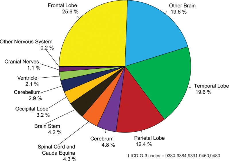
Distribution of Primary Brain and CNS Gliomas† by Site (N = 92,504), CBTRUS Statistical Report: NPCR and SEER, 2006-2010
The distribution by specific histology for glioma is illustrated in Figure 8. Glioblastoma accounts for the majority of gliomas, while astrocytoma and glioblastoma combined account for about three-fourths of all gliomas as defined by CBTRUS.
Fig. 8.
Distribution of Primary Brain and CNS Gliomas† by Histology Subtypes (N = 92,504), CBTRUS Statistical Report: NPCR and SEER, 2006-2010
Incidence of Spinal Cord Tumors
Although spinal cord tumors account for a relatively small percentage of all brain and CNS tumors, they result in significant morbidity and are, therefore, highlighted in this report. The most prevalent histologies found in the spinal cord, spinal meninges, and cauda equina are presented in Figure 9 for both children (0-19 years) and adults (20+ years). For the age group 0-19 years, the predominant histology is ependymal tumors followed by other neuroepithelial tumors (Figure 9a), whereas tumors of meninges account for the largest proportion of histologies among those ages 20 years and older (Figure 9b). ICD-O coding prevents analyses of the spinal cord tumors with additional specificity such as intramedullary and extramedullary.
Fig. 9.
(a) Distribution of Spinal Cord, Spinal Meninges and Cauda Equina Tumors by Age Group and Histology, Ages 0-19 (N = 1,067), CBTRUS Statistical Report: NPCR and SEER, 2006-2010. (b) Distribution of Spinal Cord, Spinal Meninges and Cauda Equina Tumors by Age Group and Histology, Ages 20+ (N = 14,013), CBTRUS Statistical Report: NPCR and SEER, 2006-2010
Distribution of Tumors by Site and Histology in Young Adults (Ages 20-34 Years)
About 8% of all brain and CNS tumors occurred in young adults, ages 20-34 years and the distribution of these tumors by site is shown in Figure 10a. Approximately 22% of tumors diagnosed in young adults are located within the frontal, temporal, parietal, and occipital lobes of the brain. Cerebrum, ventricle, cerebellum, and brain stem tumors combined account for about 12% of all young adult tumors. Tumors of the meninges represent 13.8%, while the cranial nerves and the spinal cord and cauda equina combined account for about 11.4%. Tumors located in the pituitary and pineal glands together account for about 33% of young adult tumors. The distribution by histology for young adults is shown in Figure 10b. Over half of reported histologies for tumors diagnosed in those 20-34 years of age are the predominately non-malignant tumors of the pituitary (29.9%), meningioma (13.7%), and nerve sheath (8.2%). The broad category glioma accounts for 30% of all brain and CNS tumors and about 81% of malignant tumors in young adults.
Fig. 10.
(a) Distribution of Primary Brain and CNS Tumors by Site in Young Adults (Ages 20-34 years) (N = 27,899), CBTRUS Statistical Report: NPCR and SEER, 2006-2010. (b) Distribution of Primary Brain and CNS Tumors by Histology in Young Adults (Ages 20-34 years) (N = 27,899), CBTRUS Statistical Report: NPCR and SEER, 2006-2010
Incidence Rates by Site and Gender
Incidence counts and average annual age-adjusted rates for brain and CNS tumors by site and gender are provided in Table 11. Incidence rates were highest for tumors located in the meninges (7.41 per 100,000), followed by tumors located in the four lobes of the brain, pituitary, other areas of the brain, cranial nerves, spinal cord/cauda equina, cerebellum, cerebrum, brain stem, ventricle, other nervous system, and pineal gland. Incidence rates were lowest for olfactory tumors of the nasal cavity (0.04 per 100,000). By gender, incidence rates were statistically significantly higher in females than in males for tumors located in the meninges, pituitary, and cranial nerves. Males had statistically significantly higher incidence rates of tumors located in the four lobes of the brain, cerebrum, ventricle, cerebellum, brain stem, other brain, spinal cord and cauda equina, other nervous system, pineal, and olfactory tumors of the nasal cavity compared to females.
Table 11.
Average Annual Age-Adjusted Incidence Rates† for Brain and Central Nervous System Tumors by Site‡ and Gender, CBTRUS Statistical Report: NPCR and SEER, 2006-2010
| ICD-O-3 Code | Site | Total |
Male |
Female |
||||||
|---|---|---|---|---|---|---|---|---|---|---|
| N | Adjusted Rate | 95% CI | N | Adjusted Rate | 95% CI | N | Adjusted Rate | 95% CI | ||
| C71.1-C71.4 | Frontal, temporal, parietal, and occipital lobes of the brain | 67,109 | 4.30 | (4.27-4.33) | 37,032 | 5.08 | (5.03-5.14) | 30,077 | 3.63 | (3.59-3.67) |
| C71.0 | Cerebrum | 6,155 | 0.40 | (0.39-0.41) | 3,218 | 0.44 | (0.43-0.46) | 2,937 | 0.37 | (0.35-0.38) |
| C71.5 | Ventricle | 3,838 | 0.26 | (0.25-0.27) | 2,101 | 0.28 | (0.27-0.30) | 1,737 | 0.23 | (0.22-0.24) |
| C71.6 | Cerebellum | 8,799 | 0.59 | (0.58-0.61) | 4,770 | 0.66 | (0.64-0.67) | 4,029 | 0.53 | (0.52-0.55) |
| C71.7 | Brain stem | 5,121 | 0.35 | (0.34-0.36) | 2,721 | 0.37 | (0.36-0.39) | 2,400 | 0.32 | (0.31-0.34) |
| C71.8-C71.9 | Other brain | 31,272 | 2.01 | (1.99-2.03) | 16,264 | 2.28 | (2.25-2.32) | 15,008 | 1.78 | (1.75-1.81) |
| C72.0-C72.1 | Spinal cord and cauda equina | 9,656 | 0.63 | (0.62-0.64) | 4,945 | 0.67 | (0.65-0.69) | 4,711 | 0.59 | (0.58-0.61) |
| C72.2-C72.5 | Cranial nerves | 22,276 | 1.42 | (1.40-1.44) | 10,427 | 1.39 | (1.36-1.42) | 11,849 | 1.45 | (1.42-1.47) |
| C72.8-C72.9 | Other nervous system | 2,052 | 0.13 | (0.13-0.14) | 1,099 | 0.15 | (0.14-0.16) | 953 | 0.12 | (0.11-0.13) |
| C70.0-C70.9 | Meninges (cerebral & spinal) | 116,574 | 7.41 | (7.37-7.46) | 30,950 | 4.44 | (4.39-4.49) | 85,624 | 9.97 | (9.90-10.04) |
| C75.1-C75.2 | Pituitary | 51,817 | 3.39 | (3.36-3.42) | 23,244 | 3.17 | (3.13-3.21) | 28,573 | 3.68 | (3.64-3.72) |
| C75.3 | Pineal | 1,444 | 0.10 | (0.09-0.10) | 842 | 0.11 | (0.11-0.12) | 602 | 0.08 | (0.07-0.09) |
| C30.0 (9522-9523) | Olfactory tumors of the nasal cavity | 598 | 0.04 | (0.04-0.04) | 348 | 0.05 | (0.04-0.05) | 250 | 0.03 | (0.03-0.04) |
| TOTAL | 326,711 | 21.03 | (20.96-21.11) | 137,961 | 19.11 | (19.00-19.21) | 188,750 | 22.79 | (22.68-22.89) | |
†Rates are per 100,000 and are age adjusted to the 2000 US standard population.
‡The sites referred to in this table are loosely based on the categories and site codes defined in the SEER site/histology validation list.
Abbreviations: CBTRUS, Central Brain Tumor Registry of the United States; NPCR, CDC's National Program of Cancer Registries; SEER, NCI's Surveillance, Epidemiology and End Results program; CI, confidence interval.
Incidence Rates by Major Histology Groupings and Specific Histologies
Tables 2 through 4 present incidence rates by major histology groupings and specific histologies. Among major histology groupings, incidence rates were highest for tumors of the meninges (7.71 per 100,000), followed by tumors of the neuroepithelial tissue (6.60 per 100,000), tumors of the sellar region (3.32 per 100,000), and tumors of the cranial and spinal nerves (1.69 per 100,000) (Table 12).
Table 12.
Distribution and Average Annual Age-Adjusted Incidence Rates† of Brain and Central Nervous System Tumors by Major Histology Groupings and Histology, CBTRUS Statistical Report: NPCR and SEER, 2006-2010
| Histology | Total |
Malignant |
Non-malignant |
||||||||
|---|---|---|---|---|---|---|---|---|---|---|---|
| N | % of All Tumors | Median Age | Rate | (95% CI) | N | Rate | (95% CI) | N | Rate | (95% CI) | |
| Tumors of Neuroepithelial Tissue | 101,825 | 31.2 | 56 | 6.60 | (6.56-6.64) | 94,874 | 6.13 | (6.09-6.17) | 6,951 | 0.47 | (0.46-0.48) |
| Pilocytic astrocytoma | 4,741 | 1.5 | 13 | 0.33 | (0.32-0.34) | 4,741 | 0.33 | (0.32-0.34) | – | – | – |
| Diffuse astrocytoma | 8,535 | 2.6 | 47 | 0.56 | (0.55-0.58) | 8,534 | 0.56 | (0.55-0.58) | – | – | – |
| Anaplastic astrocytoma | 5,621 | 1.7 | 54 | 0.37 | (0.36-0.38) | 5,621 | 0.37 | (0.36-0.38) | – | – | – |
| Unique astrocytoma variants | 943 | 0.3 | 22 | 0.06 | (0.06-0.07) | 626 | 0.04 | (0.04-0.05) | 317 | 0.02 | (0.02-0.02) |
| Glioblastoma | 50,872 | 15.6 | 64 | 3.19 | (3.16-3.21) | 50,872 | 3.19 | (3.16-3.21) | – | – | – |
| Oligodendroglioma | 4,020 | 1.2 | 43 | 0.27 | (0.26-0.28) | 4,020 | 0.27 | (0.26-0.28) | – | – | – |
| Anaplastic oligodendroglioma | 1,658 | 0.5 | 49 | 0.11 | (0.10-0.11) | 1,656 | 0.11 | (0.10-0.11) | – | – | – |
| Oligoastrocytic tumors | 3,045 | 0.9 | 42 | 0.20 | (0.20-0.21) | 3,043 | 0.20 | (0.20-0.21) | – | – | – |
| Ependymal tumors | 6,304 | 1.9 | 44 | 0.42 | (0.41-0.43) | 3,975 | 0.26 | (0.26-0.27) | 2,329 | 0.15 | (0.15-0.16) |
| Glioma malignant, NOS | 6,765 | 2.1 | 38 | 0.46 | (0.44-0.47) | 6,765 | 0.46 | (0.44-0.47) | – | – | – |
| Choroid plexus tumors | 797 | 0.2 | 19 | 0.05 | (0.05-0.06) | 123 | 0.01 | (0.01-0.01) | 674 | 0.05 | (0.04-0.05) |
| Other neuroepithelial tumors | 93 | 0.0 | 32 | 0.01 | (0.01-0.01) | 60 | 0.00 | (0.00-0.01) | 33 | 0.00 | (0.00-0.00) |
| Neuronal and mixed neuronal-glial tumors | 4,036 | 1.2 | 27 | 0.27 | (0.26-0.28) | 819 | 0.05 | (0.05-0.06) | 3,217 | 0.22 | (0.21-0.23) |
| Tumors of the pineal region | 605 | 0.2 | 33 | 0.04 | (0.04-0.04) | 340 | 0.02 | (0.02-0.03) | 265 | 0.02 | (0.02-0.02) |
| Embryonal tumors | 3,790 | 1.2 | 9 | 0.26 | (0.26-0.27) | 3,679 | 0.26 | (0.25-0.27) | 111 | 0.01 | (0.01-0.01) |
| Tumors of Cranial and Spinal Nerves | 26,564 | 8.1 | 55 | 1.69 | (1.67-1.71) | 241 | 0.02 | (0.01-0.02) | 26,323 | 1.67 | (1.65-1.70) |
| Nerve sheath tumors | 26,548 | 8.1 | 55 | 1.69 | (1.67-1.71) | 241 | 0.02 | (0.01-0.02) | 26,307 | 1.67 | (1.65-1.69) |
| Other tumors of cranial and spinal nerves | 16 | 0.0 | 62 | 0.00 | (0.00-0.00) | – | – | – | 16 | 0.00 | (0.00-0.00) |
| Tumors of Meninges | 121,110 | 37.1 | 65 | 7.71 | (7.67-7.75) | 2,525 | 0.16 | (0.16-0.17) | 118,585 | 7.55 | (7.50-7.59) |
| Meningioma | 116,986 | 35.8 | 65 | 7.44 | (7.40-7.48) | 1,802 | 0.11 | (0.11-0.12) | 115,184 | 7.33 | (7.28-7.37) |
| Mesenchymal tumors | 1,229 | 0.4 | 47 | 0.08 | (0.08-0.09) | 380 | 0.02 | (0.02-0.03) | 849 | 0.06 | (0.05-0.06) |
| Primary melanocytic lesions | 116 | 0.0 | 56 | 0.01 | (0.01-0.01) | 79 | 0.01 | (0.00-0.01) | 37 | 0.00 | (0.00-0.00) |
| Other neoplasms related to the meninges | 2,779 | 0.9 | 49 | 0.18 | (0.17-0.19) | 264 | 0.02 | (0.02-0.02) | 2,515 | 0.16 | (0.16-0.17) |
| Lymphomas and Hemopoietic Neoplasms | 7,122 | 2.2 | 65 | 0.46 | (0.45-0.47) | 7,084 | 0.46 | (0.44-0.47) | 38 | 0.00 | (0.00-0.00) |
| Lymphoma | 6,919 | 2.1 | 65 | 0.44 | (0.43-0.46) | 6,919 | 0.44 | (0.43-0.46) | – | – | – |
| Other hemopoietic neoplasms | 203 | 0.1 | 51 | 0.01 | (0.01-0.02) | 165 | 0.01 | (0.01-0.01) | 38 | 0.00 | (0.00-0.00) |
| Germ Cell Tumors and Cysts | 1,464 | 0.4 | 17 | 0.10 | (0.10-0.11) | 990 | 0.07 | (0.06-0.07) | 474 | 0.03 | (0.03-0.04) |
| Germ cell tumors, cysts and heterotopias | 1,464 | 0.4 | 17 | 0.10 | (0.10-0.11) | 990 | 0.07 | (0.06-0.07) | 474 | 0.03 | (0.03-0.04 |
| Tumors of Sellar Region | 50,709 | 15.5 | 50 | 3.32 | (3.29-3.34) | 125 | 0.01 | (0.01-0.01) | 50,584 | 3.31 | (3.28-3.34) |
| Tumors of the pituitary | 47,958 | 14.7 | 51 | 3.13 | (3.10-3.16) | 119 | 0.01 | (0.01-0.01) | 47,839 | 3.12 | (3.10-3.15) |
| Craniopharyngioma | 2,751 | 0.8 | 42 | 0.18 | (0.18-0.19) | – | – | – | 2,745 | 0.18 | (0.18-0.19) |
| Unclassified Tumors | 17,917 | 5.5 | 64 | 1.16 | (1.14-1.18) | 6,619 | 0.42 | (0.41-0.43) | 11,298 | 0.73 | (0.72-0.75) |
| Hemangioma | 3,934 | 1.2 | 49 | 0.26 | (0.25-0.27) | 24 | 0.00 | (0.00-0.00) | 3,910 | 0.26 | (0.25-0.26) |
| Neoplasm, unspecified | 13,895 | 4.3 | 70 | 0.90 | (0.88-0.91) | 6,572 | 0.42 | (0.41-0.43) | 7,323 | 0.47 | (0.46-0.49) |
| All other | 88 | 0.0 | 59 | 0.01 | (0.00-0.01) | 23 | 0.00 | (0.00-0.00) | 65 | 0.00 | (0.00-0.01) |
| TOTAL‡ | 326,711 | 100.0 | 59 | 21.03 | (20.96-21.11) | 112,458 | 7.27 | (7.22-7.31) | 214,253 | 13.77 | (13.71-13.83) |
†Rates are per 100,000 and are age-adjusted to the 2000 US standard population.
‡Refers to all brain tumors including histologies not presented in this table.
–Counts are not presented when fewer than 16 cases were reported for the specific histology category. The suppressed cases are included in the counts for totals.
Abbreviations: CBTRUS, Central Brain Tumor Registry of the United States; NPCR, CDC's National Program of Cancer Registries; SEER, NCI's Surveillance, Epidemiology and End Results program; CI, confidence interval; NOS, not otherwise specified.
Incidence rates varied significantly by specific brain and CNS histology (Table 12). Incidence rates were highest for meningiomas (7.44 per 100,000), glioblastomas (3.19 per 100,000), tumors of the pituitary (3.13 per 100,000), and nerve sheath tumors (1.69 per 100,000). The incidence rate for glioma was 6.02 per 100,000, a major contributor to the magnitude of the neuroepithelial tissue rate (data not shown). Vestibular schwannoma (acoustic neuroma), included under tumors of cranial and spinal nerves, comprise the majority (93%; 1.57 per 100,000) of nerve sheath tumors (1.69 per 100,000) and account for 8% of all primary brain and CNS tumors (data not shown).
Incidence Rates by Behavior and Histology
Brain and CNS tumor incidence rates by behavior (malignant and non-malignant) are presented in Table 12. For malignant tumors, the incidence rate was highest for glioblastoma (3.19 per 100,000) followed by diffuse astrocytoma (0.56 per 100,000), and lymphoma (0.44 per 100,000). For non-malignant tumors, meningioma (7.33 per 100,000), tumors of the pituitary (3.12 per 100,000), and nerve sheath (1.67 per 100,000) had the highest incidence rates.
Median Age at Diagnosis
The median age at diagnosis for all primary brain and CNS tumors is 59 years (Table 12). The histology-specific median ages range from 9 to 70 years. Pilocytic astrocytoma, choroid plexus tumors, neuronal and mixed neuronal-glial tumors, tumors of the pineal region, embryonal tumors, and germ cell tumors and cysts are histologies with younger median age at diagnosis onset. Meningioma and glioblastoma are primarily diagnosed at older ages (median age of 65 and 64, respectively). Unclassified tumors have a median age of 64 years, suggesting that younger individuals may receive more specific tumor identification and classification.
Incidence Rates by Gender and Histology
Incidence rates by histology and gender are presented in Table 2. Incidence rates for all primary brain and CNS tumors combined are higher among females (22.79 per 100,000) than males (19.11 per 100,000). The difference between these incidence rates is statistically significant. Incidence rates for tumors of the neuroepithelial tissue are 1.3 times greater in males as compared to females, while tumors of the meninges are 2.2 times greater in females as compared to males. Incidence rates for tumors of the neuroepithelial and tumors of the meninges are statistically significantly different between males and females. The incidence rate of gliomas is higher in males (7.14 per 100,000) than in females (5.06 per 100,000). Similar patterns were found for individual histologies with incidence rates higher in males, especially for germ cell tumors, most glial tumors, lymphomas, and embryonal tumors, or comparable between males and females, with the notable exception of meningiomas and tumors of the pituitary, which are more common in women. Incidence rate ratios (male: female) for selected histologies and groupings are shown in Figure 11.
Fig. 11.
Incidence Rate Ratios by Gender for Selected Histologies, CBTRUS Statistical Report: NPCR and SEER, 2006-2010
Incidence Rates by Race and Histology
Incidence rates by histology and race are shown in Table 3. Incidence rates for all primary brain and CNS tumors combined are substantially and statistically significantly lower for race groups AIAN (13.2 per 100,000) as compared to whites (21.23 per 100,000), blacks (20.54 per 100,000) and API (20.78 per 100,000). Incidence rates for most histologies are statistically significantly higher for whites than black, AIAN, and API race groups. An exception is observed for meningioma, tumors of the pituitary, and craniopharyngioma where the rates for blacks significantly exceed those observed for white, AIAN, and API races. It should also be noted that the average annual incidence rate for tumors of the cranial and spinal nerves in the API group is statistically significantly higher than those rates observed for black or AIAN races.
Incidence rate ratios (white: black) for selected histologies are shown in Figure 12. Incidence rates for anaplastic astrocytoma, glioblastoma, oligodendroglioma, oligoastrocytic tumors, and nerve sheath tumors are two or more times greater in whites than in blacks. Incidence rates for pilocytic astrocytoma, ependymal tumors, embryonal tumors, lymphoma, and germ cell tumors also are significantly higher among whites than blacks. In contrast, incidence rates for meningioma and tumors of the pituitary are statistically significantly higher among blacks than whites.
Fig. 12.
Incidence Rate Ratios by Race for Selected Histologies, CBTRUS Statistical Report: NPCR and SEER, 2006-2010
Incidence Rates by Hispanic Ethnicity and Histology
Incidence rates by Hispanic ethnicity and histology are shown in Table 4. The overall incidence rate for primary brain and CNS tumors is 19.77 per 100,000 among Hispanics and 21.30 per 100,000 among non-Hispanics. The difference between these two incidence rates is statistically significant, with rates among non-Hispanics exceeding those observed for Hispanics overall and for most histologies. Only the incidence rate for tumors of the pituitary is statistically significantly higher in Hispanics than non-Hispanics.
Incidence Rates by Age and Histology
The age-adjusted incidence rates by histology and age at diagnosis are presented in Table 13. The incidence for all brain and CNS tumors is highest among the 75+ year olds (80.99 per 100,000) and lowest among children ages 0-14 years (5.14 per 100,000). However, the distribution patterns of histologies within age groups differ substantially as is apparent in Table 12. For example, the incidence rates of pilocytic astrocytoma, germ cell tumors, and embryonal tumors are higher in the younger age groups and decrease with advancing age. This is in contrast to the incidence rate of meningioma, which increases progressively with age. Age-adjusted incidence rates for selected histologies by age groups for adults ages 20+ are graphically displayed in Figure 13. Table 14 shows the four most common brain and CNS tumor histologies by age at occurrence.
Table 13.
Average Annual Age-Adjusted Incidence Rates† for Brain and Central Nervous System Tumors by Major Histology Groupings, Histology and Age at Diagnosis, CBTRUS Statistical Report: NPCR and SEER, 2006-2010
| Histology | Age at Diagnosis |
Age at Diagnosis |
||||||||||||||||
|---|---|---|---|---|---|---|---|---|---|---|---|---|---|---|---|---|---|---|
| 0-14 |
0-19 |
20-34 |
35-44 |
45-54 |
55-64 |
65-74 |
75-84 |
85+ |
||||||||||
| Rate | (95% CI) | Rate | (95% CI) | Rate | (95% CI) | Rate | (95% CI) | Rate | (95% CI) | Rate | (95% CI) | Rate | (95% CI) | Rate | (95% CI) | Rate | (95% CI) | |
| Tumors of Neuroepithelial Tissue | 3.90 | (3.82-3.97) | 3.59 | (3.53-3.65) | 3.34 | (3.27-3.41) | 4.48 | (4.39-4.58) | 6.92 | (6.81-7.03) | 11.70 | (11.54-11.87) | 17.30 | (17.05-17.57) | 19.55 | (19.21-19.89) | 12.49 | (12.06-12.93) |
| Pilocytic astrocytoma | 0.90 | (0.87-0.93) | 0.82 | (0.79-0.85) | 0.24 | (0.22-0.25) | 0.12 | (0.11-0.14) | 0.09 | (0.07-0.10) | 0.09 | (0.07-0.10) | 0.06 | (0.05-0.08) | 0.07 | (0.05-0.09) | – | – |
| Diffuse astrocytoma | 0.28 | (0.26-0.30) | 0.27 | (0.26-0.29) | 0.50 | (0.48-0.53) | 0.61 | (0.58-0.65) | 0.62 | (0.58-0.65) | 0.79 | (0.75-0.83) | 1.06 | (1.00-1.13) | 1.16 | (1.08-1.25) | 0.68 | (0.58-0.79) |
| Anaplastic astrocytoma | 0.08 | (0.07-0.09) | 0.09 | (0.08-0.10) | 0.27 | (0.26-0.29) | 0.37 | (0.34-0.40) | 0.44 | (0.42-0.47) | 0.64 | (0.60-0.67) | 0.92 | (0.86-0.98) | 0.96 | (0.89-1.04) | 0.39 | (0.32-0.48) |
| Unique astrocytoma variants | 0.10 | (0.09-0.11) | 0.10 | (0.09-0.11) | 0.06 | (0.05-0.07) | 0.04 | (0.03-0.05) | 0.04 | (0.03-0.05) | 0.04 | (0.03-0.05) | 0.05 | (0.04-0.07) | 0.06 | (0.04-0.08) | 0.07 | (0.04-0.11) |
| Glioblastoma | 0.14 | (0.12-0.15) | 0.14 | (0.13-0.16) | 0.40 | (0.37-0.42) | 1.20 | (1.16-1.25) | 3.62 | (3.54-3.70) | 8.08 | (7.94-8.21) | 13.09 | (12.86-13.31) | 14.93 | (14.63-15.23) | 9.24 | (8.87-9.62) |
| Oligodendroglioma | 0.04 | (0.03-0.05) | 0.06 | (0.05-0.06) | 0.31 | (0.29-0.33) | 0.48 | (0.45-0.51) | 0.42 | (0.39-0.44) | 0.33 | (0.30-0.36) | 0.26 | (0.23-0.29) | 0.21 | (0.17-0.25) | 0.10 | (0.06-0.14) |
| Anaplastic oligodendroglioma | 0.01 | (0.00-0.01) | 0.01 | (0.01-0.01) | 0.09 | (0.08-0.10) | 0.18 | (0.16-0.19) | 0.18 | (0.16-0.20) | 0.20 | (0.18-0.22) | 0.18 | (0.15-0.20) | 0.12 | (0.09-0.15) | – | – |
| Oligoastrocytic tumors | 0.03 | (0.03-0.04) | 0.04 | (0.03-0.04) | 0.28 | (0.26-0.30) | 0.34 | (0.31-0.36) | 0.28 | (0.26-0.30) | 0.26 | (0.24-0.29) | 0.22 | (0.19-0.25) | 0.16 | (0.13-0.20) | – | – |
| Ependymal tumors | 0.28 | (0.26-0.30) | 0.28 | (0.26-0.29) | 0.37 | (0.35-0.39) | 0.48 | (0.45-0.51) | 0.59 | (0.56-0.62) | 0.57 | (0.53-0.61) | 0.54 | (0.49-0.58) | 0.39 | (0.35-0.45) | 0.15 | (0.11-0.21) |
| Glioma malignant, NOS | 0.73 | (0.70-0.76) | 0.61 | (0.59-0.64) | 0.24 | (0.22-0.26) | 0.25 | (0.23-0.27) | 0.27 | (0.25-0.30) | 0.38 | (0.35-0.41) | 0.62 | (0.57-0.67) | 1.21 | (1.12-1.29) | 1.60 | (1.44-1.76) |
| Choroid plexus tumors | 0.12 | (0.10-0.13) | 0.10 | (0.09-0.11) | 0.04 | (0.03-0.04) | 0.04 | (0.03-0.05) | 0.04 | (0.03-0.05) | 0.03 | (0.02-0.04) | 0.03 | (0.02-0.04) | 0.05 | (0.04-0.07) | – | – |
| Other neuroepithelial tumors | 0.01 | (0.01-0.01) | 0.01 | (0.01-0.01) | 0.01 | (0.00-0.01) | – | – | – | – | – | – | – | – | – | – | – | – |
| Neuronal and mixed neuronal-glial tumors | 0.33 | (0.31-0.35) | 0.36 | (0.34-0.38) | 0.31 | (0.29-0.33) | 0.24 | (0.21-0.26) | 0.21 | (0.19-0.23) | 0.21 | (0.19-0.23) | 0.20 | (0.18-0.23) | 0.16 | (0.13-0.19) | 0.07 | (0.04-0.12) |
| Tumors of the pineal region | 0.04 | (0.04-0.05) | 0.04 | (0.04-0.05) | 0.05 | (0.04-0.05) | 0.04 | (0.03-0.05) | 0.05 | (0.04-0.06) | 0.04 | (0.03-0.05) | 0.03 | (0.02-0.05) | – | – | – | – |
| Embryonal tumors | 0.80 | (0.77-0.84) | 0.66 | (0.64-0.69) | 0.18 | (0.16-0.19) | 0.10 | (0.09-0.12) | 0.08 | (0.07-0.09) | 0.05 | (0.04-0.06) | 0.05 | (0.04-0.07) | 0.05 | (0.03-0.07) | – | – |
| Tumors of Cranial and Spinal Nerves | 0.25 | (0.23-0.26) | 0.27 | (0.25-0.28) | 0.78 | (0.74-0.81) | 1.73 | (1.67-1.79) | 2.82 | (2.75-2.89) | 3.86 | (3.76-3.95) | 4.24 | (4.11-4.37) | 3.36 | (3.22-3.50) | 1.85 | (1.69-2.03) |
| Nerve sheath tumors | 0.25 | (0.23-0.26) | 0.27 | (0.25-0.28) | 0.77 | (0.74-0.81) | 1.73 | (1.67-1.79) | 2.82 | (2.75-2.89) | 3.85 | (3.76-3.95) | 4.23 | (4.11-4.36) | 3.36 | (3.22-3.50) | 1.85 | (1.68-2.02) |
| Other tumors of cranial and spinal nerves | – | – | – | – | – | – | – | – | – | – | – | – | – | – | – | – | – | – |
| Tumors of Meninges | 0.14 | (0.13-0.16) | 0.20 | (0.19-0.22) | 1.57 | (1.53-1.62) | 4.79 | (4.70-4.88) | 8.95 | (8.82-9.07) | 14.60 | (14.41-14.78) | 24.96 | (24.65-25.27) | 37.12 | (36.65-37.59) | 49.17 | (48.32-50.04) |
| Meningioma | 0.09 | (0.08-0.10) | 0.13 | (0.12-0.14) | 1.32 | (1.28-1.37) | 4.46 | (4.37-4.55) | 8.58 | (8.46-8.70) | 14.13 | (13.95-14.31) | 24.48 | (24.17-24.79) | 36.70 | (36.23-37.17) | 48.95 | (48.10-49.82) |
| Mesenchymal tumors | 0.04 | (0.03-0.05) | 0.04 | (0.03-0.04) | 0.06 | (0.05-0.07) | 0.10 | (0.09-0.12) | 0.09 | (0.08-0.11) | 0.14 | (0.12-0.16) | 0.14 | (0.12-0.16) | 0.11 | (0.09-0.14) | 0.08 | (0.05-0.12) |
| Primary melanocytic lesions | – | – | 0.00 | (0.00-0.01) | – | – | – | – | 0.01 | (0.01-0.01) | 0.01 | (0.01-0.02) | 0.02 | (0.02-0.04) | – | – | – | – |
| Other neoplasms related to the meninges | 0.01 | (0.01-0.02) | 0.03 | (0.03-0.04) | 0.18 | (0.17-0.20) | 0.23 | (0.21-0.25) | 0.26 | (0.24-0.28) | 0.31 | (0.29-0.34) | 0.32 | (0.28-0.35) | 0.28 | (0.24-0.33) | 0.13 | (0.09-0.18) |
| Lymphomas and Hemopoietic Neoplasms | 0.02 | (0.02-0.03) | 0.02 | (0.02-0.03) | 0.12 | (0.11-0.13) | 0.28 | (0.25-0.30) | 0.46 | (0.43-0.49) | 0.88 | (0.83-0.92) | 1.90 | (1.81-1.98) | 2.25 | (2.14-2.37) | 1.10 | (0.98-1.24) |
| Lymphoma | 0.01 | (0.01-0.02) | 0.01 | (0.01-0.02) | 0.11 | (0.10-0.12) | 0.27 | (0.24-0.29) | 0.45 | (0.42-0.48) | 0.85 | (0.81-0.90) | 1.86 | (1.78-1.95) | 2.24 | (2.12-2.36) | 1.08 | (0.96-1.22) |
| Other hemopoietic neoplasms | 0.01 | (0.01-0.01) | 0.01 | (0.01-0.01) | 0.01 | (0.00-0.01) | 0.01 | (0.01-0.02) | 0.01 | (0.01-0.02) | 0.03 | (0.02-0.04) | 0.03 | (0.02-0.04) | – | – | – | – |
| Germ Cell Tumors and Cysts | 0.19 | (0.17-0.21) | 0.21 | (0.20-0.23) | 0.11 | (0.10-0.12) | 0.05 | (0.04-0.06) | 0.03 | (0.02-0.04) | 0.02 | (0.01-0.03) | 0.03 | (0.02-0.04) | 0.03 | (0.02-0.05) | – | – |
| Germ cell tumors, cysts and heterotopias | 0.19 | (0.17-0.21) | 0.21 | (0.20-0.23) | 0.11 | (0.10-0.12) | 0.05 | (0.04-0.06) | 0.03 | (0.02-0.04) | 0.02 | (0.01-0.03) | 0.03 | (0.02-0.04) | 0.03 | (0.02-0.05) | – | – |
| Tumors of Sellar Region | 0.40 | (0.38-0.43) | 0.69 | (0.66-0.71) | 2.93 | (2.87-2.99) | 3.91 | (3.83-4.00) | 4.38 | (4.29-4.47) | 5.23 | (5.12-5.34) | 7.03 | (6.86-7.19) | 6.82 | (6.62-7.03) | 4.70 | (4.44-4.97) |
| Tumors of the pituitary | 0.19 | (0.18-0.21) | 0.49 | (0.47-0.52) | 2.81 | (2.75-2.87) | 3.75 | (3.67-3.84) | 4.17 | (4.09-4.26) | 4.99 | (4.89-5.10) | 6.76 | (6.60-6.93) | 6.61 | (6.41-6.82) | 4.60 | (4.34-4.87) |
| Craniopharyngioma | 0.21 | (0.19-0.23) | 0.20 | (0.18-0.21) | 0.12 | (0.11-0.13) | 0.16 | (0.14-0.18) | 0.21 | (0.19-0.23) | 0.24 | (0.21-0.26) | 0.26 | (0.23-0.30) | 0.21 | (0.17-0.25) | 0.10 | (0.07-0.15) |
| Unclassified Tumors | 0.24 | (0.22-0.26) | 0.28 | (0.26-0.29) | 0.54 | (0.51-0.57) | 0.76 | (0.73-0.80) | 1.03 | (0.99-1.08) | 1.46 | (1.40-1.52) | 2.52 | (2.42-2.62) | 5.18 | (5.01-5.36) | 11.64 | (11.22-12.06) |
| Hemangioma | 0.07 | (0.06-0.08) | 0.09 | (0.08-0.10) | 0.22 | (0.20-0.24) | 0.31 | (0.28-0.33) | 0.37 | (0.34-0.39) | 0.41 | (0.38-0.45) | 0.42 | (0.38-0.46) | 0.44 | (0.39-0.49) | 0.33 | (0.26-0.40) |
| Neoplasm, unspecified | 0.17 | (0.15-0.18) | 0.19 | (0.18-0.20) | 0.32 | (0.30-0.34) | 0.45 | (0.42-0.48) | 0.66 | (0.63-0.70) | 1.04 | (0.99-1.09) | 2.08 | (1.99-2.17) | 4.73 | (4.56-4.90) | 11.27 | (10.87-11.69) |
| All other | – | – | – | – | – | – | – | – | – | – | – | – | – | – | – | – | – | – |
| TOTAL‡ | 5.14 | (5.05-5.22) | 5.26 | (5.19-5.33) | 9.38 | (9.27-9.49) | 16.01 | (15.84-16.18) | 24.59 | (24.38-24.80) | 37.74 | (37.45-38.04) | 57.97 | (57.50-58.44) | 74.31 | (73.64-74.98) | 80.99 | (79.89-82.10) |
†Rates are per 100,000 and age-adjusted to the 2000 US. standard population.
‡Refers to all brain tumors including histologies not presented in this table.
–Counts and rates are not presented when fewer than 16 cases were reported for the specific histology category. The suppressed cases are included in the counts and rates for totals.
Abbreviations: CBTRUS, Central Brain Tumor Registry of the United States; NPCR, CDC's National Program of Cancer Registries; SEER, NCI's Surveillance, Epidemiology and End Results program; CI, confidence interval; NOS, not otherwise specified.
Fig. 13.
Age-Adjusted Incidence of Adult Brain and CNS Tumors by Selected Histologies and Age Groups (Ages 20+), CBTRUS Statistical Report: NPCR and SEER, 2006-2010
Table 14.
Most Commonly Occurring Primary Brain and CNS Tumors† and Average Age-Adjusted Incidence Rates by Age, CBTRUS Statistical Report: NPCR and SEER, 2006-2010
| Age (years) | Most Common Histology |
Second Most Common Histology |
Third Most Common Histology |
Fourth Most Common Histology |
||||||||
|---|---|---|---|---|---|---|---|---|---|---|---|---|
| Histology | Rate‡ | (95% CI) | Histology | Rate | (95% CI) | Histology | Rate | (95% CI) | Histology | Rate | (95% CI) | |
| 0-4 | Embryonal Tumors | 1.27 | (1.20-1.35) | Pilocytic Astrocytoma | 0.96 | (0.90-1.03) | Glioma Malignant, NOS | 0.90 | (0.84-0.96) | Ependymal Tumors | 0.43 | (0.39-0.48) |
| 5-9 | Pilocytic Astrocytoma | 0.91 | (0.85-0.97) | Glioma Malignant, NOS | 0.86 | (0.80-0.92) | Embryonal Tumors | 0.75 | (0.70-0.80) | Neuronal and Mixed Neuronal Glial Tumors | 0.31 | (0.28-0.35) |
| 10-14 | Pilocytic Astrocytoma | 0.83 | (0.77-0.89) | Glioma Malignant, NOS | 0.45 | (0.41-0.49) | Neuronal and Mixed Neuronal Glial Tumors | 0.42 | (0.39-0.47) | Embryonal Tumors | 0.41 | (0.38-0.46) |
| 15-19 | Tumors of the Pituitary | 1.39 | (1.32-1.46) | Pilocytic Astrocytoma | 0.58 | (0.54-0.63) | Neuronal and Mixed Neuronal Glial Tumors | 0.44 | (0.41-0.49) | Nerve Sheath Tumors | 0.33 | (0.30-0.37) |
| 20-34 | Tumors of the Pituitary | 2.81 | (2.75-2.87) | Meningioma | 1.32 | (1.28-1.37) | Nerve Sheath Tumors | 0.77 | (0.74-0.81) | Diffuse Astrocytoma | 0.50 | (0.48-0.53) |
| 35-44 | Meningioma | 4.46 | (4.37-4.55) | Tumors of the Pituitary | 3.75 | (3.67-3.84) | Nerve Sheath Tumors | 1.73 | (1.67-1.79) | Glioblastoma | 1.20 | (1.16-1.25) |
| 45-54 | Meningioma | 8.58 | (8.46-8.70) | Tumors of the Pituitary | 4.17 | (4.09-4.26) | Glioblastoma | 3.62 | (3.54-3.70) | Nerve Sheath Tumors | 2.82 | (2.75-2.89) |
| 55-64 | Meningioma | 14.13 | (13.95-14.31) | Glioblastoma | 8.08 | (7.94-8.21) | Tumors of the Pituitary | 4.99 | (4.89-5.10) | Nerve Sheath Tumors | 3.85 | (3.76-3.95) |
| 65-74 | Meningioma | 24.48 | (24.17-24.79) | Glioblastoma | 13.1 | (12.86-13.31) | Tumors of the Pituitary | 6.76 | (6.6-6.93) | Nerve Sheath Tumors | 4.23 | (4.11-4.36) |
| 75-84 | Meningioma | 36.70 | (36.23-37.17) | Glioblastoma | 14.9 | (14.63-15.23) | Tumors of the Pituitary | 6.61 | (6.41-6.82) | Nerve Sheath Tumors | 3.36 | (3.22-3.50) |
| 85+ | Meningioma | 48.95 | (48.1-49.82) | Glioblastoma | 9.24 | (8.87-9.62) | Tumors of the Pituitary | 4.60 | (4.34-4.87) | Nerve Sheath Tumors | 1.85 | (1.68-2.02) |
| Overall | Meningioma | 7.44 | (7.40-7.48) | Glioblastoma | 3.19 | (3.16-3.21) | Tumors of the Pituitary | 3.13 | (3.10-3.16) | Nerve Sheath Tumors | 1.69 | (1.67-1.71) |
†Excludes ICD-0-3 Codes 8000-8005, 8010 and 8021.
‡Rates are per 100,000 and age-adjusted to the 2000 US. standard population.
Abbreviations: CBTRUS, Central Brain Tumor Registry of the United States; NPCR, CDC's National Program of Cancer Registries; SEER, NCI's Surveillance, Epidemiology and End Results program; CI, confidence interval; NOS, not otherwise specified.
Estimated Numbers of Expected Cases of All Primary Brain and CNS Tumors by State
The estimated numbers of cases of all primary brain and CNS tumors for 2013 and 2014 by state are shown in Table 15. The estimated numbers of cases of malignant and non-malignant tumors by state were calculated by multiplying the CBTRUS age-adjusted incidence rates (2006-2010) with the 2013 and 2014 population projections for each state and the District of Columbia. The 2013 and 2014 population projections were obtained from the interim projections from 2000-2003 based on the 2000 Census.23 The total number of new cases of primary brain and CNS tumors for all 49 included states and the District of Columbia in 2013 is estimated to be 65,700 with 22,620 being malignant and 43,110 being non-malignant. For 2014, the estimates are 66,240 primary brain and CNS cases of which 22,810 and 43,430 would be expected to be malignant and non-malignant, respectively.
Table 15.
Estimated Number of Cases†‡ of Brain and Central Nervous System Tumors, Overall and by Behavior by State, 2013, 2014
| STATE | 2013 Estimated New Cases |
2014 Estimated New Cases |
||||
|---|---|---|---|---|---|---|
| All | Malignant | Non-Malignant | All | Malignant | Non-Malignant | |
| Alabama | 770 | 340 | 440 | 780 | 340 | 440 |
| Alaska | 160 | 60 | 110 | 170 | 60 | 110 |
| Arizona | 1,470 | 520 | 960 | 1,510 | 530 | 980 |
| Arkansas | 580 | 210 | 370 | 580 | 210 | 370 |
| California | 7,700 | 2,660 | 5,040 | 7,790 | 2,690 | 5,090 |
| Colorado | 1,310 | 360 | 950 | 1,320 | 370 | 960 |
| Connecticut | 690 | 280 | 410 | 690 | 280 | 410 |
| Delaware | 180 | 70 | 110 | 180 | 70 | 110 |
| District of Columbia | 100 | – | 60 | 100 | – | 60 |
| Florida | 4,560 | 1,460 | 3,100 | 4,650 | 1,490 | 3,160 |
| Georgia | 2,090 | 690 | 1,400 | 2,110 | 700 | 1,420 |
| Hawaii | 250 | 70 | 180 | 250 | 70 | 180 |
| Idaho | 310 | 120 | 190 | 310 | 120 | 190 |
| Illinois | 2,840 | 940 | 1,890 | 2,850 | 950 | 1,900 |
| Indiana | 1,330 | 490 | 840 | 1,340 | 490 | 840 |
| Iowa | 640 | 240 | 400 | 640 | 240 | 400 |
| Kansas | 530 | 200 | 330 | 540 | 200 | 330 |
| Kentucky | 1100 | 340 | 760 | 1,110 | 340 | 760 |
| Louisiana | 920 | 300 | 620 | 920 | 300 | 620 |
| Maine | 240 | 110 | 130 | 240 | 110 | 130 |
| Maryland | 1,160 | 420 | 740 | 1,170 | 420 | 740 |
| Massachusetts | 1,260 | 530 | 730 | 1,260 | 530 | 730 |
| Michigan | 2,250 | 800 | 1,450 | 2,250 | 800 | 1,450 |
| Mississippi | 580 | 200 | 380 | 580 | 200 | 380 |
| Missouri | 1,310 | 440 | 870 | 1,320 | 450 | 870 |
| Montana | 210 | 70 | 140 | 210 | 70 | 140 |
| Nebraska | 340 | 140 | 200 | 340 | 140 | 200 |
| Nevada | 460 | 180 | 280 | 480 | 190 | 290 |
| New Hampshire | 280 | 120 | 160 | 280 | 120 | 160 |
| New Jersey | 1,820 | 710 | 1,110 | 1,830 | 710 | 1,120 |
| New Mexico | 340 | 120 | 220 | 340 | 120 | 220 |
| New York | 4,590 | 1,440 | 3,150 | 4,590 | 1,440 | 3,150 |
| North Carolina | 2,050 | 700 | 1,350 | 2,070 | 710 | 1,370 |
| North Dakota | 100 | – | 60 | 100 | – | 60 |
| Ohio | 2,040 | 870 | 1,180 | 2,040 | 870 | 1,180 |
| Oklahoma | 640 | 270 | 370 | 640 | 270 | 370 |
| Oregon | 780 | 310 | 470 | 790 | 310 | 480 |
| Pennsylvania | 2,880 | 990 | 1,900 | 2,890 | 990 | 1,900 |
| Rhode Island | 220 | 80 | 150 | 220 | 80 | 150 |
| South Carolina | 920 | 320 | 590 | 920 | 330 | 600 |
| South Dakota | 140 | 50 | 80 | 140 | 50 | 80 |
| Tennessee | 1,480 | 470 | 1010 | 1,490 | 480 | 1,010 |
| Texas | 6,140 | 1,830 | 4,310 | 6,230 | 1,860 | 4,370 |
| Utah | 650 | 200 | 450 | 660 | 200 | 450 |
| Vermont | 180 | 60 | 120 | 180 | 60 | 120 |
| Virginia | 1,550 | 560 | 990 | 1,560 | 560 | 1,000 |
| Washington | 1,780 | 530 | 1250 | 1,800 | 540 | 1260 |
| West Virginia | 360 | 140 | 220 | 360 | 140 | 220 |
| Wisconsin | 1,320 | 490 | 830 | 1,320 | 490 | 840 |
| Wyoming | 100 | – | 60 | 100 | – | 60 |
| United States | 65,700 | 22,620 | 43,110 | 66,240 | 22,810 | 43,430 |
†Source: Estimation based on CBTRUS NPCR and SEER 2006-2010 data, and US Census population estimates (www.seer.cancer.gov/popdata/).
‡Rounded to the nearest 10. Numbers may not add up due to rounding.
–Estimated number is less than 50.
Childhood Primary Brain and CNS Tumors: Incidence by Site, Histology, Gender, and Age
Childhood Brain Tumors
Brain and CNS tumors are the second most common group of malignancies among children; leukemias as a group are the most common.24,25 However, brain and CNS tumors are the most common form of solid tumors in children.24 About 7% of the reported brain and CNS tumors during 2006-2010 occurred in children ages 0-19 years.
Distribution of Tumors by Site and Histology
The distribution of brain and CNS tumors for children ages 0-19 years by site is shown in Figure 14. The largest percentage of childhood tumors (16.9%) are located within the frontal, temporal, parietal, and occipital lobes of the brain. Cerebrum, ventricle, cerebellum, and brain stem tumors account for 5.5%, 5.8%, 16.3%, and 10.4% of all childhood tumors, respectively. The listing ‘other brain’ accounts for 13.8% (ICD-O-3 site codes: C71.8-C71.9) and tumors of the meninges represent 2.7% of all childhood tumors. The cranial nerves and the spinal cord and cauda equina account for 5.9% and 4.6%, respectively. Tumors located in the pituitary and pineal glands together account for about 16% of all childhood brain and CNS tumors.
Fig. 14.
Distribution of Childhood (Ages 0-19) Primary Brain and CNS Tumors by Site (N = 21,512), CBTRUS Statistical Report: NPCR and SEER, 2006-2010
Figure 15 presents the most common brain and CNS histologies in children ages 0-14 years (Figure 15a) and adolescents ages 15-19 years (Figure 15b). For children ages 0-14 years, pilocytic astrocytoma, embryonal tumors, and malignant glioma, NOS, account for 17.5%, 15.7%, and 14.2%, respectively (Figure 15a). Embryonal tumors are not commonly grouped together in clinical practice as they are within the CBTRUS histologic grouping scheme which reflects WHO Classification, as there is significant variation between the different histologies within this category. This category includes primitive neuroectodermal tumor (PNET) (ICD-O-3 Histology Code: 9473), medulloblastoma (ICD-O-3 Histology Codes: 9470-9472), atypical teratoid/rhabdoid tumor (ICD-O-3 Histology Code: 9508), and several other histologies. For more detail on specific embryonal histologies, see Figure 16 and Table 19. The most common histologies in adolescents ages 15-19 years include tumors of the pituitary (24.7%) and pilocytic astrocytoma (10.4%) (Figure 15b). The broad category glioma accounts for approximately 53% of tumors in children ages 0-14 years and 36% in adolescents ages 15-19 years.
Fig. 15.
(a) Distribution of Childhood Primary Brain and CNS Tumors by Histology and Age (Ages 0-14) (N = 15,398), CBTRUS Statistical Report: NPCR and SEER, 2006-2010. (b) Distribution of Childhood Primary Brain and CNS Tumors by Histology and Age (Ages 15-19) (N = 6,114), CBTRUS Statistical Report: NPCR and SEER, 2006-2010
Fig. 16.
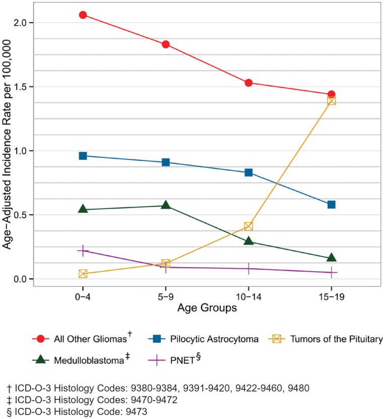
Age-Adjusted Incidence of Childhood Brain and CNS Tumors by Selected Histologies and Age Groups (Ages 0-19), CBTRUS Statistical Report: NPCR and SEER, 2006-2010
Table 19.
One-, Two-, Three-, Four-, Five-, and Ten-Year Relative Survival Rates†‡ for Selected Malignant Brain and Central Nervous System Tumors by Histology, SEER 18 Registries, 1995-2010§
| Histology | N | 1-Yr |
2-Yr |
3-Yr |
4-Yr |
5-Yr |
10-Yr |
||||||
|---|---|---|---|---|---|---|---|---|---|---|---|---|---|
| % | 95% CI | % | 95% CI | % | 95% CI | % | 95% CI | % | 95% CI | % | 95% CI | ||
| Pilocytic astrocytoma | 3,301 | 97.9 | (97.4-98.4) | 96.8 | (96.1-97.4) | 95.7 | (94.9-96.4) | 95.0 | (94.0-95.8) | 94.4 | (93.4-95.2) | 91.9 | (90.5-93.1) |
| Diffuse astrocytoma | 5,902 | 71.3 | (70.1-72.5) | 60.6 | (59.3-61.9) | 54.5 | (53.1-55.8) | 50.3 | (48.9-51.7) | 47.3 | (45.8-48.7) | 36.4 | (34.7-38.1) |
| Anaplastic astrocytoma | 3,472 | 60.1 | (58.4-61.8) | 42.1 | (40.3-43.8) | 34.1 | (32.3-35.8) | 29.7 | (28-31.4) | 26.5 | (24.8-28.2) | 18.1 | (16.4-20.0) |
| Glioblastoma | 28,212 | 35.0 | (34.4-35.6) | 13.7 | (13.3-14.1) | 8.0 | (7.6-8.3) | 5.8 | (5.5-6.2) | 4.7 | (4.4-5.0) | 2.4 | (2.1-2.7) |
| Oligodendroglioma | 3,226 | 93.8 | (92.9-94.6) | 89.5 | (88.3-90.6) | 85.7 | (84.3-86.9) | 82.2 | (80.7-83.7) | 79.1 | (77.4-80.7) | 62.6 | (60.1-64.9) |
| Anaplastic oligodendroglioma | 1,257 | 80.6 | (78.2-82.8) | 67.7 | (64.8-70.3) | 60.8 | (57.8-63.6) | 55.4 | (52.3-58.3) | 50.7 | (47.5-53.8) | 37.3 | (33.5-41.2) |
| Ependymal tumors | 2,517 | 93.9 | (92.8-94.8) | 89.7 | (88.3-90.9) | 86.7 | (85.1-88.1) | 84.8 | (83.1-86.4) | 83.4 | (81.5-85.1) | 78.8 | (76.3-81.1) |
| Oligoastrocytic tumors | 1,820 | 87.2 | (85.5-88.7) | 76.9 | (74.7-78.9) | 70.6 | (68.3-72.9) | 65.2 | (62.7-67.7) | 61.0 | (58.3-63.6) | 47.8 | (44.3-51.2) |
| Glioma malignant, NOS | 4,014 | 61.1 | (59.5-62.6) | 50.3 | (48.7-52.0) | 46.8 | (45.2-48.5) | 44.9 | (43.2-46.5) | 43.4 | (41.7-45.1) | 38.3 | (36.3-40.3) |
| Neuronal and mixed neuronal-glial tumors | 469 | 90.2 | (86.9-92.7) | 82.8 | (78.7-86.3) | 78.8 | (74.3-82.7) | 75.2 | (70.2-79.4) | 74.5 | (69.4-78.9) | 59.5 | (51.5-66.6) |
| Embryonal tumors | 2,666 | 81.5 | (80.0-83.0) | 71.9 | (70.1-73.6) | 66.7 | (64.8-68.6) | 63.7 | (61.7-65.6) | 61.3 | (59.2-63.3) | 54.2 | (51.8-56.5) |
| Medulloblastoma | 1,573 | 88.2 | (86.5-89.7) | 81.7 | (79.6-83.6) | 76.8 | (74.5-78.9) | 73.8 | (71.3-76.1) | 71.1 | (68.5-73.5) | 62.8 | (59.7-65.8) |
| Primative neuroectodermal tumor | 651 | 76.4 | (72.9-79.6) | 61.3 | (57.2-65) | 55.2 | (51.1-59.1) | 51.8 | (47.6-55.8) | 49.5 | (45.3-53.6) | 42.8 | (38.4-47.2) |
| Atypical teratoid/rhabdoid tumor | 181 | 48.1 | (40.3-55.5) | 34.6 | (27.1-42.1) | 29.2 | (21.9-36.9) | 28.0 | (20.7-35.7) | 28.0 | (20.7-35.7) | 26.2 | (18.8-34.3) |
| Other embryonal histologies | 261 | 76.3 | (70.5-81.1) | 64.4 | (58.0-70.2) | 59.7 | (52.9-65.7) | 56.8 | (50.0-63.1) | 54.2 | (47.2-60.7) | 51.6 | (44.3-58.5) |
| Meningioma | 1,099 | 82.6 | (80.0-84.8) | 75.0 | (72.0-77.8) | 70.2 | (67.0-73.3) | 68.0 | (64.6-71.2) | 64.7 | (61.0-68.1) | 56.3 | (51.6-60.8) |
| Lymphoma | 4,500 | 47.6 | (46.1-49.1) | 38.9 | (37.4-40.4) | 34.4 | (32.9-35.9) | 31.3 | (29.8-32.8) | 29.1 | (27.5-30.6) | 21.8 | (20.0-23.6) |
| Total: All Brain and Other Nervous System∥ | 66,830 | 56.9 | (56.5-57.3) | 43.1 | (42.7-43.5) | 38.2 | (37.8-38.6) | 35.6 | (35.2-36) | 33.8 | (33.4-34.2) | 28.1 | (27.7-28.6) |
†The cohort analysis of survival rates was utilized for calculating the survival estimates presented in this table. Long-term cohort-based survival estimates reflect the survival experience of individuals diagnosed over the time period, and they may not necessarily reflect the long-term survival outlook of newly diagnosed cases.
‡Rates are an estimate of the percentage of patients alive at one, two, three, four, five, and ten year, respectively.
§Estimated by CBTRUS using Surveillance, Epidemiology, and End Results (SEER) Program (www.seer.cancer.gov) SEER*Stat Database: Incidence - SEER 18 Regs Research Data + Hurricane Katrina Impacted Louisiana Cases, Nov 2012 Sub (1973-2010 varying) - Linked To County Attributes - Total U.S., 1969-2011 Counties, National Cancer Institute, DCCPS, Surveillance Research Program, Cancer Statistics Branch, released April 2013, based on the November 2012 submission.
∥Includes histologies not listed in this table.
Abbreviation: SEER, Survival, Epidemiology and End Results; CI, confidence interval; NOS, not otherwise specified.
Childhood Incidence Rates by Histology and Gender
The incidence rates of the most common childhood tumors by gender are shown in Table 5. The overall average annual incidence rate for childhood brain and CNS tumors (ages 0-19 years) is 5.26 per 100,000. Among major histology groupings, average annual incidence rates are highest for tumors of the neuroepithelial tissue (3.59 per 100,000), pilocytic astrocytoma (0.82 per 100,000), embryonal tumors (0.66 per 100,000), and glioma malignant, NOS (0.61 per 100,000). Germ cell tumors are more than twice as common in males as compared to females. Conversely, the incidence rate of tumors of the pituitary for females is more than 2.5 times the rate observed for males. Differences in incidence rates between males and females for ependymal tumors, embryonal tumors, germ cell tumors, and tumors of the pituitary are statistically significant. Due to small numbers for some histologies, caution should be used when interpreting and comparing incidence rates.
Childhood Incidence Rates by Histology and Race
Table 6 shows incidence rates by histology and race for children ages 0-19 years. Incidence rates are highest among API (5.89 per 100,000) as compared to white (5.50 per 100,000) black (4.03 per 100,000), or AIAN (2.75 per 100,000) race groups. The observed overall incidence rate differences between white, blacks and AIAN are statistically significant. Total brain and CNS tumor incidence rates between white and API races are not significantly different. However, the total average annual incidence rate for the AIAN race group is statistically significantly lower than the rate observed for black children ages 0-19 years.
Childhood Incidence Rates by Histology and Hispanic Ethnicity
Incidence rates for children ages 0-19 years by Hispanic ethnicity are shown in Table 7. The non-Hispanic rate (5.49 per 100,000) is statistically significantly higher than the observed rate for Hispanics (4.00 per 100,000). This difference is apparent for incidence rates of tumors of neuroepithelial tissue and tumors of cranial and spinal nerves. Conversely, incidence rates for tumors of the pituitary are statistically significantly higher among Hispanic children ages 0-19 years than their non-Hispanic counterparts.
Childhood Incidence Rates by Age and Histology
The detailed age-adjusted incidence rates by histology for children age groups 0-4 years, 5-9 years, 10-14 years, 15-19 years, 0-19 years, and 0-14 years are shown in Table 8. The overall incidence rates for age groups 0-4 years and 15-19 years statistically significantly exceed those observed in age groups 5-9 years and 10-14 years. It should also be noted that individual histology distributions vary substantially within these childhood age groups. The incidence rates of pilocytic astrocytoma, malignant glioma NOS, ependymal tumors, choroid plexus tumors, and embryonal tumors decrease with increasing age groups.
Age-adjusted incidence rates by age for selected histologies are graphically shown in Figure 16. Sharp declines in incidence rates between age groups for the broad gliomas and specific embryonal tumor types (PNET and medulloblastoma) are evident. The incidence decline rate for pilocytic astrocytoma is substantial from the 10-14 years to the 15-19 years age group. In contrast, PNET and medulloblastoma incidence rates are highest in the 0-4 year age group then decline and remain relatively stable across the 5-9 year, 10-14 year, and 15-19 year age groups. PNET and medulloblastoma are both histologies that are grouped into the larger embryonal category. Though the tumors in this category are histologically similar, they have different patterns of incidence and survival, so it is important to look at them individually.
Childhood Incidence Rates by Histology Defined by ICCC
Table 16 presents the CBTRUS childhood brain and CNS tumor data used for this report according to the International Classification of Childhood Cancer (ICCC) grouping system for pediatric cancers (See Appendix D for overview of grouping scheme).11 As shown, the Table 14 age group category total 0-19 age group count and age-specific and adjusted rates are equivalent to those presented throughout this report. However, the histology grouping scheme differences are apparent and reflect different approaches to the description of childhood brain and CNS tumors.
Table 16.
Age-Adjusted Incidence Rates† for Childhood Brain and Central Nervous System Tumors: Malignant and Non-Malignant by International Classification of Childhood Cancer (ICCC), CBTRUS Statistical Report: NPCR and SEER, 2006-2010
| ICCC Category | 0-19 years |
0-14 years |
< 1 year |
1-4 years |
5-9 years |
10-14 years |
15-19 years |
||||||||
|---|---|---|---|---|---|---|---|---|---|---|---|---|---|---|---|
| Count | Rate | 95% CI | Rate | 95% CI | Rate | 95% CI | Rate | 95% CI | Rate | 95% CI | Rate | 95% CI | Rate | 95% CI | |
| II Lymphomas and reticuloendothelial neoplasms | 80 | 0.02 | (0.02-0.02) | 0.02 | (0.01-0.02) | – | – | – | – | 0.02 | (0.01-0.03) | 0.02 | (0.01-0.03) | 0.03 | (0.02-0.04) |
| III CNS and misc. intracranial and intraspinal neoplasms | 18,326 | 4.49 | (4.42-4.55) | 4.46 | (4.39-4.54) | 4.91 | (4.61-5.23) | 5.11 | (4.95-5.27) | 4.32 | (4.20-4.46) | 4.02 | (3.90-4.15) | 4.56 | (4.44-4.69) |
| III(a) Ependymomas and choroid plexus tumor | 1,539 | 0.37 | (0.36-0.39) | 0.40 | (0.38-0.42) | 0.95 | (0.82-1.10) | 0.65 | (0.59-0.71) | 0.26 | (0.23-0.30) | 0.24 | (0.22-0.28) | 0.3 | (0.27-0.33) |
| III(b) Astrocytomas | 6,465 | 1.59 | (1.55-1.63) | 1.71 | (1.66-1.75) | 1.45 | (1.29-1.63) | 2.04 | (1.94-2.14) | 1.68 | (1.60-1.76) | 1.53 | (1.45-1.61) | 1.25 | (1.18-1.32) |
| III(c) Intracranial and intraspinal embryonal tumors | 2,478 | 0.61 | (0.59-0.64) | 0.74 | (0.71-0.77) | 1.25 | (1.10-1.41) | 1.10 | (1.03-1.18) | 0.71 | (0.66-0.77) | 0.4 | (0.36-0.44) | 0.23 | (0.20-0.26) |
| III(d) Other gliomas | 2,264 | 0.56 | (0.54-0.58) | 0.61 | (0.58-0.64) | 0.31 | (0.23-0.39) | 0.64 | (0.59-0.7) | 0.75 | (0.70-0.81) | 0.5 | (0.46-0.55) | 0.41 | (0.37-0.45) |
| III(e) Other specified intracranial and intraspinal neoplasms | 4,805 | 1.16 | (1.13-1.19) | 0.83 | (0.80-0.87) | 0.62 | (0.52-0.74) | 0.53 | (0.48-0.58) | 0.78 | (0.72-0.83) | 1.17 | (1.10-1.23) | 2.13 | (2.04-2.22) |
| III(f) Unspecified intracranial and intraspinal neoplasms | 775 | 0.19 | (0.18-0.20) | 0.17 | (0.15-0.18) | 0.33 | (0.26-0.42) | 0.15 | (0.12-0.18) | 0.14 | (0.12-0.17) | 0.18 | (0.16-0.21) | 0.25 | (0.22-0.28) |
| IV Neuroblastoma and other peripheral nervous cell tumors | 171 | 0.04 | (0.04-0.05) | 0.05 | (0.04-0.06) | 0.26 | (0.19-0.34) | 0.08 | (0.06-0.11) | 0.02 | (0.01-0.03) | – | – | 0.02 | (0.01-0.03) |
| IX Soft tissue and other extraosseous sarcomas | 77 | 0.02 | (0.01-0.02) | 0.02 | (0.01-0.02) | – | – | 0.02 | (0.01-0.03) | 0.02 | (0.01-0.03) | 0.02 | (0.01-0.03) | 0.02 | (0.01-0.03) |
| X(a) Intracranial & intraspinal germ cell tumors | 148 | 0.04 | (0.03-0.04) | 0.05 | (0.04-0.05) | 0.26 | (0.19-0.34) | 0.08 | (0.06-0.1) | 0.02 | (0.01-0.03) | 0.01 | (0.00-0.01) | 0.01 | (0.00-0.01) |
| All other categories | 30 | 0.01 | (0.00-0.01) | 0.01 | (0.00-0.01) | – | – | – | – | – | – | – | – | – | – |
| Not classified by ICCC | 1,962 | 0.48 | (0.45-0.50) | 0.39 | (0.37-0.41) | 6.14 | (5.80-6.50) | 5.68 | (5.52-5.85) | 4.89 | (4.75-5.03) | 4.78 | (4.64-4.91) | 5.64 | (5.50-5.78) |
| TOTAL§ | 21,512 | 5.26 | (5.19-5.33) | 5.14 | (5.05-5.22) | 6.14 | (5.80-6.50) | 5.68 | (5.52-5.85) | 4.89 | (4.75-5.03) | 4.78 | (4.64-4.91) | 5.64 | (5.50-5.78) |
†Rates are per 100,000 and are age adjusted to the 2000 U.S. standard population.
§Refers to all brain tumors including histologies not presented in this table.
-Counts and rates are not presented when fewer than 16 cases were reported for the specific ICCC category. The suppressed cases are included in the counts and rates for totals.
Abbreviations: ICCC, International Classification of Childhood Cancer; CBTRUS, Central Brain Tumor Registry of the United States; NPCR, CDC's National Program of Cancer Registries; SEER, NCI's Surveillance, Epidemiology and End Results program; CI, confidence interval.
Mortality Rates, Expected Incidence, and Survival
Estimated Mortality Rates from Malignant Brain and CNS Tumors by State and Gender
Table 17 shows average annual age-adjusted mortality rates from primary malignant brain and CNS tumors in the United States during 2006-2010 by state and gender. The aggregate total rate observed is 4.25 deaths per 100,000. However, there is considerable variation by individual state, which ranges from a low of 2.29 deaths per 100,000 to a high of 5.37 deaths per 100,000 (Figure 17). Males have statistically significantly higher mortality rates from brain and CNS tumors than females in the United States population, 5.19 per 100,000 as compared to 3.46 per 100,000 (Table 17).
Table 17.
Average Annual Age-Adjusted Mortality Rates† for Malignant Brain and Central Nervous System Cancer Overall and by State and Gender, United States, 2006-2010‡
| State | TOTAL |
Males |
Females |
||||||
|---|---|---|---|---|---|---|---|---|---|
| N | Rate | 95% CI | N | Rate | 95% CI | N | Rate | 95% CI | |
| Alabama | 1,229 | 4.78 | (4.51, 5.06) | 666 | 5.69 | (5.26, 6.15) | 563 | 3.99 | (3.66, 4.34) |
| Alaska | 114 | 4.29 | (3.47, 5.24) | 65 | 5.09 | (3.74, 6.73) | 49 | 3.70 | (2.68, 4.96) |
| Arizona | 1,332 | 4.05 | (3.83, 4.28) | 745 | 4.83 | (4.48, 5.19) | 587 | 3.36 | (3.09, 3.65) |
| Arkansas | 786 | 4.93 | (4.58, 5.29) | 431 | 5.96 | (5.4, 6.56) | 355 | 4.07 | (3.65, 4.53) |
| California | 7,509 | 4.24 | (4.15, 4.34) | 4,210 | 5.17 | (5.01, 5.33) | 3,299 | 3.46 | (3.34, 3.58) |
| Colorado | 1,056 | 4.48 | (4.20, 4.76) | 606 | 5.49 | (5.04, 5.96) | 450 | 3.61 | (3.28, 3.97) |
| Connecticut | 842 | 4.24 | (3.95, 4.54) | 464 | 5.20 | (4.73, 5.70) | 378 | 3.44 | (3.09, 3.81) |
| Delaware | 203 | 4.20 | (3.64, 4.83) | 118 | 5.46 | (4.50, 6.57) | 85 | 3.20 | (2.55, 3.98) |
| Washington DC | 103 | 3.62 | (2.94, 4.41) | 56 | 4.49 | (3.36, 5.87) | 47 | 2.93 | (2.13, 3.92) |
| Florida | 4,470 | 3.91 | (3.79, 4.03) | 2,526 | 4.82 | (4.63, 5.01) | 1,944 | 3.13 | (2.99, 3.27) |
| Georgia | 1,730 | 3.96 | (3.77, 4.15) | 944 | 4.79 | (4.47, 5.12) | 786 | 3.31 | (3.08, 3.55) |
| Hawaii | 173 | 2.29 | (1.96, 2.67) | 100 | 2.86 | (2.32, 3.49) | 73 | 1.83 | (1.42, 2.31) |
| Idaho | 375 | 4.92 | (4.42, 5.45) | 214 | 5.81 | (5.04, 6.65) | 161 | 4.06 | (3.45, 4.74) |
| Illinois | 2,623 | 3.99 | (3.84, 4.15) | 1,451 | 4.88 | (4.63, 5.14) | 1,172 | 3.27 | (3.08, 3.46) |
| Indiana | 1,526 | 4.51 | (4.28, 4.74) | 832 | 5.40 | (5.03, 5.79) | 694 | 3.77 | (3.49, 4.07) |
| Iowa | 911 | 5.37 | (5.02, 5.73) | 523 | 6.71 | (6.14, 7.32) | 388 | 4.24 | (3.82, 4.70) |
| Kansas | 713 | 4.84 | (4.48, 5.21) | 409 | 5.99 | (5.42, 6.61) | 304 | 3.79 | (3.37, 4.25) |
| Kentucky | 1,072 | 4.61 | (4.34, 4.90) | 583 | 5.46 | (5.01, 5.93) | 489 | 3.88 | (3.54, 4.24) |
| Louisiana | 983 | 4.33 | (4.06, 4.62) | 538 | 5.18 | (4.74, 5.65) | 445 | 3.58 | (3.25, 3.93) |
| Maine | 408 | 5.03 | (4.54, 5.55) | 231 | 6.20 | (5.41, 7.08) | 177 | 4.06 | (3.47, 4.73) |
| Maryland | 1,144 | 3.86 | (3.64, 4.09) | 629 | 4.73 | (4.36, 5.13) | 515 | 3.17 | (2.89, 3.46) |
| Massachusetts | 1,520 | 4.22 | (4.01, 4.44) | 848 | 5.30 | (4.95, 5.68) | 672 | 3.34 | (3.09, 3.61) |
| Michigan | 2,629 | 4.86 | (4.68, 5.06) | 1,489 | 6.02 | (5.71, 6.34) | 1,140 | 3.91 | (3.68, 4.15) |
| Minnesota | 1,231 | 4.45 | (4.20, 4.71) | 686 | 5.34 | (4.94, 5.76) | 545 | 3.69 | (3.38, 4.02) |
| Mississippi | 672 | 4.42 | (4.09, 4.77) | 344 | 5.14 | (4.60, 5.73) | 328 | 3.88 | (3.47, 4.33) |
| Missouri | 1,473 | 4.52 | (4.29, 4.76) | 806 | 5.41 | (5.03, 5.80) | 667 | 3.77 | (3.49, 4.08) |
| Montana | 250 | 4.44 | (3.89, 5.04) | 147 | 5.37 | (4.52, 6.34) | 103 | 3.60 | (2.92, 4.39) |
| Nebraska | 473 | 4.95 | (4.51, 5.42) | 255 | 5.79 | (5.09, 6.55) | 218 | 4.13 | (3.59, 4.74) |
| Nevada | 528 | 4.03 | (3.69, 4.40) | 341 | 5.47 | (4.88, 6.12) | 187 | 2.76 | (2.38, 3.20) |
| New Hampshire | 351 | 4.86 | (4.36, 5.41) | 212 | 6.31 | (5.47, 7.24) | 139 | 3.63 | (3.04, 4.31) |
| New Jersey | 1,735 | 3.66 | (3.49, 3.84) | 980 | 4.62 | (4.33, 4.93) | 755 | 2.88 | (2.67, 3.09) |
| New Mexico | 391 | 3.68 | (3.32, 4.07) | 221 | 4.36 | (3.80, 4.99) | 170 | 3.06 | (2.61, 3.57) |
| New York | 3,884 | 3.73 | (3.61, 3.85) | 2,185 | 4.73 | (4.53, 4.93) | 1,699 | 2.93 | (2.79, 3.08) |
| North Carolina | 1,978 | 4.09 | (3.91, 4.27) | 1,096 | 5.06 | (4.76, 5.38) | 882 | 3.30 | (3.08, 3.52) |
| North Dakota | 162 | 4.47 | (3.80, 5.23) | 86 | 5.12 | (4.08, 6.35) | 76 | 3.79 | (2.97, 4.78) |
| Ohio | 2,864 | 4.48 | (4.32, 4.65) | 1,575 | 5.41 | (5.14, 5.69) | 1,289 | 3.68 | (3.48, 3.89) |
| Oklahoma | 908 | 4.61 | (4.31, 4.92) | 498 | 5.47 | (4.99, 5.98) | 410 | 3.83 | (3.46, 4.22) |
| Oregon | 1,020 | 4.91 | (4.60, 5.22) | 595 | 6.07 | (5.59, 6.59) | 425 | 3.90 | (3.53, 4.29) |
| Pennsylvania | 3,086 | 4.12 | (3.97, 4.27) | 1,671 | 4.95 | (4.71, 5.20) | 1,415 | 3.43 | (3.25, 3.62) |
| Rhode Island | 241 | 4.01 | (3.51, 4.56) | 132 | 4.91 | (4.09, 5.83) | 109 | 3.28 | (2.68, 3.98) |
| South Carolina | 1,047 | 4.32 | (4.06, 4.60) | 606 | 5.66 | (5.20, 6.14) | 441 | 3.29 | (2.98, 3.61) |
| South Dakota | 231 | 5.36 | (4.68, 6.11) | 129 | 6.28 | (5.23, 7.47) | 102 | 4.53 | (3.68, 5.53) |
| Tennessee | 1,628 | 4.83 | (4.59, 5.07) | 892 | 5.84 | (5.45, 6.25) | 736 | 4.01 | (3.72, 4.31) |
| Texas | 4,524 | 4.08 | (3.96, 4.20) | 2,465 | 4.84 | (4.65, 5.04) | 2,059 | 3.46 | (3.31, 3.62) |
| Utah | 474 | 4.37 | (3.98, 4.79) | 270 | 5.15 | (4.54, 5.82) | 204 | 3.63 | (3.14, 4.17) |
| Vermont | 173 | 4.77 | (4.07, 5.55) | 92 | 5.27 | (4.22, 6.51) | 81 | 4.16 | (3.28, 5.20) |
| Virginia | 1,563 | 3.87 | (3.67, 4.06) | 868 | 4.74 | (4.42, 5.07) | 695 | 3.15 | (2.91, 3.39) |
| Washington | 1,738 | 5.10 | (4.86, 5.35) | 1,022 | 6.38 | (5.99, 6.80) | 716 | 3.96 | (3.67, 4.27) |
| West Virginia | 497 | 4.35 | (3.97, 4.77) | 260 | 4.92 | (4.32, 5.58) | 237 | 3.90 | (3.41, 4.46) |
| Wisconsin | 1,480 | 4.82 | (4.58, 5.08) | 849 | 5.99 | (5.59, 6.41) | 631 | 3.82 | (3.53, 4.14) |
| Wyoming | 131 | 4.55 | (3.79, 5.42) | 73 | 5.14 | (3.99, 6.51) | 58 | 3.93 | (2.97, 5.10) |
| United States | 68,184 | 4.25 | (4.22, 4.28) | 38,034 | 5.19 | (5.14, 5.25) | 30,150 | 3.46 | (3.42, 3.50) |
†Rates are per 100,000 and are age-adjusted to the 2000 US standard population.
‡Estimated by CBTRUS using Surveillance, Epidemiology, and End Results (SEER) Program (www.seer.cancer.gov) SEER*Stat Database: Mortality - All COD, Aggregated With State, Total U.S. (1969-2010) <Katrina/Rita Population Adjustment>, National Cancer Institute, DCCPS, Surveillance Research Program, Surveillance Systems Branch, released May 2013 Underlying mortality data provided by NCHS (www.cdc.gov/nchs). Database: Mortality - All COD, Aggregated With State, Total U.S. (1969-2010) <Katrina/Rita Population Adjustment>
Abbreviations: SEER, Survival, Epidemiology and End Results; NCHS, National Center for Health Statistics; CI, confidence interval.
Fig. 17.
Average Annual Age-Adjusted Mortality Rates for Malignant Primary Brain and CNS Tumors by Central Cancer Registry, CBTRUS Statistical Report: SEER, 2006-2010
Relative Survival Rates for Malignant Brain and CNS Tumors by Tumor Location (Site)
Relative survival estimates by brain and CNS tumor location (site) are presented in Table 18. Individuals diagnosed from 1995 through 2010 with tumors located in the cerebrum, the frontal, temporal, parietal and occipital lobes of the brain, other brain, and other nervous system have poor short- and long-term survival rates. In contrast, those with tumor locations in the cerebellum, spinal cord and cauda equina, cranial nerves, pituitary and pineal glands, and nasal cavity are observed to have better survival outcomes with ten-year survival rates ranging from 13.4% (parietal lobe) to as high as more than 90.4% (cranial nerves).
Table 18.
One-, Two-, Five-, and Ten-Year Relative Survival Rates† for Malignant Brain and Central Nervous System Tumors by Site‡, SEER 18 Registries, 1995-2010§
| ICD-O-3 CODE | SITE‡ | N | 1-Yr |
2-Yr |
5-Yr |
10-Yr |
||||
|---|---|---|---|---|---|---|---|---|---|---|
| % | 95% CI | % | 95% CI | % | 95% CI | % | 95% CI | |||
| C71.1 | Frontal lobe of the brain | 14,973 | 59.6 | (58.8-60.5) | 45.3 | (44.4-46.1) | 34.4 | (33.6-35.3) | 25.9 | (24.9-26.9) |
| C71.2 | Temporal lobe of the brain | 10,748 | 54.1 | (53.1-55.1) | 33.8 | (32.8-34.7) | 22.3 | (21.4-23.2) | 16.9 | (16.0-17.9) |
| C71.3 | Parietal lobe of the brain | 7,265 | 47.0 | (45.8-48.2) | 28.9 | (27.8-30.1) | 18.9 | (17.9-20.0) | 13.4 | (12.3-14.5) |
| C71.4 | Occipital lobe of the brain | 1,807 | 49.1 | (46.7-51.5) | 30.5 | (28.2-32.7) | 20.8 | (18.7-23.0) | 17.2 | (15.0-19.6) |
| C71.0 | Cerebrum | 3,288 | 48.2 | (46.4-49.9) | 34.3 | (32.6-36.0) | 25.8 | (24.2-27.5) | 22.4 | (20.7-24.2) |
| C71.5 | Ventricle | 1,205 | 74.4 | (71.5-76.6) | 66.9 | (64.3-69.8) | 61.3 | (58.2-64.3) | 57.1 | (53.5-60.4) |
| C71.6 | Cerebellum | 3,689 | 84.5 | (83.2-85.7) | 78.3 | (76.8-79.6) | 70.8 | (69.2-72.4) | 66.0 | (64.0-67.9) |
| C71.7 | Brain stem | 3,004 | 68.7 | (66.9-70.4) | 56.2 | (54.3-58.0) | 47.7 | (45.7-49.7) | 42.6 | (40.4-44.8) |
| C71.8-C71.9 | Other brain | 14,718 | 41.5 | (40.6-42.3) | 29.7 | (28.9-30.5) | 21.3 | (20.6-22.1) | 16.9 | (16.1-17.7) |
| C72.0-C72.1 | Spinal cord and cauda equina | 2,257 | 88.8 | (87.3-90.1) | 84.4 | (82.7-86.0) | 79.7 | (77.6-81.7) | 75.4 | (72.6-78.0) |
| C72.2-C72.5 | Cranial nerves | 676 | 96.2 | (94.3-97.4) | 94.3 | (92.1-95.9) | 91.9 | (89.1-94.0) | 90.4 | (87.0-93.0) |
| C72.8-C72.9 | Other nervous system | 631 | 59.4 | (55.3-63.2) | 50.6 | (46.4-54.7) | 43.5 | (38.9-47.9) | 38.8 | (33.7-43.9) |
| C70.0-C70.9 | Meninges (cerebral and spinal) | 1,257 | 81.6 | (79.7-84.2) | 74.4 | (71.6-77.0) | 63.8 | (60.4-67.0) | 56.0 | (51.6-60.1) |
| C75.1-C75.2 | Pituitary and craniopharyngeal duct | 265 | 84.2 | (79.0-88.2) | 80.1 | (74.3-84.8) | 71.2 | (64.2-77.0) | 62.7 | (54.2-70.1) |
| C75.3 | Pineal | 681 | 87.5 | (84.7-89.8) | 81.1 | (77.8-84.0) | 73.8 | (69.9-77.4) | 68.3 | (63.2-72.9) |
| C30.0 (9522-9523) | Olfactory tumors of the nasal cavity | 366 | 89.7 | (85.8-92.6) | 83.1 | (78.4-86.9) | 75.7 | (69.9-80.5) | 61.4 | (52.5-69.1) |
| All Codes | All Sites | 66,830 | 56.9 | (56.5-57.3) | 43.1 | (42.7-43.5) | 33.8 | (33.4-34.2) | 28.1 | (27.7-28.6) |
†The cohort analysis of survival rates was utilized for calculating the survival estimates presented in this table. Long-term cohort-based survival estimates reflect the survival experience of individuals diagnosed over the time period, and they may not necessarily reflect the long-term survival outlook of newly diagnosed cases.
‡The sites referred to in this table are loosely based on the categories and site codes defined in the SEER Site/Histology Validation List.
§Estimated by CBTRUS using Surveillance, Epidemiology, and End Results (SEER) Program (www.seer.cancer.gov) SEER*Stat Database: Incidence - SEER 18 Regs Research Data + Hurricane Katrina Impacted Louisiana Cases, Nov 2012 Sub (1973-2010 varying) - Linked To County Attributes - Total U.S., 1969-2011 Counties, National Cancer Institute, DCCPS, Surveillance Research Program, Cancer Statistics Branch, released April 2013, based on the November 2012 submission.
Abbreviation: SEER, Survival, Epidemiology and End Results; CI, confidence interval.
Survival Rates for Malignant Brain and CNS Tumors by Histology and Age
Survival estimates for malignant brain tumors by histology and age at diagnosis are presented in Tables 19 and 20. The one- through ten-year relative survival rates by histology are shown in Table 19. The estimated five- and ten-year relative survival rates for malignant brain and CNS tumors are 33.8% and 28.1%, respectively. However, there is a large variation in survival estimates depending upon tumor histologies. For example, five-year survival rates are 94.4% for pilocytic astrocytoma but are less than 5% for glioblastoma. Survival generally decreases with older age at diagnosis (Table 20). Children and young adults generally have better survival outcomes for most histologies.
Table 20.
One-, Two-, Five-, and Ten-Year Relative Survival Rates†‡ for Selected Malignant Brain and Central Nervous System Tumors by Age Groups, SEER 18 Registries, 1995-2010§
| Histology | Age Group | N | 1-Yr |
2-Yr |
5-Yr |
10-Yr |
||||
|---|---|---|---|---|---|---|---|---|---|---|
| % | 95% CI | % | 95% CI | % | 95% CI | % | 95% CI | |||
| Pilocytic astrocytoma | 0-14 yr | 1,988 | 98.7 | (98.1-99.1) | 98.5 | (97.8-99.0) | 97.2 | (96.3-98.0) | 96.2 | (94.9-97.2) |
| 0-19 yr | 2,386 | 98.5 | (98.0-99.0) | 98.3 | (97.7-98.8) | 96.8 | (95.9-97.6) | 96.0 | (94.8-96.9) | |
| 20-44 yr | 680 | 96.7 | (95.0-97.9) | 94.8 | (92.7-96.3) | 90.4 | (87.5-92.6) | 84.6 | (80.5-88.0) | |
| 45-54 yr | 124 | 94.4 | (88.1-97.4) | 86.4 | (78.2-91.7) | 78.7 | (68.8-85.7) | 73.8 | (61.8-82.5) | |
| 55-64 yr | 69 | 97.5 | (86.8-99.5) | 90.2 | (76.6-96.1) | 84.1 | (68.7-92.3) | 66.4 | (45.2-81.0) | |
| 65-74 yr | 26 | 85.6 | (63.1-94.9) | – | – | – | – | – | – | |
| 75+ yr | – | – | – | – | – | – | – | – | – | |
| Diffuse astrocytoma | 0-14 yr | 706 | 91.2 | (88.8-93.1) | 87.0 | (84.1-89.4) | 82.7 | (79.4-85.5) | 80.7 | (77.0-83.9) |
| 0-19 yr | 890 | 92.4 | (90.3-94.0) | 87.3 | (84.8-89.5) | 82.7 | (79.8-85.3) | 80.4 | (77.1-83.3) | |
| 20-44 yr | 2,088 | 92.2 | (90.9-93.3) | 84.9 | (83.2-86.4) | 64.8 | (62.4-67.1) | 45.3 | (42.3-48.4) | |
| 45-54 yr | 932 | 73.3 | (70.2-76.1) | 58.5 | (55.1-61.8) | 42.5 | (38.9-46.1) | 30.0 | (25.7-34.4) | |
| 55-64 yr | 812 | 53.4 | (49.8-56.9) | 33.4 | (29.9-36.9) | 21.0 | (17.7-24.4) | 12.5 | (9.0-16.6) | |
| 65-74 yr | 630 | 35.8 | (31.9-39.7) | 22.1 | (18.7-25.6) | 12.9 | (10.0-16.3) | 7.3 | (3.9-11.9) | |
| 75+ yr | 550 | 19.8 | (16.4-23.5) | 9.5 | (7.0-12.5) | 4.7 | (2.7-7.6) | 0.8 | (0.1-3.9) | |
| Anaplastic astrocytoma | 0-14 yr | 203 | 58.7 | (51.3-65.4) | 39.7 | (32.6-46.8) | 30.4 | (23.6-37.3) | 25.9 | (19.3-33.0) |
| 0-19 yr | 273 | 62.9 | (56.6-68.5) | 41.9 | (35.7-48.0) | 31.0 | (25.1-37.1) | 26.5 | (20.6-32.8) | |
| 20-44 yr | 1,127 | 86.4 | (84.3-88.4) | 71.5 | (68.6-74.2) | 48.9 | (45.5-52.3) | 34.5 | (30.6-38.4) | |
| 45-54 yr | 617 | 70.0 | (66.4-73.8) | 47.7 | (43.5-51.9) | 29.3 | (25.2-33.5) | 17.6 | (13.4-22.4) | |
| 55-64 yr | 586 | 48.5 | (44.7-53.0) | 25.1 | (21.4-28.9) | 10.4 | (7.6-13.7) | 4.4 | (1.8-8.8) | |
| 65-74 yr | 489 | 30.8 | (27.2-35.8) | 13.5 | (10.4-17.0) | 5.3 | (3.2-8.1) | – | – | |
| 75+ yr | 380 | 15.0 | (12.3-20.2) | 6.4 | (3.9-9.7) | 0.6 | (0.1-3.2) | – | – | |
| Glioblastoma | 0-14 yr | 246 | 50.9 | (44.2-57.2) | 29.1 | (23.1-35.4) | 22.2 | (16.6-28.4) | 15.2 | (9.7-21.8) |
| 0-19 yr | 357 | 57.2 | (51.7-62.3) | 32.5 | (27.3-37.8) | 19.2 | (14.7-24.2) | 12.6 | (8.3-17.9) | |
| 20-44 yr | 2,763 | 66.5 | (64.7-68.3) | 35.7 | (33.8-37.6) | 16.9 | (15.3-18.6) | 9.9 | (8.3-11.6) | |
| 45-54 yr | 5,022 | 52.7 | (51.2-54.1) | 20.8 | (19.6-22) | 5.9 | (5.1-6.8) | 2.7 | (1.9-3.6) | |
| 55-64 yr | 7,311 | 40.7 | (39.6-41.9) | 14.2 | (13.3-15.1) | 3.8 | (3.3-4.4) | 1.2 | (0.6-2.2) | |
| 65-74 yr | 6,908 | 23.7 | (22.6-24.7) | 7.2 | (6.5-7.9) | 1.7 | (1.3-2.2) | 0.5 | (0.2-1.1) | |
| 75+ yr | 5,851 | 9.2 | (8.4-10.0) | 2.6 | (2.2-3.1) | 0.8 | (0.5-1.2) | – | – | |
| Oligodendroglioma | 0-14 yr | 146 | 95.8 | (90.9-98.1) | 95.1 | (89.9-97.6) | 92.7 | (86.7-96.0) | 90.1 | (82.4-94.6) |
| 0-19 yr | 251 | 96.8 | (93.6-98.4) | 94.6 | (90.8-96.9) | 92.2 | (87.7-95.1) | 90.1 | (84.6-93.7) | |
| 20-44 yr | 1,655 | 98.1 | (97.3-98.7) | 95.5 | (94.3-96.4) | 85.3 | (83.2-87.1) | 66.4 | (63.1-69.6) | |
| 45-54 yr | 707 | 94.1 | (91.9-95.7) | 89.2 | (86.4-91.4) | 78.5 | (74.7-81.8) | 61.1 | (55.4-66.4) | |
| 55-64 yr | 367 | 87.3 | (83.1-90.5) | 78.2 | (73.1-82.4) | 63.9 | (57.6-69.5) | 46.7 | (38.7-54.3) | |
| 65-74 yr | 159 | 77.0 | (69.0-83.1) | 68.1 | (59.3-75.4) | 48.6 | (38.8-57.6) | 34.5 | (22.2-47.1) | |
| 75+ yr | 87 | 58.2 | (46.2-68.4) | 46.3 | (34.1-57.6) | 35.7 | (22.4-49.3) | – | – | |
| Anaplastic oligodendroglioma | 0-14 yr | – | – | – | – | – | – | – | – | – |
| 0-19 yr | 33 | 84.5 | (66.5-93.2) | 62.3 | (43.2-76.6) | – | – | – | – | |
| 20-44 yr | 509 | 93.1 | (90.5-95.0) | 82.8 | (79.0-85.9) | 66.5 | (61.7-70.9) | 49.2 | (42.7-55.4) | |
| 45-54 yr | 301 | 84.5 | (79.8-88.3) | 71.2 | (65.4-76.3) | 54.5 | (47.9-60.7) | 42.0 | (34.4-49.4) | |
| 55-64 yr | 242 | 74.5 | (68.2-79.7) | 59.5 | (52.6-65.8) | 38.3 | (31.1-45.5) | 25.8 | (17.9-34.4) | |
| 65-74 yr | 123 | 49.1 | (39.6-57.9) | 34.2 | (25.4-43.1) | 16.2 | (9.5-24.6) | 8.2 | (2.2-19.6) | |
| 75+ yr | 49 | 33.3 | (20.0-47.1) | – | – | – | – | – | – | |
| Ependymal tumors | 0-14 yr | 602 | 93.6 | (91.3-95.4) | 86.0 | (82.8-88.7) | 72.2 | (67.9-76.1) | 62.8 | (57.5-67.7) |
| 0-19 yr | 716 | 94.0 | (91.9-95.5) | 86.9 | (84.1-89.3) | 74.7 | (70.9-78.1) | 64.9 | (60.0-69.3) | |
| 20-44 yr | 813 | 96.4 | (94.9-97.5) | 95.0 | (93.1-96.4) | 91.4 | (88.9-93.4) | 89.0 | (85.7-91.6) | |
| 45-54 yr | 451 | 93.9 | (91.1-95.9) | 91.2 | (87.9-93.7) | 85.7 | (81.2-89.1) | 84.3 | (79.5-88.1) | |
| 55-64 yr | 312 | 92.8 | (88.9-95.3) | 88.6 | (83.9-92.0) | 84.9 | (78.9-89.3) | 84.9 | (78.9-89.3) | |
| 65-74 yr | 149 | 89.3 | (82.4-93.7) | 79.4 | (70.6-85.8) | 76.6 | (67.0-83.8) | 70.9 | (56.5-81.3) | |
| 75+ yr | 76 | 76.7 | (64.5-85.2) | 72.5 | (58.2-82.6) | 67.8 | (51.3-79.7) | 36.0 | (12.7-60.3) | |
| Oligoastrocytic tumors | 0-14 yr | 77 | 94.6 | (86.1-98.0) | 86.5 | (75.5-92.8) | 78.7 | (65.6-87.3) | 72.5 | (56.9-83.3) |
| 0-19 yr | 126 | 93.4 | (87.1-96.7) | 86.8 | (79.0-91.9) | 81.2 | (72.1-87.6) | 76.1 | (65.0-84.1) | |
| 20-44 yr | 975 | 95.8 | (94.3-97.0) | 89.4 | (87.1-91.2) | 71.2 | (67.7-74.5) | 55.8 | (50.9-60.4) | |
| 45-54 yr | 356 | 86.8 | (82.7-90.0) | 74.7 | (69.5-79.2) | 59.3 | (53.0-65.0) | 40.8 | (32.2-49.2) | |
| 55-64 yr | 203 | 70.7 | (63.5-76.7) | 46.9 | (39.1-54.3) | 29.7 | (22.1-37.7) | 22.9 | (14.6-32.3) | |
| 65-74 yr | 111 | 62.6 | (52.2-71.4) | 40.9 | (30.6-50.9) | 27.9 | (18.2-38.4) | 19.3 | (7.7-34.8) | |
| 75+ yr | – | – | – | – | – | – | – | – | – | |
| Glioma malignant, NOS | 0-14 yr | 1,322 | 73.6 | (71.0-75.9) | 60.5 | (57.6-63.2) | 56.9 | (54-59.7) | 55.6 | (52.6-58.5) |
| 0-19 yr | 1,463 | 75.1 | (72.8-77.3) | 62.6 | (59.9-65.1) | 58.4 | (55.6-61.0) | 57.0 | (54.2-59.8) | |
| 20-44 yr | 779 | 87.0 | (84.3-89.2) | 77.3 | (74.0-80.2) | 64.4 | (60.4-68.1) | 48.7 | (43.5-53.6) | |
| 45-54 yr | 405 | 70.5 | (65.6-74.9) | 56.9 | (51.7-61.9) | 47.1 | (41.5-52.4) | 38.7 | (32-45.5) | |
| 55-64 yr | 349 | 47.9 | (42.3-53.2) | 35.6 | (30.2-40.9) | 25.1 | (19.9-30.6) | 22.6 | (16.8-29.1) | |
| 65-74 yr | 363 | 33.2 | (28.3-38.3) | 20.5 | (16.3-25.1) | 14.5 | (10.6-19.0) | 11.7 | (7.9-16.4) | |
| 75+ yr | 655 | 15.2 | (12.5-18.3) | 10.8 | (8.3-13.6) | 6.9 | (4.4-10.2) | 5.5 | (2.5-10.4) | |
| Neuronal and mixed neuronal-glial tumors | 0-14 yr | 39 | 97.1 | (80.9-99.6) | 90.6 | (73.3-96.9) | 87.2 | (69.2-95.1) | 87.2 | (69.2-95.1) |
| 0-19 yr | 62 | 96.3 | (85.8-99.1) | 86.3 | (73.2-93.3) | 84.1 | (70.5-91.8) | 72.4 | (41.2-88.9) | |
| 20-44 yr | 132 | 94.4 | (88.3-97.3) | 91.0 | (84.0-95.0) | 78.3 | (68.7-85.3) | 56.9 | (41.8-69.4) | |
| 45-54 yr | 117 | 92.4 | (85.5-96.1) | 88.9 | (80.9-93.6) | 77.9 | (67.5-85.4) | 69.2 | (53.8-80.3) | |
| 55-64 yr | 84 | 88.5 | (78.8-93.9) | 72.1 | (59.9-81.2) | 59.2 | (45.6-70.4) | 44.2 | (27.5-59.6) | |
| 65-74 yr | 42 | 78.8 | (61.5-89.0) | 74.9 | (56.3-86.4) | 73.1 | (53.5-85.5) | 49.0 | (21.4-71.9) | |
| 75+ yr | 32 | 70.9 | (50.1-84.3) | 56.1 | (34.2-73.2) | 56.1 | (34.2-73.2) | – | – | |
| Embryonal tumors | 0-14 yr | 1,781 | 79.9 | (77.9-81.7) | 70.3 | (68.0-72.5) | 62.1 | (59.6-64.5) | 55.5 | (52.7-58.3) |
| 0-19 yr | 1,955 | 80.9 | (79.1-82.6) | 71.0 | (68.9-73.0) | 61.9 | (59.5-64.2) | 55.8 | (53.1-58.4) | |
| 20-44 yr | 557 | 86.3 | (83.0-88.9) | 79.1 | (75.3-82.4) | 64.6 | (60.0-68.9) | 56.0 | (50.7-61.1) | |
| 45-54 yr | 72 | 78.8 | (67.2-86.8) | 66.3 | (53.3-76.4) | 51.6 | (37.2-64.3) | 31.0 | (14.9-48.6) | |
| 55-64 yr | 45 | 67.6 | (51.1-79.6) | 48.8 | (32.2-63.5) | – | – | – | – | |
| 65-74 yr | – | – | – | – | – | – | – | – | – | |
| 75+ yr | – | – | – | – | – | – | – | – | – | |
| Meningioma | 0-14 yr | – | – | – | – | – | – | – | – | – |
| 0-19 yr | – | – | – | – | – | – | – | – | – | |
| 20-44 yr | 150 | 96.7 | (91.9-98.7) | 96.0 | (90.9-98.3) | 91.6 | (84.7-95.4) | 85.2 | (76.2-91.0) | |
| 45-54 yr | 182 | 92.4 | (87.3-95.6) | 86.2 | (79.9-90.7) | 76.9 | (69.1-82.9) | 72.0 | (62.4-79.6) | |
| 55-64 yr | 252 | 87.4 | (82.4-91.1) | 78.3 | (72.2-8.32) | 66.9 | (59.6-73.1) | 48.9 | (38.6-58.5) | |
| 65-74 yr | 227 | 83.0 | (76.9-87.6) | 72.7 | (65.6-78.6) | 55.6 | (47.1-63.3) | 52.0 | (42.7-60.5) | |
| 75+ yr | 276 | 63.5 | (56.8-69.5) | 54.3 | (46.9-61.2) | 45.9 | (37.0-54.3) | 29.1 | (16.1-43.5) | |
| Lymphoma | 0-14 yr | 41 | 87.6 | (72.6-94.6) | 84.7 | (68.9-92.8) | 78.2 | (60.7-88.6) | 72.7 | (52.3-85.5) |
| 0-19 yr | 67 | 83.2 | (71.6-90.3) | 77.9 | (65.4-86.3) | 74.0 | (60.8-83.3) | 66.5 | (49.9-78.6) | |
| 20-44 yr | 1,054 | 40.1 | (37.1-43.1) | 34.3 | (31.4-37.3) | 29.0 | (26.1-31.9) | 23.0 | (19.8-26.2) | |
| 45-54 yr | 724 | 56.5 | (52.7-60.1) | 48.2 | (44.4-51.9) | 38.2 | (34.3-42.1) | 30.2 | (25.6-35.0) | |
| 55-64 yr | 855 | 60.1 | (56.6-63.4) | 49.4 | (45.8-52.9) | 37.0 | (33.2-40.7) | 27.4 | (23.0-31.8) | |
| 65-74 yr | 983 | 47.5 | (44.2-50.7) | 37.5 | (34.2-40.7) | 23.5 | (20.3-26.9) | 12.9 | (9.0-17.4) | |
| 75+ yr | 817 | 33.6 | (30.2-37.0) | 23.2 | (20.0-26.5) | 13.4 | (10.3-16.8) | 9.7 | (6.5-13.6) | |
| Total: All Brain and Other Nervous System∥ | 0-14 yr | 7,935 | 85.2 | (84.4-86.0) | 78.0 | (77.0-78.9) | 72.3 | (71.2-73.3) | 68.2 | (66.9-69.4) |
| 0-19 yr | 9,690 | 86.4 | (85.7-87.1) | 79.1 | (78.3-80.0) | 73.0 | (72.0-74.0) | 69.0 | (67.9-70.1) | |
| 20-44 yr | 14,100 | 83.3 | (82.7-83.9) | 72.2 | (71.4-72.9) | 58.1 | (57.1-59.0) | 46.0 | (44.9-47.1) | |
| 45-54 yr | 10,400 | 65.7 | (64.8-66.7) | 44.9 | (43.9-46.0) | 31.8 | (30.8-32.8) | 24.9 | (23.8-26.1) | |
| 55-64 yr | 11,856 | 49.9 | (49.0-50.9) | 28.2 | (27.3-29.1) | 17.4 | (16.6-18.2) | 12.9 | (12.0-13.9) | |
| 65-74 yr | 10,694 | 32.1 | (31.1-33.0) | 17.3 | (16.5-18.1) | 10.2 | (9.5-10.9) | 7.2 | (6.3-8.1) | |
| 75+ yr | 10,090 | 16.0 | (15.2-16.8) | 9.1 | (8.5-9.8) | 5.8 | (5.2-6.5) | 3.7 | (2.9-4.6) | |
†The cohort analysis of survival rates was utilized for calculating the survival estimates presented in this table. Long-term cohort-based survival estimates reflect the survival experience of individuals diagnosed over the time period, and they may not necessarily reflect the long-term survival outlook of newly diagnosed cases.
‡Rates are an estimate of the percentage of patients alive at one, two, five, and ten year, respectively. Rates were not presented for categories with 50 or less cases and were suppressed for rates where less than 16 cases were surviving within a category.
§Estimated by CBTRUS using Surveillance, Epidemiology, and End Results (SEER) Program (www.seer.cancer.gov) SEER*Stat Database: Incidence - SEER 18 Regs Research Data + Hurricane Katrina Impacted Louisiana Cases, Nov 2012 Sub (1973-2010 varying) - Linked To County Attributes - Total U.S., 1969-2011 Counties, National Cancer Institute, DCCPS, Surveillance Research Program, Cancer Statistics Branch, released April 2013, based on the November 2012 submission.
∥Includes histologies not listed in this table
Abbreviation: SEER, Survival, Epidemiology and End Results; CI, confidence interval; NOS, not otherwise specified.
Descriptive Summary of Meningioma and Glioblastoma
The data in the CBTRUS Statistical Report 2006-2010 are synthesized to describe the two most common histologies, meningioma and glioblastoma. Meningiomas are the most frequently reported tumors and account for more than 35% of tumors reported to CBTRUS (Table 12; Figure 5). Approximately 98% of meningiomas reported to CBTRUS had a non-malignant behavior code (Table 12). Of the non-malignant meningiomas, 46% were histologically confirmed, while 51% were radiographically confirmed. For non-malignant brain tumors, diagnostic confirmation through radiographic scans is becoming increasing more important and acceptable within the central cancer registries.
Meningiomas are more common in older adults (Table 13) than in children. The incidence of meningiomas increases with increasing age. The rates for meningiomas increase dramatically after age 65 and continue to be high even among the population aged 85 years and older (Table 13). Meningiomas are more than twice as common in females as compared to males (Figure 11). The incidence in meningiomas is statistically significantly higher in blacks than whites (Figure 12). Ten year relative survival for malignant meningioma was 56.3% (Table 19). Age had a large effect on relative survival: 10 year survival was 85.2% for ages 24-44, and 29.1% for 75+ (Table 20). As only malignant meningiomas were reported in the SEER database prior to 2004, insufficient time has passed to estimate 5-year survival for non-malignant meningiomas, and therefore, survival estimates were not generated for this report.
Glioblastomas are the second most frequently reported histology and the most common malignancy. They account for 15.6% of all primary brain tumors (Table 12; Figure 5) and 45.2% of primary malignant brain tumors (Figure 6b). Glioblastomas are more common in older adults (Table 13) and are uncommon in children. Glioblastomas comprise approximately 3% of all brain and CNS tumors reported among 0-19 year olds (Table 5). The incidence of glioblastoma increases with increasing age, with rates highest in the 75 to 84 years olds (Table 12). Glioblastomas are 1.57 times more common in males (Figure 11). Glioblastomas are about two to three times higher among whites as compared to blacks (Figure 12). The relative survival estimates for glioblastoma are quite low; less than 5% of patients survived five years post diagnosis (Table 19). Glioblastoma survival estimates are somewhat higher for the small number of patients who are diagnosed under age 20 (Table 20).
Concluding Comment
The CBTRUS Statistical Report: Primary Brain and Central Nervous System Tumors Diagnosed in the United States in 2006-2010 presents a comprehensive view of current population-based incidence and related surveillance measures on primary malignant and non-malignant brain and CNS tumors collected and reported by central cancer registries that cover approximately 98% of the United States population. This report aims to serve as a useful resource for researchers, patient families, as well as for brain tumor clinicians. In keeping with its mission, CBTRUS continually revises its reports mindful of the broader surveillance community in which it works while balancing the input it receives from the clinical and research community, especially those comments from neuropathologists. In this way, the CBTRUS facilitates communication between the cancer surveillance and the brain tumor research and clinical communities and contributes meaningful insight into the descriptive epidemiology of all primary brain and CNS tumors in the United States.
Appendix A:
2000 U.S. Standard Population
| Age Group | 2000 U.S. | Age Group | 2000 U.S. | Age Group | 2000 U.S. |
|---|---|---|---|---|---|
| 0-4 | 18,986,520 | 45-49 | 19,805,793 | Total | 274,633,642 |
| 5-9 | 19,919,840 | 50-54 | 17,224,359 | ||
| 10-14 | 20,056,779 | 55-59 | 13,307,234 | ||
| 15-19 | 19,819,518 | 60-64 | 10,654,272 | ||
| 20-24 | 18,257,225 | 65-69 | 9,409,940 | ||
| 25-29 | 17,722,067 | 70-74 | 8,725,574 | ||
| 30-34 | 19,511,370 | 75-79 | 7,414,559 | ||
| 35-39 | 22,179,956 | 80-84 | 4,900,234 | ||
| 40-44 | 22,479,229 | 85 + | 4,259,173 |
Appendix B:
Average Annual Populations† for 2006-2010, By Age, Gender and Race
| Male | |||||
|---|---|---|---|---|---|
| Age Group | White | Black | AIAN | API | Total |
| 0-4 | 7,704,319 | 1,666,608 | 177,834 | 568,767 | 10,117,528 |
| 5-9 | 7,673,938 | 1,631,064 | 170,615 | 533,167 | 10,008,783 |
| 10-14 | 8,005,004 | 1,745,921 | 177,066 | 527,798 | 10,455,790 |
| 15-19 | 8,489,499 | 1,884,377 | 192,320 | 565,381 | 11,131,576 |
| 20-24 | 8,293,773 | 1,608,581 | 177,773 | 625,174 | 10,705,301 |
| 25-29 | 8,023,850 | 1,408,351 | 159,655 | 663,595 | 10,255,451 |
| 30-34 | 7,531,629 | 1,275,314 | 143,303 | 654,541 | 9,604,788 |
| 35-39 | 8,029,489 | 1,310,118 | 137,549 | 650,234 | 10,127,390 |
| 40-44 | 8,426,157 | 1,345,580 | 134,151 | 576,813 | 10,482,701 |
| 45-49 | 9,003,812 | 1,365,169 | 129,963 | 532,774 | 11,031,718 |
| 50-54 | 8,549,064 | 1,225,188 | 111,275 | 471,156 | 10,356,683 |
| 55-59 | 7,560,394 | 977,094 | 86,060 | 392,125 | 9,015,673 |
| 60-64 | 6,186,395 | 687,511 | 61,798 | 294,104 | 7,229,809 |
| 65-69 | 4,592,714 | 484,412 | 41,089 | 214,426 | 5,332,640 |
| 70-74 | 3,473,536 | 349,867 | 26,783 | 155,753 | 4,005,940 |
| 75-79 | 2,761,388 | 236,193 | 16,789 | 104,713 | 3,119,083 |
| 80-84 | 1,985,600 | 145,665 | 9,779 | 65,845 | 2,206,888 |
| 85+ | 1,476,134 | 100,958 | 6,326 | 48,308 | 1,631,727 |
| Total | 117,766,696 | 19,447,970 | 1,960,128 | 7,644,675 | 146,819,470 |
|
Female | |||||
| Age Group | White | Black | AIAN | API | Total |
| 0-4 | 7,350,092 | 1,612,527 | 172,967 | 548,505 | 9,684,091 |
| 5-9 | 7,308,715 | 1,578,664 | 165,717 | 531,102 | 9,584,198 |
| 10-14 | 7,600,173 | 1,686,196 | 173,511 | 512,799 | 9,972,679 |
| 15-19 | 8,000,625 | 1,825,637 | 181,373 | 536,822 | 10,544,458 |
| 20-24 | 7,808,263 | 1,628,358 | 160,674 | 611,859 | 10,209,155 |
| 25-29 | 7,728,520 | 1,540,244 | 148,493 | 721,841 | 10,139,098 |
| 30-34 | 7,270,847 | 1,429,718 | 138,212 | 720,911 | 9,559,688 |
| 35-39 | 7,858,362 | 1,479,055 | 135,769 | 718,943 | 10,192,129 |
| 40-44 | 8,337,261 | 1,512,502 | 134,062 | 640,932 | 10,624,757 |
| 45-49 | 9,048,839 | 1,539,915 | 133,962 | 599,266 | 11,321,981 |
| 50-54 | 8,730,875 | 1,402,193 | 117,780 | 547,031 | 10,797,879 |
| 55-59 | 7,879,048 | 1,158,988 | 92,067 | 471,140 | 9,601,243 |
| 60-64 | 6,582,943 | 848,210 | 66,024 | 352,250 | 7,849,427 |
| 65-69 | 5,091,513 | 635,157 | 45,435 | 253,187 | 6,025,292 |
| 70-74 | 4,081,538 | 495,032 | 32,453 | 192,945 | 4,801,968 |
| 75-79 | 3,568,561 | 382,175 | 22,629 | 146,435 | 4,119,801 |
| 80-84 | 3,010,168 | 278,441 | 14,936 | 100,439 | 3,403,983 |
| 85+ | 3,109,806 | 264,165 | 12,645 | 81,526 | 3,468,142 |
| Total | 120,366,150 | 21,297,175 | 1,948,709 | 8,287,935 | 151,899,969 |
†Population data source for 50 population-based geographic regions: Estimates from the United States. Bureau of the Census <http://seer.cancer.gov/popdata/index.html>.
Abbreviations: AIAN, American Indian Alaskan Native; API, Asian Pacific Islander.
Appendix C:
Average Annual Populations† for 2006-2010 By Age, Gender, and Hispanic Ethnicity
| Male | |||||
|---|---|---|---|---|---|
| Age Group | Hispanic | Non-Hispanic | Total | ||
| 0-4 | 2,524,048 | 7,593,480 | 10,117,528 | ||
| 5-9 | 2,254,938 | 7,753,845 | 10,008,783 | ||
| 10-14 | 2,197,898 | 8,257,892 | 10,455,790 | ||
| 15-19 | 2,211,025 | 8,920,551 | 11,131,576 | ||
| 20-24 | 2,207,996 | 8,497,305 | 10,705,301 | ||
| 25-29 | 2,217,253 | 8,038,198 | 10,255,451 | ||
| 30-34 | 2,052,774 | 7,552,013 | 9,604,788 | ||
| 35-39 | 1,884,223 | 8,243,166 | 10,127,390 | ||
| 40-44 | 1,668,156 | 8,814,545 | 10,482,701 | ||
| 45-49 | 1,408,246 | 9,623,472 | 11,031,718 | ||
| 50-54 | 1,092,289 | 9,264,394 | 10,356,683 | ||
| 55-59 | 806,238 | 8,209,435 | 9,015,673 | ||
| 60-64 | 566,014 | 6,663,795 | 7,229,809 | ||
| 65-69 | 389,476 | 4,943,164 | 5,332,640 | ||
| 70-74 | 281,654 | 3,724,286 | 4,005,940 | ||
| 75-79 | 200,116 | 2,918,967 | 3,119,083 | ||
| 80-84 | 126,028 | 2,080,860 | 2,206,888 | ||
| 85+ | 82,369 | 1,549,358 | 1,631,727 | ||
| Total | 24,170,742 | 122,648,728 | 146,819,470 | ||
| Female | |||||
| Age Group | Hispanic | Non-Hispanic | Total | ||
| 0-4 | 2,423,748 | 7,260,344 | 9,684,091 | ||
| 5-9 | 2,162,117 | 7,422,081 | 9,584,198 | ||
| 10-14 | 2,105,032 | 7,867,647 | 9,972,679 | ||
| 15-19 | 2,040,089 | 8,504,368 | 10,544,458 | ||
| 20-24 | 1,905,160 | 8,303,994 | 10,209,155 | ||
| 25-29 | 1,954,478 | 8,184,620 | 10,139,098 | ||
| 30-34 | 1,889,179 | 7,670,509 | 9,559,688 | ||
| 35-39 | 1,785,003 | 8,407,125 | 10,192,129 | ||
| 40-44 | 1,591,464 | 9,033,293 | 10,624,757 | ||
| 45-49 | 1,382,800 | 9,939,181 | 11,321,981 | ||
| 50-54 | 1,116,037 | 9,681,842 | 10,797,879 | ||
| 55-59 | 866,689 | 8,734,554 | 9,601,243 | ||
| 60-64 | 642,980 | 7,206,446 | 7,849,427 | ||
| 65-69 | 472,440 | 5,552,853 | 6,025,292 | ||
| 70-74 | 366,208 | 4,435,760 | 4,801,968 | ||
| 75-79 | 280,775 | 3,839,026 | 4,119,801 | ||
| 80-84 | 193,730 | 3,210,253 | 3,403,983 | ||
| 85+ | 157,184 | 3,310,958 | 3,468,142 | ||
| Total | 23,335,113 | 128,564,856 | 151,899,969 | ||
†Population data source for 50 population-based geographic regions: Estimates from the U.S. Census Bureau <http://seer.cancer.gov/popdata/index.html>.
Appendix D:
Main Classification Table from the ICCC-3 based on ICD-O-3†
| Histology | ICD-O-3† Histology Code |
|---|---|
| II Lymphomas and reticuloendothelial neoplasms | |
| II(a) Hodgkin lymphomas | 9650-9655, 9659, 9661-9665, 9667 |
| II(b) Non-Hodgkin lymphomas (except Burkitt's lymphoma) | 9591, 9670, 9671, 9673, 9675, 9678-9680, 9684, 9689-9691, 9695, 9698-9702, 9705, 9708, 9709, 9714, 9716-9719, 9727-9729, 9731-9734, 9760-9762, 9764-9769, 9970 |
| II(c) Burkitt lymphoma | 9687 |
| II(d) Miscellaneous lymphoreticular neoplasms | 9740-9742, 9750, 9754-9758 |
| II(e) Unspecified lymphomas | 9590, 9596 |
| III CNS and miscellaneous intracranial and intraspinal neoplasms | |
| III(a) Ependymomas and choroid plexus tumor | 9383, 9390-9394 |
| III(b) Astrocytomas | 9380 (Only Site C723), 9384, 9400-9411, 9420, 9421-9424, 9440-9442 |
| III(c) Intracranial and intraspinal embryonal tumors | 9470-9474, 9480, 9501-9504 (Only Site C700-C729), 9508 |
| III(d) Other gliomas | 9380 (Only Sites C700-C722, C724-C729, C751, C753), 9381, 9382, 9430, 9444, 9450, 9451, 9460 |
| III(e) Other specified intracranial/intraspinal neoplasms | 8270-8281, 8300, 9350-9352, 9360-9362, 9412, 9413, 9492, 9493, 9505-9507, 9530-9539, 9582 |
| III(f) Unspecified intracranial and intraspinal neoplasms | 8000-8005 |
| IV Neuroblastoma and other peripheral nervous cell tumors | |
| IV(a) Neuroblastoma and ganglioneuroblastoma | 9490, 9500 |
| IV(b) Other peripheral nervous cell tumors | 8680-8683, 8690-8693, 8700, 9520-9523, 9501-9504 (Only Site C000-C699, C739-C768) |
| IX Soft tissue and other extraosseous sarcomas | |
| IX(a) Rhabdomyosarcomas | 8900-8905, 8910, 8912, 8920, 8991 |
| IX(b) Fibrosarcomas, peripheral nerve & other fibrous | 8810, 8811, 8813-8815, 8821, 8823, 8834-8835 (All Prior Only On Sites C000-C399, C440-C768, C809), 8820, 8822, 8824-8827, 9150, 9160, 9491, 9540-9571, 9580 |
| IX(c) Kaposi sarcoma | 9140 |
| IX(d) Other specified soft tissue sarcomas | 8587, 8710-8713, 8806, 8830 (Only Sites C000-C399, C440-C768, C809), 8831-8833, 8836, 8840-8842, 8850-8858, 8860-8862, 8870, 8880, 8881, 8890-8898, 8921, 8963 (Only Sites C000-C639, C659-C699, C739-C768, C809), 8982, 8990, 9040-9044, 9120-9125, 9130-9133, 9135, 9136, 9141, 9142, 9161, 9170-9175, 9180 (Only Sites C490-C499), 9210 (Only Sites C490-C499), 9220 (Only Sites C490-C499), 9240 (Only Sites C490-C499) 9231, 9251, 9252, 9260 (C000-C399, C470-C759), 9364 (Only Sites C000-C399, C470-C639, C659-C699, C739-C768, C809), 9365 (Only Sites C000-C399, C470-C639, C659-C768, C809), 9373, 9581 |
| IX(e) Unspecified soft tissue sarcomas | 8800-8805 |
| XII Other and unspecified malignant neoplasms | |
| XII(a) Other specified malignant tumors | 8930-8936, 8950, 8951, 8971-8981, 9050-9055, 9110, 9363 (Only Sites C000-C399, C470-C759) |
| XII(b) Other unspecified malignant tumors | 8000-8005 |
†International Classification of Diseases for Oncology, 3rd Edition, 2000. World Health Organization, Geneva, Switzerland.
References
- 1.Centers for Disease Control and Prevention (CDC) National Program of Cancer Registries Cancer Surveillance System Rationale and Approach. 1999 http://www.cdc.gov/cancer/npcr/pdf/npcr_css.pdf . [Google Scholar]
- 2.Cancer Registries Amendment Act. 1992 http://www.cdc.gov/cancer/npcr/npcrpdfs/publaw.pdf . [Google Scholar]
- 3.Benign Brain Tumor Cancer Registries Amendment Act. 2002 http://www.gpo.gov/fdsys/pkg/PLAW-107publ260/pdf/PLAW-107publ260.pdf . [Google Scholar]
- 4.Fritz A, Percy C, Jack A, Shanmugaratnam K, Sobin L, Perkin DM, Whelan S, editors. International Classification of Diseases for Oncology, Third edition. World Health Organization; 2000. [Google Scholar]
- 5.National Cancer Institute. Overview of the SEER Program. http://seer.cancer.gov/about/overview.html . [Google Scholar]
- 6.Louis DN, Ohgaki H, Wiestler OD, Cavanee WK, editors. WHO Classification of Tumours of the Central Nervous System. Lyon, France: International Agency for Research on Cancer; 2007. [Google Scholar]
- 7.Institute Surveillance Research Program - National Cancer. SEER … as a Research Resource. 2010 http://seer.cancer.gov/about/factsheets/SEER_Research_Brochure.pdf . [Google Scholar]
- 8.Kleihues P., Burger P. C., Scheithauer B. W. The new WHO classification of brain tumours. Brain pathology. Jul. 1993;3(3):255–268. doi: 10.1111/j.1750-3639.1993.tb00752.x. [DOI] [PubMed] [Google Scholar]
- 9.McCarthy B. J., Surawicz T., Bruner J. M., Kruchko C., Davis F. Consensus Conference on Brain Tumor Definition for registration. November 10. Neuro Oncol. 2000;4(2):134–145. doi: 10.1215/15228517-4-2-134. Apr 2002. [DOI] [PMC free article] [PubMed] [Google Scholar]
- 10.Surveillance Research Program - National Cancer Institute. ICCC Recode ICD-O-3/WHO. 2008 http://seer.cancer.gov/iccc/iccc-who2008.html . [Google Scholar]
- 11.Steliarova-Foucher E., Stiller C., Lacour B., Kaatsch P. International Classification of Childhood Cancer, third edition. Cancer. Apr 1. 2005;103(7):1457–1467. doi: 10.1002/cncr.20910. [DOI] [PubMed] [Google Scholar]
- 12.Surveillance Research Program - National Cancer Institute. ICD-0-3 SEER Site/Histology Validation List. 2012 http://seer.cancer.gov/icd-o-3/sitetype.icdo3.d20121205.pdf . [Google Scholar]
- 13.R Core Team. Vienna, Austria: R Foundation for Statistical Computing; 2013. R: A language and environment for statistical computing. [Google Scholar]
- 14.Surveillance Epidemiology, and End Results (SEER) Program. SEER*Stat software version 8.04. 2013 www.seer.cancer.gov/seerstat . [Google Scholar]
- 15.Surveillance Epidemiology, and End Results (SEER) Program. SEER*Stat Database: Populations - Total U.S. (1990-2011) - Linked To County Attributes - Total U.S., 1969-2011 Counties, National Cancer Institute, DCCPS, Surveillance Research Program, Surveillance Systems Branch, released October 2012. http://seer.cancer.gov/popdata/ [Google Scholar]
- 16.Tiwari R. C., Clegg L. X., Zou Z. Efficient interval estimation for age-adjusted cancer rates. Statistical methods in medical research. 2006;15(6):547–569. doi: 10.1177/0962280206070621. Dec. [DOI] [PubMed] [Google Scholar]
- 17.Thornton ML, editor. Standards for Cancer Registries Vol. II: Data Standards and Data Dictionary, Record Layout Version 13. 17th ed. Springfield, IL: North American Association of Central Cancer Registries; 2013. [Google Scholar]
- 18.NAACCR Race and Ethnicity Work Group. NAACCR Guideline for Enhancing Hispanic/Latino Identification: Revised NAACCR Hispanic/Latino Identification Algorithm [NHIA v2.2.1]. . September. 2012 [Google Scholar]
- 19.Surveillance Epidemiology, and End Results (SEER) Program. SEER*Stat Database: Mortality - All COD, Aggregated With State, Total U.S. (1969-2010) <Katrina/Rita Population Adjustment>, National Cancer Institute, DCCPS, Surveillance Research Program, Surveillance Systems Branch, released April 2013 [Google Scholar]
- 20.Surveillance Epidemiology, and End Results (SEER) Program. SEER*Stat Database: Incidence - SEER 18 Regs Research Data + Hurricane Katrina Impacted Louisiana Cases, Nov 2012 Sub (1973-2010 varying) - Linked To County Attributes - Total U.S., 1969-2011 Counties, National Cancer Institute, DCCPS, Surveillance Research Program, Surveillance Systems Branch, released April 2013, based on the November 2012 submission [Google Scholar]
- 21.Clegg L. X., Feuer E. J., Midthune D. N., Fay M. P., Hankey B. F. Impact of reporting delay and reporting error on cancer incidence rates and trends. J Natl Cancer Inst. 2002;94(20):1537–1545. doi: 10.1093/jnci/94.20.1537. Oct 16. [DOI] [PubMed] [Google Scholar]
- 22.Howlader N., Ries L. A., Stinchcomb D. G., Edwards B. K. The impact of underreported Veterans Affairs data on national cancer statistics: analysis using population-based SEER registries. J Natl Cancer Inst. Apr 1. 2009;101(7):533–536. doi: 10.1093/jnci/djn517. [DOI] [PMC free article] [PubMed] [Google Scholar]
- 23.United States Census Bureau, Population Division. State Population Projections: Interim Projections 2000–2030 based on Census 2000. United States Census Bureau, released 2005. http://www.census.gov/population/projections/ [Google Scholar]
- 24.Gurney JG, Smith MA, Bunin GR. Chapter III: CNS and miscellaneous intracranial and intraspinal neoplasms. In: Ries LAG SM, Gurney JG, Linet M, Tamra T, Young JL, Bunin GR, editors. Cancer Incidence and Survival among Children and Adolescents: United States SEER Program 1975-1995, National Cancer Institute, SEER Program. . NIH Pub. No. 99-4649. Bethesda, MD1999. [Google Scholar]
- 25.Howlader N, Noone AM, Krapcho M, Garshell J, Neyman N, Altekruse SF, Kosary CL, Yu M, Ruhl J, Tatalovich Z, Cho H, Mariotto A, Lewis DR, Chen HS, Feuer EJ, Cronin KA. SEER Cancer Statistics Review, 1975-2010, National Cancer Institute. Bethesda, MD, http://seer.cancer.gov/csr/1975_2010/ , based on November 2012 SEER data submission, posted to the SEER web site, April 2013. [Google Scholar]


