Abstract
Pheochromocytoma of the urinary bladder is often misdiagnosed as it is a rare tumor. In this report, we described a case with primary pheochromocytoma of the urinary bladder. We specifically conversed the diagnostic role of X-ray computed tomography and sonography to identify the location of tumor within urinary bladder compared to other malignant or benign tumors in the bladder, and exclude other ectopic pheochromocytoma. Histopathological report from bladder tissue biopsy was confirmative of extra adrenal pheochromocytoma of the urinary bladder finally. Importance in careful management of hypertensive crisis during cystoscope and partial cystectomy was addressed.
Keywords: Urinary bladder, pheochromocytoma, diagnosis, immunohistochemical tests
Introduction
Pheochromocytoma is a neuroendocrine tumour that usually develops ahead of the chromaffin cells of the adrenal medulla. Embryonic rests of chromaffin tissue arising from sympathetic plexus within bladder wall are believed to be the source of these tumors [1]. Extra-adrenal pheochromocytomas usually are located within the abdomen in association with the celiac, superior mesenteric, inferior mesenteric ganglia and Organ of Zuckerkandl (chromaffin body derived from neural crest located at the bifurcation of the aorta or at the origin of the inferior mesenteric artery). Approximately 10% are in the thorax, 1% is within the urinary bladder, and less than 3% are in the neck, usually in association with the sympathetic ganglia or the extracranial branches of the ninth cranial nerves.
The primary pheochromocytoma of the urinary bladder constitutes less than 0.06% of all vesical neoplasms [2]. The typical signs are hematuria, hypertension during micturition together with generalized symptoms due to raised catecholamines (headache, blurred vision, heart palpitation, flushing). However, 27% of pheochromocytomas of the urinary bladder do not feature any hormonal activity [3].
In this report, we described a case with primary pheochromocytoma of the urinary bladder. The expression of CD56, chromogranin A, synaptophysin and S-100 protein was assayed by immunohistochemical staining. We specifically conversed the diagnostic role of X-ray computed tomography and sonography to identify the location of tumor within urinary bladder compared to other malignant or benign tumors in the bladder, and excludes other ectopic pheochromocytoma, finally diagnosed definitely by pathology. In addition, we discussed the importance in management of hypertensive crisis during partial cystectomy.
Patients
A 47-year-old lady visited our hospital with complaints of one progressive increase mass in the right anterior superior spine, and presented with a 3 years history of repeated episodes of dizziness and palpitations immediately following micturition. No hematuria, dysuria (pain during urination) or other local or general symptomatology was present. Her medical history and physical examination were unremarkable. Further the pulse rate and blood pressure of the patient on admission were found to be 88/min and 122/85 mmHg, and ECG report was within normal limits, no family history of hypertension. Admission laboratory values were within normal ranges, these include the complete blood count, blood urea nitrogen, creatinine, catecholamine, alpha-fetoprotein, carcinoembryonic antigen and serum electrolytes. The initial diagnosis was Simple subcutaneous mass, Vertigo of unknown origin.
Results
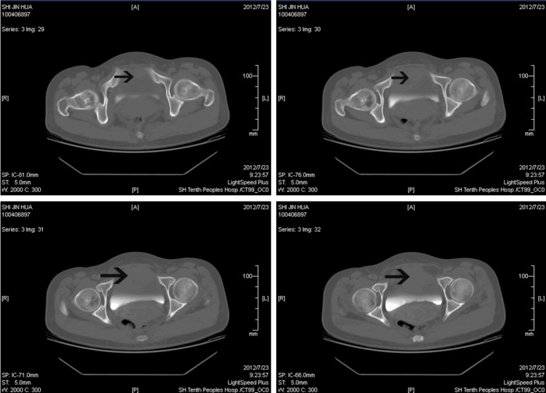
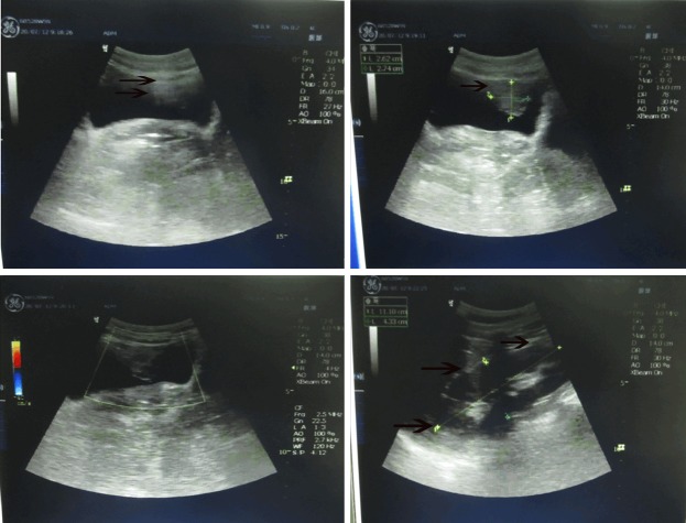
Ultrasonography (USG) revealed a (26 mm × 26 mm × 27 mm) size heterogeneous mass located in the bladder dome. Kidney and ureter were normal on USG. There was no significant enlargement of pelvic lymph nodes. (Figure 1). X-ray Computed tomography (CT) of the abdomen showed a well defined 2.5 cm size tumour located in the anterior front of the bladder, bladder wall is thickened 5 cm approximately, CT values 51, CT enhancement scanning of shows a diameter of 2.5 cm high-density nodules broke into the cavity of the bladder, CT values 90-95 Hu, with a heterogeneous increase in density after injection of iodine-containing contrast solution (Figure 2). Adrenal CT scan shows the bilateral adrenal have no obvious abnormalities.
Figure 1.
Ultrasonography showing a heterogeneous mass (26×26×27 mm) located in the bladder dome.
Figure 2.

Computerized tomography scan of the pelvis showing tumor located in the anterior front of the bladder with well defined bounds, bladder wall is thickened 5 cm approximately, shows a diameter of 2.5 cm high-density nodules broke into the cavity of the bladder, a heterogeneous increase in density after injection of iodine-containing contrast solution, having clear outline.
On cystoscopy a solid oval shape tumor at size of 3.0 cm × 2.5 cm arising from anterior wall and roof of the bladder. Mucosa over tumor has shown angiogenesis and old signs of hemorrhage. Tissue biopsy was not taken for histopathological examination of tumor. Blood pressure of the patient increased rapidly during the surgery, and then decreased rapidly after drainage. Blood pressure increases to 240/120 mmHg. Hypertensive crisis was revealed during the cystoscopy.
After adequate improvement of general condition, patient’s blood pressure was under control. Partial cystectomy, including mass and pelvic lymph node dissection, was performed under general anesthesia. Pelvic lymph nodes (Internal iliac, external iliac and common iliac) were examined to rule out secondaries in lymph nodes. No other tumour or para-aortic lymph node enlargement was found. Biopsies of pelvic lymph nodes were taken for HPE on both sides. Biopsy was negative on histopathological examination. Post cystectomy specimen of bladder was again confirmative of extra adrenal pheochromocytoma with minimal invasion in bladder muscle in a few places. During surgery, blood pressure and heart rate were stable despite pressurization by the fingers. The surgical margins were free of the tumor.
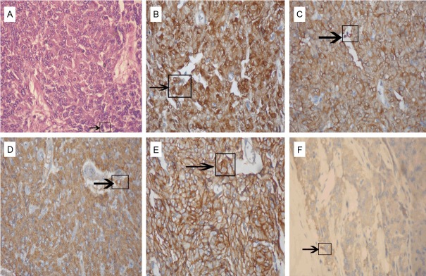
Histopathological report from bladder tissue biopsy was confirmative of extra adrenal pheochromocytoma of the urinary bladder finally (Figure 3). Tumor cells were large and rich in granular matrix and were arranged in nests separated by a prominent fibrovascular stroma resembling normal adrenal medulla. Immunohistochemical tests revealed that the tumor cells were strongly positive for the neuroendocrine markers such as chromogranin A and synaptophysin. The findings were similar to those obtained for the carcinoid tumors, except that the sustentacular cells were positive for S-100 protein. The disease was histopathologically diagnosed as pheochromocytoma.
Figure 3.

Pathological findings with histological and immunocytochemical staining. A: Timorous proliferation composed of small cells associated to endocrine visualization (arrows) (H&E ×400); B: The sustentacular cells of pheochromocytomas have immunoreactivity to the S-100 protein (arrows) (×400); C-F: The chief cells of the tumor have immunoreactivity to neuroendocrine markers such as NSE, chromogranin A, CD56 and synaptophysin (×400).
The postoperative course was uneventful. Blood pressure came down to normal on the first day after surgery and the patient was discharged without antihypertensive drug on the 7th post-operative day. Three months after surgery, cystoscopy showed no tumor recurrence in the bladder lumen. In one year follow-up, patient’s previous symptoms completely disappeared, and his blood pressure, urinary VMA, epinephrine level remained normal.
Discussion
Imaging has an ancillary but important role in the detection, staging, and follow-up of urological disease, including bladder cancer [4]. CT has widely replaced intravenous urography (IVU) and is currently the imaging modality most commonly used for the initial evaluation of patients with or suspected of having bladder tumors, also CTU allows a fast and comprehensive evaluation of the urinary tract in a single exam [5,6]. Magnetic resonance imaging (MRI) affords better soft tissue contrast, which allows for more accurate staging than can be achieved with other imaging modalities; the role of MRI in bladder cancer is expected to grow. Despite myriad technical advances, imaging of the bladder has several limitations and technical challenges [7,8]. The performance of the common and some promising newer imaging modalities in the evaluation of bladder cancer are discussed. The techniques available urinary bladder tumors include cystoscopy, KUB+IVU, CT, MRI, and USG. Each technique has its own advantages and limitations (Table 1).
Table 1.
Comparison of diagnosis approaches for urinary bladder tumors
| Diagnosis approaches | Manifestation | Advantages | Disadvantages |
|---|---|---|---|
| USG | Submucosal homogeneous mass, having clear outline, continuous mucosa, and abundant blood | Most commonly used and noninvasive | USG is commonly used, but it may miss urothelial tumors of the upper tract and small stones or tumors |
| KUB+IVU | Abnormal density; filling defect | Simple and intuitive | Unclear boundaries; get less information |
| CT | A heterogeneous increase in density after injection of iodine-containing contrast solution | Being sensitive for calcification or lithangiuria | Invasive, Radiation injure, soft tissue resolution is not high enough for diagnosis |
| MRI | The relationship between the mass and the bladder mucosa, muscular and peri-tissue | High-resolution and multiplanar capability | Time and cost consuming and not convenient |
| Cystoscopyt | Delineate the exact location of the lesion, especially with regard to the depth of invasion and the involvement of the ureters | more important in locating and qualitative diagnosis | Invasive, cost consuming and not convenien |
Notes: USG, Ultrasonographic examination; IVU, Intravenous Pyelograph; MRI, magnetic resonance imaging.
Pheochromocytoma of the urinary bladder arises from the chromaffin tissues of the sympathetic nervous system within all layers of the bladder wall. It is a rare neoplasm, accounting for less than 0.06% of all bladder tumors and less than 1% of all pheochromocytomas [9]. Extra-adrenal Pheochromocytoma may arise in any position of the paraganglionic system and accounts for less than 10% of all Pheochro-mocytoma. Extra adrenal Pheochromocytoma probably represents in, at least 15% of adults and 30% of childhood [3]. Urinary bladder pheochromocytoma is similar to adrenal pheochromocytoma, most of which can secrete catecholamines. They usually present two symptoms of the pheochromocytoma and bladder tumors, which are continuous or paroxysmal, and are especially associated with urination and hematuria. In recent years, as we know more and more about the disease, misdiagnosis decreased apparently, although asymptomatic bladder pheochromocytoma is almost impossible to diagnose preoperatively. The most common presentation is hypertension, which may be sustained or paroxysmal. Fifty percent of patients may have painful hematuria. Other symptoms include severe headache, excessive sweating, palpitation, tachycardia anxiety, constipation, and an orthostatic hypotension [10]. Most of the pheochromocytomas of the urinary bladder occur in young and middle-aged adults, and are more common in women [11]. Pheochromocytomas usually have a benign clinical course, with only 10% being malignant. The cytologic features of being and malignant tumors overlap, thus, there are no reliable features of malignancy [12].
This case presented with dizziness and palpitations immediately following micturition but with no hematuria or other symptoms. Although urinary bladder pheochromocytomas are often hormonally active with elevated catecholamine metabolites [13], typical signs and symptoms in this patient showed only when the bladder was full. Reduction of the sensitivity of catecholamine receptors is considered the cause of this phenomenon. Several case reports of bladder pheochromocytoma were reviewed, and most of them had symptoms of hypertension, and a drop in blood pressure occurred during the operation [14,15]. Two case reports revealed that patients treated with transurethral resection of the bladder tumour did not experience a hypertensive crisis during the operation, but a drop in the systolic pressure to 50 mmHg was noted in one case during the operation. In those patients, the bladder pheochromocytoma was larger than 2 cm in diameter and all of them received a partial cystectomy [16,17].
Some reports have demonstrated that metastasis is possible even after surgery. Other reports have suggested that bladder pheochromocytomas may be malignant, and patients should be aggressively treated and should receive long-term followup after initial surgery. In this patient, histopathological examination reported a well-circumferential tumor without muscle invasion. However the patient still requires regular follow-up. Malignancy is determined more by the clinical behavior than histological features of the tumor. Partial cystectomy is the treatment of choice in bladder tumor but TURBT is also recommended in some study without any complication [18]. CT scan should evaluate regional lymph nodes preoperatively, operatively by gross inspection and biopsy of suspicious lymph nodes. Lymph node involvement is marked by hypo vascular-lesion on arteriography. Pelvic lymphoidectomy is indicated in case of lymph node involvement by tumor.
The most helpful diagnostic laboratory test available is the determination of blood or urinary levels of epinephrine and nor epinephrine and their metabolic byproducts. Neither urinary VMA > 9.0 mg/24 hours, nor epinephrine > 80 μg/24 hours and epinephrine > 20 μg/24 hours have been found in most of the patients. Preoperative location and qualitative analysis are very important in confirming the diagnosis.
Although USG, CT, and cystoscopy are not able to provide the specific image features of pheochromocytomas, these diagnostic imaging can help locate the tumors, and exclude multifocal and metastatic diseases. On USG, pheochromocytomas appear as sharply demarcated soft tissue masses that may be purely solid or may contain foci of hemorrhage and necrosis that appear cystic, having clear outline, continuous mucosa, and abundant blood [19]. CT can show the relationship between the mass and the bladder mucosa, muscular and peri-tissue. CT scan is more sensitive than USG in detecting adrenal (94%) and extra-adrenal pheochromocytomas. I131 meta-iodo-benzyl-guanidine (MI-BG) scan serves as a complementary functional diagnostic tool, but its use is limited because of its expense and restricted availability [20]. One study reported that CT has high sensitivity for pheochromocytomas in the adrenal gland but lower sensitivity for detecting extra-adrenal pheochromocytomas. The MIBG scan has high sensitivity (77–90%) and high specificity (88–99%) for localizing pheochromocytomas, and it can provide information complementary to CT and MRI [21]. One study reported that the tests of choice to diagnose pheochromocytomas are urinary nor metanephrine and platelet nor epinephrine estimation. In combination with an MIBG scan, the diagnostic sensitivity can be improved. When a pheochromocytoma is suspected and catecholamine measurements are within a normal range, an MIBG scan should be done. So, an MIBG scan may be necessary during follow-up if the tumor recurs or there is any residual tumour [22].
Cystoscopy is more important in locating and qualitative diagnosis. The blood pressure fluctuates because of the irrigation of the flush water and the sheath, which is determinative for the diagnosis. Some people suggested that to confirm the diagnosis preoperatively, we can irrigate the mass during cystoscopy; if the blood pressure fluctuates we can confirm the diagnosis. But we think that if there are no necessary medicines and instruments for salvage, we should not irrigate the mass repeatedly in case there is danger. The tumors in cystoscopy appear as globular submucosa mass protruding into the bladder, which has smooth surface, continuous mucosa, and abundant blood.
The histopathological findings are very similar to those of the normal adrenal medulla. Further, the immunohistological findings are important for diagnosis. The chief cells of the tumor have immunoreactivity to neuroendocrine markers such as CD56, chromogranin A and synaptophysin, whereas the sustentacular cells of pheochromocytomas have immunoreactivity to the S-100 protein [23]. It is difficult to histopathologically diagnose pheochromocytoma as malignant or benign. The clinical staging of paraganglioma of the urinary bladder can be classified in a similar manner as that of urothelial cancer. The TNM staging can be used for this purpose. Pathological T classification is an important prognostic factor of pheochromocytoma. Zhou et al. reported that patients who suffer from pheochromocytoma of the advanced classification (≥ T3) are at a risk of recurrence, metastasis and death due to the disease, whereas patients with the T1 or T2 classification of the disease had favorable outcomes after complete tumor resection [24].
Pheochromocytoma in the urinary bladder requires partial or total cystectomy. Preoperative confirmed diagnosis and adequate preparation are very important to make the surgery safe. Total cystectomy is often performed in the case of pelvic lymphadenopathy. In addition, pelvic lymph node dissection has been recommended to exclude metastatic disease for all cases of pheochromocytomas in the urinary bladder. Therefore, preoperative pharmacological preparation by administering α and β receptors blocking agents, attentive intraoperative monitoring and aggressive surgical therapy has an important role in achieving the safest and most successful outcome. We mainly suggested open operation. Because transurethral resection can irrigate the tumors to induce the blood pressure to fluctuate, to increase the dangers and it is very difficult to control its depth and extent. Transurethral resection is an optional form of treatment, but in the case of invasion of muscular layer witnessed in the present case, transurethral resection cannot be recommended because of the risk of local recurrence of the residual tumor.
Life-long follow up is required because of late endocrinal manifestations and metastasis in this tumor. Extra-adrenal pheochromocytomas are more likely to recur and metastasize than their adrenal counterparts making lifetime follow up with annual determination of catecholamine production essential. From a histological perspective, the usual criteria of malignancy have no ground, but the appearance of a metastasis is a sign of malignancy. This case is reported because of rarity of this disease in urinary bladder. Suspected cases should be thoroughly investigated by imaging studies such as CT and USG scans before venturing to surgery.
In conclusion, a combination of specific symptoms, laboratory tests, histology from tissue biopsy and image investigation is critical in diagnosis of this rare disease. Image study including CT and USG can help identify the location of tumor in the bladder and exclude the ectopic pheochromocytoma tumor for surgery. Careful control of blood pressure and of risk factors for developing systemic hypertension is critical during cystoscopy and bladder surgery.
Disclosure of conflict of interest
The authors declared no potential conflicts of interests with respect to the authorship and/or publication of this article.
References
- 1.Whalen RK, Althausen AF, Daniels GH. Extra-adrenal pheochromocytoma. J Urol. 1992;147:1–10. doi: 10.1016/s0022-5347(17)37119-7. [DOI] [PubMed] [Google Scholar]
- 2.Leestma JE, Price EB Jr. Paraganglioma of the urinary bladder. Cancer. 1971;28:1063–1073. doi: 10.1002/1097-0142(1971)28:4<1063::aid-cncr2820280433>3.0.co;2-r. [DOI] [PubMed] [Google Scholar]
- 3.Naqiyah I, Rohaizak M, Meah FA, Nazri MJ, Sundram M, Amram AR. Phaeochromocytoma of the urinary bladder. Singapore Med J. 2005;46:344–346. [PubMed] [Google Scholar]
- 4.Purysko AS, Leao Filho HM, Herts BR. Radiologic imaging of patients with bladder cancer. Semin Oncol. 2012;39:543–558. doi: 10.1053/j.seminoncol.2012.08.010. [DOI] [PubMed] [Google Scholar]
- 5.Zhang Y, Tong Z, Zhang Y. X-ray computed tomography and sonography features of continuous splenogonadal fusion. J Xray Sci Technol. 2013;21:303–308. doi: 10.3233/XST-130378. [DOI] [PubMed] [Google Scholar]
- 6.Lu X, Wu R, Huang X, Zhang Y. Noncontrast multidetector-row computed tomography scanning for detection of radiolucent calculi in acute renal insufficiency caused by bilateral ureteral obstruction of ceftriaxone crystals. J Xray Sci Technol. 2012;20:11–16. doi: 10.3233/XST-2012-0315. [DOI] [PubMed] [Google Scholar]
- 7.Sa YL, Xu YM, Feng C, Ye XX, Song LJ. Three-dimensional spiral computed tomographic cysto-urethrography for post-traumatic complex posterior urethral strictures associated with urethral-rectal fistula. J Xray Sci Technol. 2013;21:133–139. doi: 10.3233/XST-130360. [DOI] [PubMed] [Google Scholar]
- 8.Kappers MH, van den Meiracker AH, Alwani RA, Kats E, Baggen MG. Paraganglioma of the urinary bladder. Neth J Med. 2008;66:163–165. [PubMed] [Google Scholar]
- 9.Bonacruz Kazzi G. Asymptomatic bladder phaeochromocytoma in a 7-year-old boy. J Paediatr Child Health. 2001;37:600–602. doi: 10.1046/j.1440-1754.2001.00716.x. [DOI] [PubMed] [Google Scholar]
- 10.Siatelis A, Konstantinidis C, Volanis D, Leontara V, Thoma-Tsagli E, Delakas D. Pheochromocytoma of the urinary bladder: report of 2 cases and review of literature. Minerva Urol Nefrol. 2008;60:137–140. [PubMed] [Google Scholar]
- 11.Deklerk DP, Catalona WJ, Nime FA, Freeman C. Malignant pheochromocytoma of the bladder: the late development of renal cell carcinoma. J Urol. 1975;113:864–868. doi: 10.1016/s0022-5347(17)59601-9. [DOI] [PubMed] [Google Scholar]
- 12.Punekar S, Gulanikar A, Sobti MK, Sane SY, Pardanani DS. Pheochromocytoma of the urinary bladder (report of 2 cases with review of literature) J Postgrad Med. 1989;35:90–92. [PubMed] [Google Scholar]
- 13.Pinto KJ, Jerkins GR. Bladder pheochromocytoma in a 10-year-old girl. J Urol. 1997;158:583–584. [PubMed] [Google Scholar]
- 14.Onishi T, Sakata Y, Yonemura S, Sugimura Y. Pheochromocytoma of the urinary bladder without typical symptoms. Int J Urol. 2003;10:398–400. doi: 10.1046/j.1442-2042.2003.00645.x. [DOI] [PubMed] [Google Scholar]
- 15.Westphal SA. Diagnosis of a pheochromocytoma. Am J Med Sci. 2005;329:18–21. doi: 10.1097/00000441-200501000-00004. [DOI] [PubMed] [Google Scholar]
- 16.Ishida K, Tsuchiya K, Kamei S, Taniguchi M, Tada K, Takahashi Y, Iwata H. [Extra-adrenal pheochromocytoma (paraganglioma) of the urinary bladder without typical symptoms: a case report] . Hinyokika Kiyo. 2006;52:883–886. [PubMed] [Google Scholar]
- 17.Snavely MD, Mahan LC, O’Connor DT, Insel PA. Selective down-regulation of adrenergic receptor subtypes in tissues from rats with pheochromocytoma. Endocrinology. 1983;113:354–361. doi: 10.1210/endo-113-1-354. [DOI] [PubMed] [Google Scholar]
- 18.Crecelius SA, Bellah R. Pheochromocytoma of the bladder in an adolescent: sonographic and MR imaging findings. AJR Am J Roentgenol. 1995;165:101–103. doi: 10.2214/ajr.165.1.7785565. [DOI] [PubMed] [Google Scholar]
- 19.Gittes RF, Mahoney EM. Pheochromocytoma. Urol Clin North Am. 1977;4:239–252. [PubMed] [Google Scholar]
- 20.Guller U, Turek J, Eubanks S, Delong ER, Oertli D, Feldman JM. Detecting pheochromocytoma: defining the most sensitive test. Ann Surg. 2006;243:102–107. doi: 10.1097/01.sla.0000193833.51108.24. [DOI] [PMC free article] [PubMed] [Google Scholar]
- 21.Mizuta E, Hamada T, Taniguchi S, Shimoyama M, Nawada T, Miake J, Kaetsu Y, Peili L, Ishiguro K, Ishiguro S, Igawa O, Shigemasa C, Hisatome I. Small extra-adrenal pheochromocytoma causing severe hypertension in an elderly patient. Hypertens Res. 2006;29:635–638. doi: 10.1291/hypres.29.635. [DOI] [PubMed] [Google Scholar]
- 22.Kliewer KE, Cochran AJ. A review of the histology, ultrastructure, immunohistology, and molecular biology of extra-adrenal paragangliomas. Arch Pathol Lab Med. 1989;113:1209–1218. [PubMed] [Google Scholar]
- 23.Zhou M, Epstein JI, Young RH. Paraganglioma of the urinary bladder: a lesion that may be misdiagnosed as urothelial carcinoma in transurethral resection specimens. Am J Surg Pathol. 2004;28:94–100. doi: 10.1097/00000478-200401000-00011. [DOI] [PubMed] [Google Scholar]
- 24.Das S, Bulusu NV, Lowe P. Primary vesical pheochromocytoma. Urology. 1983;21:20–25. doi: 10.1016/0090-4295(83)90116-4. [DOI] [PubMed] [Google Scholar]


