Abstract
Background and Aims: A variety of treatment modalities have been used to reduce the size of en-larged pores. The 1064 nm Nd:YAG laser, in addition to its role in removal of tattoos and age-related dyschromia, depilation and skin rejuvenation, may also play a role in reducing the size of enlarged pores. The present split-face controlled study assessed and compared the efficacy between the quasi long-pulsed (micropulsed) and the Q-switched modes of the Nd:YAG laser in the treatment of enlarged pores.
Subjects and Methods: Twenty subjects with enlarged pores were recruited for the micropulsed vs Q-switched study, all treated with the same 1064 nm Nd:YAG laser system. Ten subjects were treated with the 300 µs micropulsed mode and the other ten subjects were treated with the 5 ns Q-switched mode. All subjects were treated on the right half of the face, the left half serving as an untreated control. Five laser sessions were performed. The pore sizes were measured using an image analysis program and the sebum level was measured with a Sebumeter® before and after the treatments.
Results: The pore size and sebum level significantly decreased with treatment on the treated side (right cheek and right half of nose) in both the micropulsed and Q-switched modes compared to the control side (p<0.05), but without any statistically significant difference between the modes.
Conclusions: The micropulsed and Q-switched Nd:YAG laser treatments reduced pore size and sebum levels with more or less equal efficacy and with no adverse side effects.
Keywords: Enlarged pores, Q-switched 1064 nm Nd:YAG laser, quasi long-pulsed 1064 nm Nd:YAG laser, micropulsed laser, sebum level, split-face study
INTRODUCTION
‘Skin pores’ are visible topographic features at the skin surface corresponding to the openings of the pilosebaceous follicles. Under certain conditions, these can become enlarged and even more visible.1,2) These funnel-shaped pores are physiologically present in all individuals, but in a growing number of patients the pores may be perceived as enlarged. Recently, affected patients have complained of these pores as a cosmetic problem, the so called ‘orange peel skin’ effect, and seek treatment. Various factors such as sex, genetic predisposition, aging, chronic ultraviolet exposure, comedogenic xenobiotics, acne, and seborrhea are known to be responsible for enlarged pores. In an unpublished observation by our group, the degree of sebum production was the most significant factor responsible for enlarged pores. Therefore, we suggested that a treatment which focused on reducing the amount of sebum produced while increasing dermal collagen production might have an effect in reducing pore size. Enlarged pores have been considered in the literature as one of the sequelae of photoaging3–7) and a variety of modalities such as intense pulsed light,4) tazarotene cream,5) radiofrequency,6) isotretinoin,7) isotretinoin iontophoresis8) and glycolic acid peeling9) have all been reported to treat this aspect of the aging face, however an increasing number of young age group adults are presenting at the dermatology clinic, requiring improvement of their enlarged pores without there being any association with other sequelae accompanying aging skin.
The 1064 nm Nd:YAG laser has been widely used in cosmetic laser dermatology for the removal of unwanted hair, tattoos, pigmented and vascular lesions and more recently in dermal remodeling for treatment of wrinkles.10–11) Many authors reported that laser treatment at this wavelength increased homogenization of the collagen through the penetration of this wavelength into the deeper dermis and its stimulation of the wound healing mechanisms with the formation of controlled zones of mild photothermal damage.11–14) This was argued to trigger wound healing-related neocollagenesis from the dermal fibroblasts in the mid- to upper dermis, followed by the remodeling phase to give a tight, well-organized dermal matrix under a younger looking epidermis. The present study was designed to evaluate and compare the efficacy between the micropulsed and Q-switched modes of the 1064 nm Nd:YAG laser in the treatment of enlarged pores.
MATERIALS AND METHODS
Patient Characteristics
Twenty female subjects with enlarged pores were enrolled in the study, ages ranging from 23 to 41 years (mean age, 32.4 years) with Fitzpatrick skin types III-IV, who underwent treatments with a dual-mode 1064 nm Nd:YAG laser (Spectra VRM, Lutronic, Goyang, Korea). After having the purpose and regimen of the study explained to them, all subjects gave written informed consent to participate and for the use of their clinical photography. The study was approved by the Ethics Committee of Yonsei University School of Medicine and carried out in accordance with the principles of Good Clinical Practice (GCP) originating from the World Medical Association's Declaration of Helsinki.
Ten subjects received 5 treatments at a 3-week interval with the 1064 nm Nd:YAG laser in the micropulsed mode with a pulse fluence of 3.0 J/cm2 and a pulse width of 300 µsec using a 7 mm collimated handpiece. The other 10 subjects received 5 treatments at a 3-week interval with the same 1064 nm Nd:YAG laser in the Q-switched mode at the same pulse fluence and handpiece, but with a pulse width of 5 nsec. In all subjects only the right cheek and right half of the nose were exposed to the laser, and the left side was left untreated for comparison. Two laser passes with a 10–20% overlap, were used for both modes in all subjects. After the treatment, a cold compression pack was applied to the whole face following which 1% hydrocortisone lotion was applied to both the treated and untreated areas.
Measurement of Pore Size and Sebum Level
Photographs were taken using a digital camera (EOS 300D, Canon, Tokyo, Japan) prior to each treatment and 8 weeks after the last treatment. In addition, magnified pictures (x 100) of the nose and cheek surfaces were taken with a dermoscopic video camera (Coscam CCL-205, Sometech Cosmetic, Seoul, Korea) at a fixed distance. Using the magnified images, randomly selected enlarged pores from each side were measured and the area of the pores was calculated with an image analysis program (Simple PCIp®, Compix Inc., C-Imaging Systems, PA, USA). The area was therefore used as an indicator of pore size. Sebum levels were also checked with a Sebumeter® (SM 815, Courage-Khazaka, Koln, Germany) prior to each treatment and 8 weeks after the last treatment.
Statistical Analysis
A repeated measur ANOVA and the student's t-test were used to compare pore size and sebum level changes at baseline, after the third and fifth treatment, and at 8 weeks after the last treatment. Values for p of 0.05 or less were considered significant.
RESULTS
All 20 subjects completed the study. As for baseline values, there was no statistical difference in the pore size and sebum level between the left and right sides for either group (p<0.05). The baseline pore size and sebum level of the nose were significantly higher than those of the cheeks in both groups (p<0.05).
Treatment in the 300 µs Micropulsed Mode
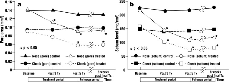
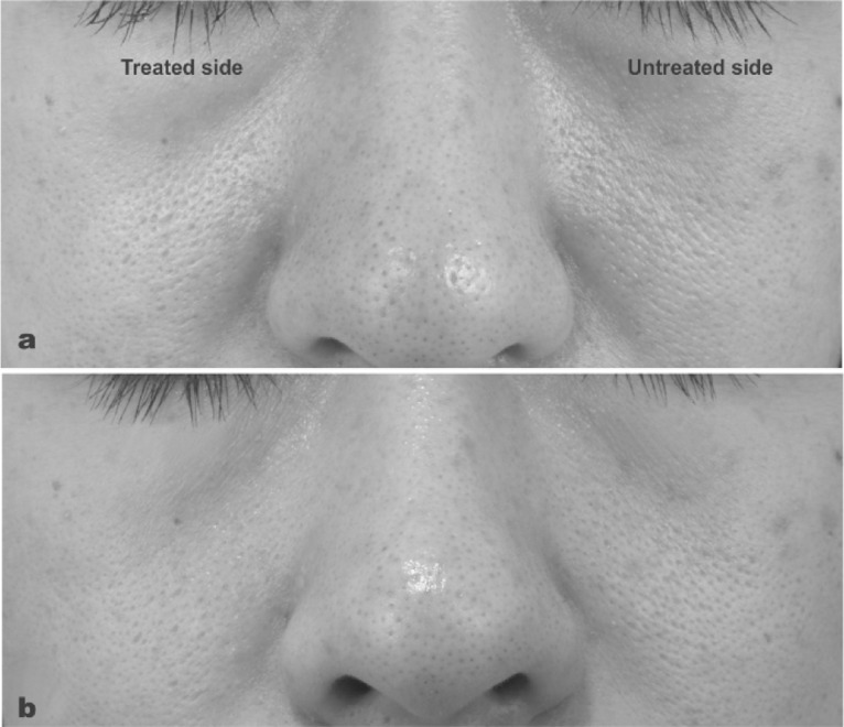
Figure 1a shows the difference in pore size over time comparing the treated and control sides. The mean pore size was based on the pore area in mm2 calculated from the × 300 microphotography by the image analysis software as described above. The size was derived by computing the average of the 10 subjects in each group at baseline, after the 3rd and 5th treatments and at 8 weeks after the last treatment. The pore size significantly decreased with treatment on the treated side (right cheek and right half of nose) compared to the control (p<0.05), with significance appearing at treatment week 3. The decrease in pore size was maintained at 8 weeks after the last treatment. Figure 1b shows the difference in sebum level over time between the treated and control sides, as measured objectively with the Sebumeter®. The sebum level also significantly decreased with treatment (p<0.05). Figure 2 shows a typical clinical example.
Fig. 1:
Measurement of (a) mean pore size and (b) mean sebum levels compared between the treated and untreated sides at baseline (before the 1st treatment session), after 3 and 5 treatments, and at 8 weeks after the last treatment using the 300 µs micropulsed 1064 nm Nd:YAG laser.
Fig. 2:
Typical example of the clinical photography showing significant macroscopically visible improve-ment in pore size before treatment (a) and after 5 treatments (b) using the Nd:YAG laser in micro-pulsed mode.
Treatment in the 5 ns Q-Switched Mode
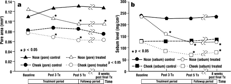
Figure 3a shows the difference in pore size over time compared between the treated and untreated sides. The mean pore size calculated as described above decreased significantly with treatment on the treated side (right cheek and right half of nose) compared to the control (p<0.05). The maintenance of the treatment effect at 8 weeks after the last treatment and the reduction of the sebum level with treatment (p<0.05; Figure 3b) were similar to the results observed with the micropulsed laser. A typical clinical example is illustrated in Figure 4.
Fig. 3:
Measurement of (a) mean pore size and (b) mean sebum levels compared between the treated and untreated sides at baseline (before the 1st treatment session), after 3 and 5 treatments, and at 8 weeks after the last treatment using the 5 ns Q-switched 1064 nm Nd:YAG laser.
Fig. 4:
Significant visual improvement in pore size seen in a typical clinical example, before treatment (a) and after 5 treatments (b) using the 5 ns Q-switched 1064 nm Nd:YAG laser
Comparison of the Two Treatment Methods
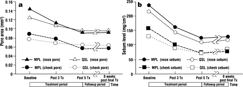
No statistically significant difference in the reduction of pore size or sebum level was seen between the micropulsed and Q-switched modes, with the improvements being more or less equal (Figure 5a, 5b).
Fig. 5:
Comparative measurements of the improvements in (a) mean pore size and (b) mean sebum levels between the groups treated with the micropulsed and Q-switched 1064 nm Nd:YAG laser. Although significant improvements in both pore size and sebum level were seen for both modes, there was no significant difference in improvement between the modes themselves. Note the slight falling-off in efficacy for both modes at the 8-week assessment.
Complications
The subjects reported mild erythema and swelling on the treated side, but these symptoms generally resolved within 12–48 hours after the treatment. The side effects were so mild that subjects did not experience any limitations in their normal activities.
Discussion
Prolonged exposure to the sun causes significant changes in the skin and is clinically manifested as mottled pigmentation and other dyschromias, erythema, telangiectasia, wrinkles, textural changes and enlargement of pores. Solar elastotic collagen damage produces a sallow skin tone, dilated pore structure and an appearance and elasticity similar to crepe paper.15) In addition, the degree of sebum production, age, sex and hormonal factors are known to contribute to the increase of pore size. In an unpublished study by our group, we found a significant correlation among the pore size and sebum level, sex, age and hormonal factors in women. Among the independent variables, sebum level was the most significant factor that correlated to the pore size. Therefore, treatment focusing on reducing sebum production along with dermal remodeling may be beneficial in decreasing the size of enlarged pores. Interestingly, with an average age of only 32 yr., the subjects in both groups in our study were in a younger age group than those normally associated with age-related pore enlargement, so this was an indication of the growing cosmetic significance of enlarged pores in a younger age group than those usually exhibiting the facial sequelae of photoaging.
The micropulsed and Q-switched modes of the Nd:YAG have potentially different bioeffects due to the difference in exposure time, pulse energy and peak power. With a pulse width of 5 ns and a pulse fluence of 3.0 J/cm2, the irradiance per pulse is 600 MW/cm2 giving a peak power per pulse of 2.3 × 10⁸ W, whereas the peak power at a pulse width of 300 µs and the same fluence is 3.8 × 10³ W, a difference of around some 5 orders of magnitude. The present study was thus designed to assess which set of parameters was more effective for pore size and related sebum reduction, given the same wavelength of 1064 nm and spot size of 7 mm in diameter. Pulse duration is known to be related to the dermal coagulation depth and in theory, therefore, the micropulsed mode of the 1064 nm Nd:YAG laser should penetrate more deeply into the dermis compared with the Q-switched mode and deliver potentially better skin remodeling. However, the present study showed no difference in the clinical effect of pore size reduction when the two methods were compared, although both modes gave significant improvement in both pore size and sebum levels.
The effect of treatment with both modes was assessed objectively with accurate measurement of changes between baseline and post-treatment conditions using a dermoscopic camera to take magnified views of the pores (x 100) and an image analysis program to calculate the pore area. We also measured the sebum levels before and after the treatments. In both modes, only one side of the subject's face was treated, thus providing both an intrapatient and intergroup comparison. The results showed that both the micropulsed and Q-switched modes of the 1064 nm Nd:YAG laser were more or less equally effective in reducing pore size and sebum level with no statistically significant difference seen between the modes. In both modes, the decreased levels were well maintained until the final assessment point, 8 weeks after the last treatment session. However, the slight fall-off seen in Figure 5 in the difference between baseline and the final assessment point would suggest that some form of maintenance treatment would need to be structured in to help maintain good results over the long term. The minor side effects noted by the subjects would suggest good compliance with such a maintenance program.
The Nd:YAG laser wavelength of 1064 nm is associated with increased collagen deposition in the papillary and upper reticular dermis11–14) and it has also been suggested that this wavelength may lead to a deeper dermal wound that can be utilized for nonablative dermal remodeling.10) In addition, heat-induced protein (hsp) 70 and procollagen 1 have been reported to be expressed in dermal dendritic cells and we suggest that these cells may participate in the deposition of dermal collagen after the treatment.12)
Although the exact mechanism of the effect of both the 300 µs micropulsed and 5 ns Q-switched modes of the 1064 nm Nd:YAG laser on pore size reduction is not clear, we can hypothesize that dermal collagen deposition and remodeling in the perifollicular area may result in reducing the size of the pores. We also suggest that a direct photothermal effect of the laser energy resulting in some shrinkage of the sebaceous gland may be responsible for the long-term maintenance of both the reduced pore size and sebum level. However, further studies with histologic confirmation should be performed to explain the exact mechanism.
CONCLUSION
This study objectively showed that the use of both the micropulsed and Q-switched 1064 nm Nd:YAG laser modes was effective in reducing the pore size and sebum level in patients complaining of enlarged pores, thereby improving the cosmetic appearance of the patients and maintaining that improvement over a reasonable followup period.
REFERENCES
- 1.Pierard GE, Eisner P, Marks R. et al (2003): EEMCO guidance for the efficacy assessment of antiperspirants and deodorants. Skin Pharmacol Appl Skin Physiol; Skin Pharmacol Appl Skin Physiol;: 324–342 [DOI] [PubMed] [Google Scholar]
- 2.Pierard GE, Pierard-Franchimont C, Marks R. et al (2000): EEMCO guidance for the in vivo assessment of skin greasiness. Skin Pharmacol Appl Skin Physiol; Skin Pharmacol Appl Skin Physiol;: 372–389 [DOI] [PubMed] [Google Scholar]
- 3.Brazil J, Owens P. (2003): Long-term clinical results of IPL photorejuvenation. J Cosmet Laser Ther; J Cosmet Laser Ther;: 168–174 [DOI] [PubMed] [Google Scholar]
- 4.Bitter PH. (2000): Noninvasive rejuvenation of photodamaged skin using serial, full-face intense pulsed light treatments. Dermatol Surg; 26: 835–842 [DOI] [PubMed] [Google Scholar]
- 5.Phillips TJ, Gottlieb AB, Leyden JJ. et al (2002): Efficacy of 0.1% tazarotene cream for the treatment of photodamage: a 12-month multicenter, randomized trial. Arch Dermatol; Arch Dermatol;: 1486–1493 [DOI] [PubMed] [Google Scholar]
- 6.Abraham M, Chiang S, Keller G. et al (2004): Clinical evaluation of non-ablative radiofrequency facial rejuvenation. Cosmet Laser Ther; Cosmet Laser Ther;: 136–144 [DOI] [PubMed] [Google Scholar]
- 7.Hernandez-Perez E, Khawaja HA, Alvarez TY. (1999): Oral isotretinoin as part of the treatment of cutaneous aging. Dermatol Surg 2000; 26: 649–652 [DOI] [PubMed] [Google Scholar]
- 8.Schmidt JB, Donath P, Hannes J. et al (1999): Tretinoin-iontophoresis in atrophic acne scars. Int J Dermatol; Int J Dermatol;: 149–153 [DOI] [PubMed] [Google Scholar]
- 9.Grimes PE. The safety and efficacy of salicylic acid chemical peels in darker racial-ethnic groups. Dermatol Surg; Dermatol Surg;: 18–22 [DOI] [PubMed] [Google Scholar]
- 10.Goldberg DJ, Whitworth J. (1997): Laser skin resurfacing with the Q-switched Nd:YAG laser. Dermatol Surg; Dermatol Surg;: 903–907 [DOI] [PubMed] [Google Scholar]
- 11.Goldberg DJ, Silapunt S. (2001): Histologic evaluation of a Q-switched Nd:YAG laser in the nonablative treatment of wrinkles. Dermatol Surg; Dermatol Surg;: 744–746 [DOI] [PubMed] [Google Scholar]
- 12.Prieto VG, Diwan AH, Shea CR. et al (2005): Effects of intense pulse light and the 1064 nm Nd:YAG laser on sun-damaged human skin: Histologic and Immunohistochemical analysis. Dermatol Surg; Dermatol Surg;: 522–525 [DOI] [PubMed] [Google Scholar]
- 13.Dang Y, Ren Q, Liu H. et al (2005): Comparison of histologic, biochemical, and mechanical properties of murine skin treated with the 1064-nm and 1320-nm Nd:YAG lasers. Exp Dermatol; Exp Dermatol;: 876–882 [DOI] [PubMed] [Google Scholar]
- 14.Lee MC. (2003): Combination 532-nm and 1064-nm lasers for noninvasive skin rejuvenation and toning. Arch Dermatol; 139: 1265–1276 [DOI] [PubMed] [Google Scholar]
- 15.Yaar M, Gilchrest BA. Aging of skin. In: Freedberg IM, Eisen AZ, Wolff K. et al, editors. Fitz-patrick's Dermatology in General Medicine. New York: McGraw Hill; New York: McGraw Hill;: 1386–1398 [Google Scholar]


