Abstract
Background and Aims: The limited available effective treatments make the management of keloids challenging. Intralesional triamcinolone and pulsed dye laser have been used for the treatment of keloids. We sought to assess the efficacy of a treatment regime using rotational intralesional triamcinolone and pulsed dye laser in the management of keloids.
Materials (Subjects) and Methods: Case notes and photographic records of 99 patients with keloids treated with pulsed dye laser (PDL) alone or rotational PDL and intralesional triamcinolone (ILT) at our centre between 2005 and 2010 were reviewed. Patients with raised, erythematous and/or symptomatic keloids unresponsive to ILT alone (usually 6 treatments) are referred for consideration of PDL. Patients are offered repeated rotational treatments of three PDL (4–15 J/cm2, 7 mm spot, 1.5 msec pulse duration, 595 nm wavelength, DCD, 30 msec spray: 20 msec delay; spaced at 6–8 weeks intervals) followed by one ILT (10 mg or 40 mg/dl). ILT-treated flat but erythematous and/or symptomatic keloids are treated with PDL alone at 6–8 week intervals. Response after each laser treatment is documented as a percentage improvement from baseline. Based on the improvement in redness, thickness and pruritus the operator classified the response to treatment as mild (0–49%), moderate (50–75%) or excellent (>75%). Patients are reviewed at 6 months following last treatment. A patient satisfaction questionnaire was also sent out to all patients who received treatments between 2005 and 2010 and this was compared with the information gathered from the notes.
Results: Of the 99 patients, 58 were women and 41 were men and most were Caucasian (n=84). A total of 755 keloids were treated. The average number of PDL treatments to achieve a moderate-excellent result was 14 in male and 12 in females. Moderate-excellent improvement was seen in 76% patients. The average number of ILT required to achieve a moderate-excellent result was 5 in males and 4 in females. All patients were sent a satisfaction questionnaire and 33 responses were received wherein patients reported an average of 70% improvement in the redness and thickness of the keloids. Localised cutaneous atrophy, self-limiting erythema and injection site discomfort were noted in 3 female patients whilst no side-effects noted in the male cohort.
Conclusions: Pulsed dye laser with or without intralesional triamcinolone is a moderately effective treatment of keloid scars with a very good side-effect profile and high patient satisfaction.
Keywords: Keloids, Laser, Pulsed dye laser, Scars, Triamcinolone
INTRODUCTION
Aberrant scars were first described in 1806 and thought to be a type of cancerous tissue.1) Further research into these lesions showed that keloids were a result of abnormal deposition of collagen during the wound healing process.2) Keloids present at around 3 months post-injury and show no signs of growth cessation or regression without treatment. They can develop in any location but are more common on the earlobes, sternum and deltoid regions without predisposition to either gender. They commonly invade adjacent tissues beyond the confines of the original wound.1, 3–5)
The pathogenic mechanisms in the formation of keloids are still unclear, but it is thought that the process begins early in the wound healing process. Cytokines such as transforming growth factor—beta (TGF-ß) are expressed in higher concentrations, which is thought to contribute to the lack of growth regulation exhibited by these scars. Fibroblasts present in these scars show an increased production and, in the case of keloids, heightened sensitivity to growth factors.6, 7)
Many genetic links have been investigated and conflicting conclusions have been reached. One well-described feature is the increased incidence of the scars in ethnicities with darker skin tones, particularly in Black and Hispanic populations with incidence rates as high as 16%.1, 6, 8)
Treatment options available for the management of keloids range from silicone gels and pressure treatments to surgical interventions. Many of these can be combined into treatment regimes to suit the individual patient. Pulsed dye laser (PDL) was first used to treat keloid scars in 1993. Selective photothermolysis leads to high temperatures within cutaneous vasculature, inducing localised hypoxia and consequently degradation of collagen. The main aim of PDL is to reduce the redness and restore skin texture. 9)
Intralesional triamcinolone is the mainstay of treatment for keloids. It results in a decreased action of fibroblasts and consequently collagen production. Side effects include localised skin atrophy, hypopigmentation and ulceration.1, 6)
MATERIALS AND METHODS
Ninety-nine individuals with keloids were treated with PDL alone or in conjunction with ILT between 2005 and 2010. The case notes of these patients were reviewed and they were all sent patient satisfaction questionnaires. The objective outcomes recorded in the notes were gathered along with those given by the patients in the questionnaire.
Treatment Regime
In our centre, the treatment of hypertrophic and keloid scars is split into two pathways; one for scars which are raised and red with or without symptoms; the other for those which are flat but still red with or without symptoms. As part of the former pathway, patients are offered a course of six ILT treatments (Adcortyl® injections, 10 mg ml−1). Those that are unresponsive to ILT will then be considered for 3 PDL treatments (4–15 J/cm2, 7 mm spot, 1.5 msec pulse duration, 595 nm wavelength, DCD, 30 msec spray: 20 msec delay) at 6–8 week intervals accompanied by a post-laser ILT treatment. Patients on the latter pathway are considered for PDL without initial ILT use.
Case Notes (Appendix 1)
The case notes and photographic records of ninetynine patients were reviewed. The information gathered included the age and sex of the patient along with the site and number of the patient's scars. The numbers of PDL and ILT treatments received were recorded. For PDL treatment the laser parameters were also collected. Any improvement in appearance and symptoms of keloids documented in the notes were recorded along with any of the side effects experienced after either of the treatments. If any other treatments had been attempted to treat the keloids, this was also recorded. Based on any improvement in thickness, pruritis or redness of the scar, the response to treatment was classified as mild (0–49%), moderate (50–75%) or excellent (>75%) improvement. Patients are reviewed 6 months following final laser treatment for monitoring of response.
Satisfaction Questionnaire (Appendix 2)
All the patients in the study were sent a patient satisfaction questionnaire. They were then asked to rate their response to treatment using a scale of 0–10 (0 indicating no improvement and 10 indicating complete clearance) and to address the symptoms including redness, thickness, discomfort and itching that improved the most. There was also an opportunity to state whether they had experienced any side effects from the treatments received.
RESULTS
Ninety-nine patients received treatment with PDL alone or in combination with ILT from 2005–2010. Fifty-eight (59%) of these patients were female. The majority of these patients were Caucasian (90%), 8% were Asian and 2% Polynesian or Chinese.
The commonest sites of the keloids varied between men and women. In females, the shoulders (46%) were the commonest site of presentation, followed by the chest (19%), head and neck (12%), back (10%) the extremities (7%) and finally the trunk (6%). The commonest site of keloids in the male cohort was the back (29%), followed by the chest (28%), extremities (20%), shoulders (18%), face/scalp/neck (4%) and trunk (1%) (Table 1).
Table 1. Location of keloids (n=755).
| Situation of | ||||||
|---|---|---|---|---|---|---|
| keloids | ||||||
| Chest, n (%) | Back, n (%) | Face/Scalp/Neck, | Trunk, | Shoulders, | Extremities, | |
| n (%) | n (%) | n (%) | n (%) | |||
| Females | 35 (19) | 18 (10) | 22 (12) | 10 (6) | 82 (46) | 13 (7) |
| Males | 161 (28) | 169 (29) | 19 (4) | 5 (1) | 104 (18) | 117 (20) |
IntralesionalTriamcinolone (Table 2)
Table 2. Mean number of treatments used to achieve treatment outcomes.
|
Improvement after treatment | |||
| Excellent >75% | Moderate 50–75% | Mild 0–49% | |
| Mean no. ILT treatments in females | 2 | 4 | 2 |
| Mean no. ILT treatments in males | 5 | 5 | 2 |
| Mean no. PDL treatments in females | 12 | 12 | 6 |
| Mean no. PDL treatments in males | 8 | 17 | 7 |
The average number of injections used to achieve an excellent or moderate result varied between males and females. An excellent outcome (>75%) was accomplished in females after an average of 2 (0–9) treatments and in males after an average of 5 treatments (0–17). Moderate improvements (50–75%) were achieved after an average of 4 injections (0–29) in females and after 5 treatments (0–18) in males. Both males and females required an average of 2 treatments (female 0–14, male 0–7) to achieve a mild (0–49%) improvement.
Pulsed Dye Laser Treatments (Table 2)
The average number of PDL treatments needed to achieve an excellent or moderate result in males and females varied significantly. An average of 8 treatments (1–18) were needed in males to achieve an excellent result and 12 (2–14) were needed in females. Moderate improvement was accomplished in males after mean average of 17 treatments (2–80) and after 12 treatments (1–54) in females. Men needed 7 treatments (3–13) to achieve a mild improvement, whereas women needed an average of 6 (2–11).
Treatment Outcomes (Table 3)
Table 3. Treatment outcomes.
| Documented Improvement | |||
| Excellent >75% > n (%) | Moderate 50–75%), n (%>) | Mild 0–49%, n(%) | |
| Females | 8 (8) | 36 (36) | 14 (14) |
| Males | 9 (9) | 23 (23) | 10 (10) |
The outcomes were categorized into excellent, moderate and mild improvements (see Methods).
In total 76% of our patients reported a moderate to excellent result after combination therapy (ILT + PDL). Most of our patients however reported a moderate improvement.
Eight percent of females and 9% of males achieved an excellent result after treatment. Moderate improvement was achieved by 36% of females and 23% of males. Fourteen percent of females achieved a mild improvement compared with 10% of males. Of interest the total percentage of females and males achieving a moderate to excellent result was equal.
Patient Satisfaction (Table 4)
Table 4. Most significant improvements noted by patients (Results from patient survey, n=32).
|
Features | ||||
| Redness | Thickness | Discomfort | Pruritus | |
| Female | 12 | 9 | 3 | 2 |
| Male | 2 | 2 | 1 | 1 |
As shown in Table 3, 33 patients (33%) replied to the satisfaction survey, of these 27 were females and 6 were males. One patient who provided a response included no specific details as requested by the questionnaire but thanked us for his successful treatment. When asked to rate their response to treatment from 0–10 (0 being no response and 10 being complete resolution) the average response from the females was 8 and the average response from the males was 7. Patients were also asked to provide an estimation of the duration of their treatment. This was 44 months for the female patients and 37 months for the males.
Patients were asked which aspect of the keloid improved the most. Of the 30 patients who replied to this question, 50% stated that redness was improved and 70% reported an improvement in the thickness of the keloids. 13% patients reported an improvement in discomfort. An improvement in pruritus was reported by 64% patients. Three female patients experienced side-effects from the treatment. These were redness (n=1), discomfort (n=1) and pruritus (n=1).
DISCUSSION
The treatment of keloid and hypertrophic scars is one that has challenged clinicians for generations and continues to do so today. Even with the bank of knowledge that continuing research into the pathogenesis of these scars has provided, there is still a lack of treatment options that are infallible. Management must be tailored to each individual in turn and take into account several important factors such as the age of the scar, its size, location and thickness.2, 9) However, even taking into account these factors, it is impossible to predict an individual's response to a certain treatment. With so many treatments currently available, it is common for more than one modality to be used in the treatment of one patient's scars. Treatments in the arsenal of the clinician include steroids, silicone treatments, pressure therapy, surgery, laser therapy, radiation and the use of anti-proliferative agents.2, 6)
The use of corticosteroids is well documented in the treatment of keloids and hypertrophic scars. Although the use of topical corticosteroid treatment has yielded mixed results, intralesional use has shown very promising outcomes. Several different steroids have been utilised but the most widely used is triamcinolone. ILT can be used alone or in conjunction with other therapies. In one study conducted by Ketchum et al, keloids were injected with 120 mg of triamcinolone and compared to a control group who received injections of saline. Although the follow-up period was only three weeks, 40% of the keloids had achieved a 75% reduction in thickness. The control group showed no improvement. In another study Darzi treated keloids and hypertrophic scars with a dose of triamcinolone dependent on the surface area of the lesion every 1–2 weeks. Patients were reviewed regularly and followed up for a period of ten years. The results showed a full flattening in 64% of the scars and partial flattening in 28%.6, 10–12)
The use of ILT as a part of combination therapy in the treatment of keloids has also been described. For example in one study, the scars were first excised and then intraoperatively injected with triamcinolone. This was followed by weekly post-operative injections for 2–5 weeks and monthly injections for 4–6 months. Total relief of symptoms was achieved within 5 weeks of surgery. At a mean follow-up time of 30.5 months, there was a less than 9% recurrence rate in keloid scars and less than 5% in hypertrophic scars. Other studies have been conducted evaluating the efficacy of combinations with cryotherapy, PDL, 5-FU.10, 13)
Even though steroids have become a mainstay of treatment, their use has been linked with side-effects such as localised atrophy and hypopigmentation. Apart from the side-effects, another disadvantage of ILT is that it can be painful to administer and several treatments are usually required.10)
ILT works by decreasing the abnormal proliferation of fibroblasts and halting the growth of existing fibroblasts. This leads to a reduction in collagen synthesis and production of inflammatory cytokines, which are two of the factors involved in the pathogenesis of the scars. It has been shown to be most effective on young scars but still has a place in the treatment of older ones, especially in eliminating symptoms.1, 6, 10)
The use of lasers in the management of abnormal scars has evolved greatly in the last twenty years. Several different laser types have been used beginning with the carbon dioxide (CO2), argon and Nd:YAG lasers. These lasers are now very rarely used. CO2 laser was found to have high recurrence rates ranging from 75–95% and in many of the studies involving the use of Nd:YAG laser, the outcomes are not well documented. For example in one study by Abergel et al, using Nd:YAG laser in the treatment of keloids present on the skin of 8 patients. Although one patient clearly achieved a very impressive outcome, the outcomes in remaining patients were not discussed in any significant detail and conclusions cannot be drawn.9, 15)
Alster et al. were the first to demonstrate the use of PDL in the treatment of keloids and hypertrophic scars in 1993 and 1995, showing improvements in redness, thickness and texture of the scars. As part of the first study in 1993, skin texture was measured using optical prolifometry and showed that 100% of patients involved in the trial showed flattening with or without the restoration of surface markings to the skin. This is thought to be due to the effect of PDL on cutaneous blood vessels; causing them to break down and local hypoxia to occur leading to the degradation of collagen. Although both these studies showed significant improvements in scarring in response to PDL, recurrence rates were not assessed. The improvements noted in the 1995 study were said to have remained at follow-up six months later, but there was no long-term follow-up.9, 16, 17)
In our centre, the treatment of hypertrophic and keloid scars is split into two pathways; one for scars which are raised and red with or without symptoms; the other for those which are flat but still red with or without symptoms. As part of the former pathway, patients are offered a course of six ILT treatments. Those that are unresponsive to ILT will then be considered for PDL treatment at 6–8 week intervals accompanied by a post-laser ILT treatment. Patients on the latter pathway are considered for PDL without initial ILT use.
Seventy-six percent of patients treated between 2005 and 2010 achieved a moderate-excellent outcome. This demonstrates that the combination of PDL and ILT has very encouraging results. A possible reason for this has been outlined in one particular study conducted by Manuskiatti et al. in which keloid scars were split into 5 segments and each segment was randomly assigned a different treatment as follows: 1) ILT, 2) ILT plus 5-FU, 3) 5-FU alone, 4) PDL and 5) a control segment which received no treatment. The results of this study showed that ILT seemed to flatten the scar whereas PDL did not flatten the scar as effectively, but did normalize the skin texture.18) This indicates that the two modalities both have very different but complimentary effects on aberrant scars.
Other studies have been conducted into the efficacy of combined treatment with ILT and PDL. One such study conducted by Alster et al. in 2003 showed mixed results. The patient cohort consisted of 22 female patients with bilateral post-surgery inframammary scars. On each patient, one scar was randomly assigned to receive treatment with PDL alone and the other to receive PDL in combination with ILT. A 10–20 mg dose of triamcinolone was used per injection and PDL administered every 6 weeks. The scars were evaluated after each treatment and also 6 weeks after the final (second) treatment. Results showed that PDL had a significant effect both clinically and histologically on the scars but this effect was not enhanced by the used of ILT. It has been suggested that this may be due to the low dosages of triamcinolone used as it has been suggested that the ideal dose is in fact 10–40 mg depending on the site of the lesion.10)
Tranilast, N (3, 4-dimethoxycinnamoyl) anthranilic acid (Rizaben®, Kissei Pharmaceuticals and Co. Matsumo City, Japan.) was founded as an anti-allergic drug used in bronchial asthma also used commonly in the treatment of both keloid and hypertrophic scars in Japan. Systemic use of Tranilast in the form of tablets (300 mg. orally three times a day) and topical delivery have been trialled with success.19) Tranilast inhibits the production of transforming growth factor beta 1 (TGF-ß1) which has been shown to be causal in the formation of keloids and hypertrophic scars by stimulating the proliferation of fibroblasts and in turn the synthesis of collagen I and III.
The release of prostaglandin E2 (PGE2) and histamine also stimulate fibroblast proliferation. Tranilast has also been shown to inhibit the release of PGE2 and histamine from mast cells which in turn improves keloid/hypertrophic scar formation.
There is no comparative trial data for the efficacy of the use of tranilast in the treatment of keloid scars against other treatment modalities and reported side effects include hyperbilirubinaemia especially with a background of Gilbert's Syndrome.
The use of Tranilast has been explored in various other proliferative conditions i.e. neurofibromatosis21) and desmoids tumours20) with success and less success fully in the treatment of coronary artery restenosis.22) Its effect on other conditions associated with the overproduction of TGF-ß1, i.e. Scleroderma is still under investigation.23)
Despite the promising results displayed in our study, it is important to discuss its limitations. This was a retrospective case series and as a result recording bias can not be excluded. Also, the patient cohort in the study was mainly caucasian even though keloids and hypertrophic scars are much more common in those with darker skin. This may mean that the results shown in this study may not be applicable to all patient populations. The third factor to consider is that the use of patient feedback allows for the possibility of recall bias as some patients may have received treatment some time ago. Finally, although most patients demonstrated moderate improvement, long-term follow-up is lacking. It is difficult to predict the sustainability of response to this treatment based on patient's feedback alone.
CONCLUSIONS
Results from our retrospective study have shown that there is clearly a place for both PDL and ILT treatments alone and in combination in the management of keloids. This is coupled with very good side-effect profile and patient satisfaction levels. Most noticeably our patient cohort reported considerable improvement in the thickness and erythema of their keloids complimented by an improvement itch and discomfort. However, as most patients show a moderate response, the treatment of aberrant scarring still remains challenging. Although more research must be conducted into the long-term recurrence rates, it is clear that PDL and ILT offer another alternative in the treatment of keloids.
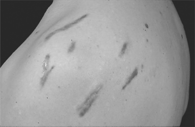
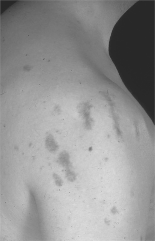
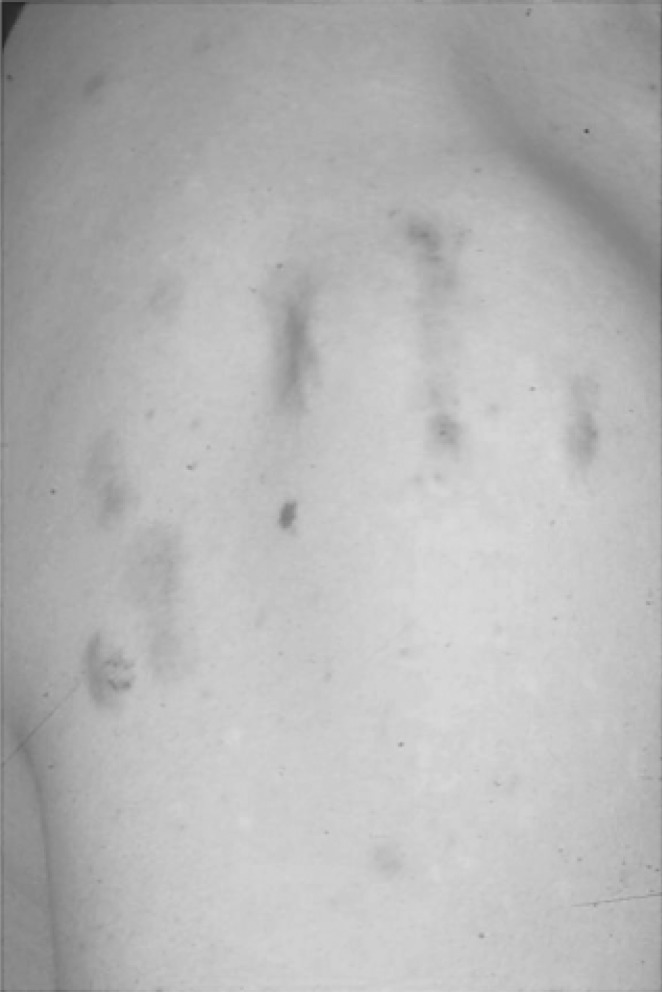
Fig. 1a:
Keloid scars on the shoulder before treatment
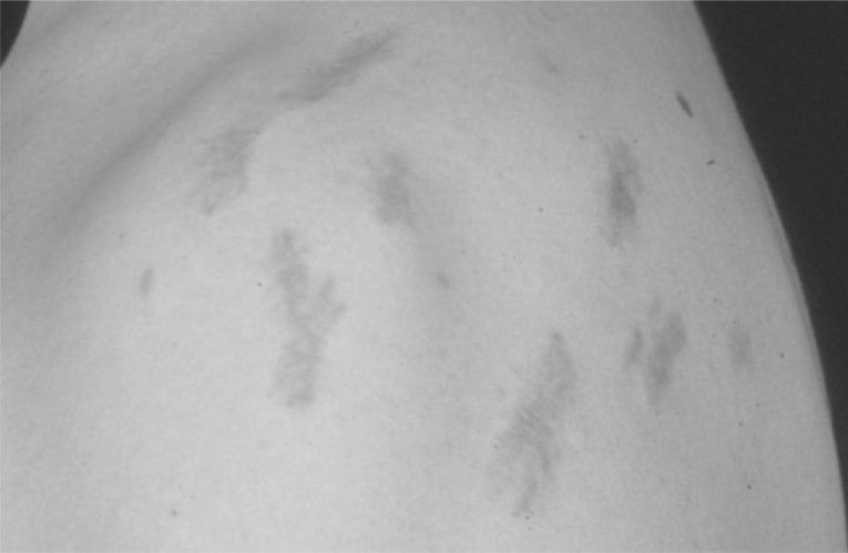
Fig. 1b:
Keloid scars on the shoulder 12 months after combination therapy (rotational ILT and PDL). Improvement in the thickness of the keloid with some remanant erythema is demonstrated.
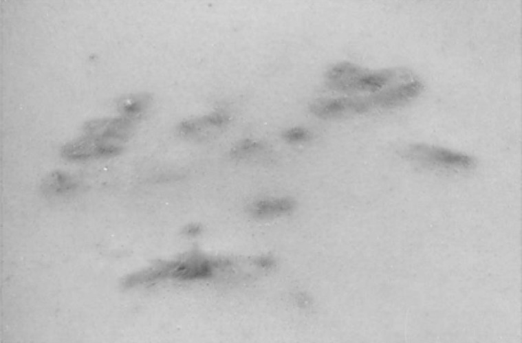
Fig. 2a:
Keloid scars on the anterior chest wall before treatment
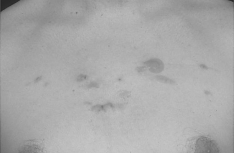
Fig. 2b:
Keloid scars immediately after treatment with intralesional triamcinolone in to the individual keloid scars. Spot plasters are applied to the areas of bleeding if required after the appropriate application of pressure for haemostasis.
Fig. 3a:
Keloid scars on the left shoulder before treatment
Fig. 3b:
Keloid scars on the left shoulder 1 year after combination therapy (rotational ILT and PDL). Considerable improvement in both the thickness and erythema related to the scar was observed.
REFERENCES
- 1. Niessen FB, Spauwen PH, Schalkwijk J, Kon M. (1999): On the nature of hypertrophic scars and keloids: a review. Plastic Reconstructive Surgery. 104: 1435–1458 [DOI] [PubMed] [Google Scholar]
- 2. Slemp AE, Kirschner RE. (2006): Keloids and scars: a review of keloids and scars, their pathogenesis. risk factors, and management. Current Opinion in Pediatrics, 18: 396–402 [DOI] [PubMed] [Google Scholar]
- 3. Alster TS, Handrick C. (2000): Laser treatment of hypertrophic scars, keloids and striae. Seminars in Cutaneous Medicine and Surgery, 19: 287–92 [DOI] [PubMed] [Google Scholar]
- 4. English RS, Shenefelt PD. (1999): Keloids and Hypertrophic Scars. Dermatologic Surgery, 25: 631–638 [DOI] [PubMed] [Google Scholar]
- 5. Rockwell WB, Cohen IK, Ehrlich HP. (1989): Keloids and hypertrophic scars: A comprehensive review. Plastic Reconstructive Surgery, 84: 827–837 [DOI] [PubMed] [Google Scholar]
- 6. Wolfram D, Tzankov A, Pulzi P, Piza-Katzer H. (2009): Hypertrophic scars and keloids—a review of their pathophysiology, risk factors and therapeutic management. Dermatologic Surgery, 35: 171–181 [DOI] [PubMed] [Google Scholar]
- 7. Matsuoka LY, Uitto J, Wortsman J, Abergel RP, Dietrich J. (1988): Ultrastructural characteristics of keloid fibroblasts. The American Journal of Dermatopathology, 10 (6): 505–508 [DOI] [PubMed] [Google Scholar]
- 8. Shih B, Bayat A. (2010): Genetics of keloid scarring. Archives of Dermatology Research, 302: 319–339 [DOI] [PubMed] [Google Scholar]
- 9. Alster TS, Tanzi E. (2003): Hypertrophic scars and keloids - Etiology and management. American Journal of Clinical Dermatology, 4: 235–243 [DOI] [PubMed] [Google Scholar]
- 10. Roques C, Teot L. (2008): The use of corticosteroids to treat keloids: A review. The International Journals of Lower Extremity Wounds, 7: 137–145 [DOI] [PubMed] [Google Scholar]
- 11. Ketchum LD, Smith J, Robinson DW, Masters FW. (1966): The treatment of hypertrophic scar, keloid and scar contracture by triamcinolone acetonide. Plastic and Reconstructive Surgery, 38: 209–218 [DOI] [PubMed] [Google Scholar]
- 12. Darzi MA, Chowdri NA, Kaul SK, Khan M. (1992): Evaluation of various methods of treating keloids and hypertrophic scars: a 10-year follow-up study. British Journal of Plastic Surgery, 45: 374–379 [DOI] [PubMed] [Google Scholar]
- 13. Chowdri NA, Mattoo MMA, Darzi MA. (1999): Keloids and hypertrophic scars: Results with intraopertive and serial post-operative corticosteroid injection therapy. Australian and New Zealand Journal of Surgery, 69: 655–659 [DOI] [PubMed] [Google Scholar]
- 14. Shaffer JJ, Taylor SC, Cook-Bolden F. (2002): Keloidal scars: A review with a critical look at therapeutic options. Journal of the American Academy of Dermatology, 46: 63–97 [DOI] [PubMed] [Google Scholar]
- 15. Abergel RP, Dwyer RM, Meeker CA, Lask G, Kelly AP, Uitto J. (1984): Laser treatment of keoids: A clinical trial and an in vitro study with Nd:YAG laser. Lasers in Surgery and Medicine, 4: 291–195 [DOI] [PubMed] [Google Scholar]
- 16. Alster TS, Kurban AK, Grove GL, Grove MJ, Tan OT. (1993): Alteration of argon laser-induced scars by the pulsed dye laser. Lasers in Surgery and Medicine, 13: 368–373 [DOI] [PubMed] [Google Scholar]
- 17. Alster TS, Williams CM. (1995): Treatment of keloid sternotomy scars with the 585nm flashlamp-pumped pulsed dye laser. Lancet, 345: 1198–1200 [DOI] [PubMed] [Google Scholar]
- 18. Manuskiatti W, Fitzpatrick RE. (2002): Treatment response of keloidal and hypertrophic sternotomy scars: comparison among intralesional corticosteroid, 5-fluorouracil, and 585-nm flashlamp-pumped pulsed-dye laser treatments. Archives of Dermatology, 138: 1149–55 [DOI] [PubMed] [Google Scholar]
- 19. Goto T. et al; Successful treatment of desmoid tumor of the chest wall with tranilast: a case report; J Med Case Reports. 2010 Nov 29; 4: 384. [DOI] [PMC free article] [PubMed] [Google Scholar]
- 20. Yamada H. et al; Tranilast, a selective inhibitor of collagen synthesis in human skin fibroblasts; J Biochem. 1994 Oct; 116 (4): 892–7 [DOI] [PubMed] [Google Scholar]
- 21. Yamamoto M. et al; Tranilast, an anti-allergic drug, down-regulates the growth of cultured neurofibroma cells derived from neurofibromatosis type 1; Tohoku J Exp Med. 2009 Mar; 217 (3): 193–201 [DOI] [PubMed] [Google Scholar]
- 22. Singh M. et al; Geographical differences in the rates of angiographic restenosis and ischemia-driven target vessel revascularization after percutaneous coronary interventions: results from the Prevention of Restenosis With Tranilast and its Outcomes (PRESTO) Trial; J Am Coll Cardiol. 2006 Jan 3; 47 (1): 34–9 [DOI] [PubMed] [Google Scholar]
- 23. Prud'homme GJ; Pathobiology of transforming growth factor beta in cancer, fibrosis and immunologic disease, and therapeutic considerations; Lab Invest. 2007 Nov; 87 (11): 1077–91 Epub 2007 Aug 27 [DOI] [PubMed] [Google Scholar]


