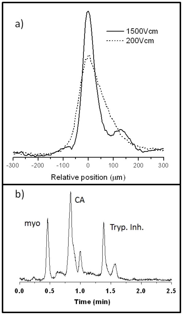
Figure 6.
cIEF in a capillary packed with micrometer-sized silica particles with polyacrylamide coatings, using carrier ampholytes. a) Image data for a focused zone of trypsin inhibitor, showing increased resolution at very high electric field. b) Remobilized protein peaks after cIEF using pressure driven flow in the absence of electric field. Figures are adapted from Hua et al. [45].

