Abstract
Introduction
Insulin resistance in obesity is decreased after successful diet and exercise. Aerobic exercise training alone was evaluated as an intervention in subjects with the metabolic syndrome.
Methods
Eighteen non-diabetic, sedentary subjects, eleven with the metabolic syndrome, participated in eight weeks of increasing intensity stationary cycle training.
Results
Cycle training without weight loss did not change insulin resistance in metabolic syndrome subjects or sedentary control subjects. Maximal oxygen consumption (VO2max), activated muscle AMP-dependent kinase, and muscle mitochondrial marker ATP synthase all increased. Strength, lean body mass, and fat mass did not change. Activated mammalian target of rapamycin was not different after training. Training induced a shift in muscle fiber composition in both groups but in opposite directions. The proportion of 2x fibers decreased with a concomitant increase in 2a mixed fibers in the control subjects, but in metabolic syndrome, 2x fiber proportion increased and type 1 fibers decreased. Muscle fiber diameters increased in all three fiber types in metabolic syndrome subjects. Muscle insulin receptor expression increased in both groups and GLUT4 expression increased in the metabolic syndrome subjects. Excess phosphorylation of insulin receptor substrate-1 (IRS-1) at Ser337 in metabolic syndrome muscle tended to increase further after training in spite of a decrease in total IRS-1.
Conclusion
In the absence of weight loss, cycle training of metabolic syndrome subjects resulted in enhanced mitochondrial biogenesis, and increased expression of insulin receptors and GLUT4 in muscle, but did not decrease the insulin resistance. The failure for the insulin signal to proceed past IRS-1 tyrosine phosphorylation may be related to excess serine phosphorylation at IRS-1 Ser337 and this is not ameliorated by eight weeks of endurance exercise training.
Keywords: insulin resistance, metabolic syndrome, euglycemic clamp, exercise training
Introduction
Obesity and diabetes have increased in prevalence in the United States over the past twenty years at a near epidemic rate (24, 25, 37). More than half of all adults are now overweight (BMI > 25 kg/m2), with some regions having in excess of two thirds of the population being overweight or obese (BMI > 30 kg/m2). Diabetes has been diagnosed in as many as 13% of adults in several states. Insulin resistance and hyperinsulinemia are key elements of the metabolic syndrome that is marked by visceral obesity, hypertension, hyperlipidemia, hyperglycemia, and coronary heart disease (1, 19, 26). The metabolic syndrome is a pre-diabetes condition until fasting hyperglycemia reaches 126 mg/dL (20).
Regular exercise is beneficial to the prevention (21) and management of type 2 diabetes (15, 34). Whether the exercise consists predominantly of endurance training or strength training, glycemic control improves (6, 12, 13). A dose-response relationship for the amount of regular exercise was apparent in a recent report of diabetes incidence among male health professionals (18). Grontved and coworkers evaluated data from biennially administered detailed questionnaires from more than 32,000 participants over an average of 16 years (18). During the time of follow-up, there were 2,278 new cases of type 2 diabetes. Regular aerobic exercise or weight training of at least 150 minutes per week resulted in 34% and 52% less diabetes, respectively. Men who reported 150 minutes or more of combined weight training and aerobic exercise had a 59% lower incidence of type 2 diabetes (18). As little as 20 minutes of weight training per week significantly decreased the diabetes incidence compared to no exercise.
Even though there is evidence in healthy individuals for activation of the mammalian target of rapamycin (mTOR) being crucial for muscle remodeling after weight training and AMP-dependent kinase (AMPK) activation after endurance training (2, 36), it is unclear how the muscle hypertrophy and mitochondrial biogenesis cellular pathways are involved in response to either type of exercise alone in persons at high risk for type 2 diabetes. In the absence of weight loss, the impact of stationary cycle training on muscle remodeling and whole body insulin responsiveness is presented in this report. This study of supervised, increasing intensity of cycle training of metabolic syndrome participants for eight weeks, was designed as a follow up to a protocol of purely strength training of the same duration and similar energy expenditure that demonstrated no improvement in insulin resistance (22). The hypothesis driving these studies was that predominantly endurance training (with no weight loss) might have more impact than strength training on insulin responsiveness in the metabolic syndrome, and the cellular mechanisms of muscle adaptation would differ from those seen in strength training.
Materials and Methods
Materials
Monoclonal antibodies directed against slow-twitch myosin heavy chain were purchased from Millipore (Billerica, MA). ATP synthase antibodies (ab110273) were purchased from Abcam (Cambridge, MA). An alkaline phosphatase-conjugated fast myosin antibody (Sigma clone MY-32 alkaline phosphatase conjugate) was purchased from Sigma-Aldrich (St. Louis, MO). A peroxidase-conjugated rabbit anti-mouse IgG antibody (315-035-045) was purchased from Jackson ImmunoResearch Laboratories (West Grove, PA). GLUT4 antibodies (AB1049, goat anti-human) were purchased from Chemicon (Temecula, CA). Monoclonal antibodies directed at the human insulin receptor beta subunit (05-1104) were purchased from Millipore. Rabbit polyclonal antibodies directed at the human insulin receptor beta subunit (07-724) were purchased from Millipore. Rabbit polyclonal antibodies directed against human insulin receptor substrate-1 (IRS-1) (#2382) were purchased from Cell Signaling (Danvers, MA). Rabbit polyclonal antibodies specific for human IRS-1 phosphorylated at Tyr896 (#3070), Ser307 (#2491), Ser337 (#2580), Ser636 (#2388), and Ser1101 (#2385) were purchased from Cell Signaling.
Subject Selection
Eighteen sedentary subjects were recruited. None of the subjects had performed regular exercise for at least one year. The research protocol and the consent documents were approved by the East Tennessee State University Institutional Review Board. Each subject provided written informed consent. Sedentary subjects were recruited into two groups: high risk for type 2 diabetes (BMI ≥ 30 kg/m2 and a family history of type 2 diabetes) and low risk for type 2 diabetes (BMI < 30 kg/m2, no family history of type 2 diabetes). The eleven subjects at high risk for diabetes qualified for the designation “metabolic syndrome,” as set forward by the International Diabetes Federation (IDF) (1). Each of these had BMI greater than 30 kg/m2, waist circumference greater than 102 cm (40 inches), insulin resistance by euglycemic clamp, and dyslipidemia (triglycerides > 150 mg/dL, HDL < 40 mg/dL in males or 50 mg/dL in females) and/or hypertension (systolic blood pressure > 130 mm Hg).
The exercise intervention
All subjects performed their exercise training in groups of two to six, typically side-by-side, on SciFit cycle ergometers (model ISO1000, SCIFIT Systems Inc, Tulsa, OK) in the ETSU Sport and Exercise Science Laboratory under the direct supervision of an exercise science graduate student. Subjects were instructed to maintain a cadence of 75-85 RPMs. After five minutes of light cycling warm up (50W), four five-minute sets at the weekly target intensity setting were alternated with one-minute light cycling periods at reduced intensity (50W). The target intensities (in watts) for weeks one through eight were 100, 110, 120, 100, 125, 135, 145, and 120. These sessions took place on Monday and Tuesday, and Thursday and Friday in assigned afternoon time slots with Wednesday being designated for mid-section training. Participants performed mid-section exercises consisting of Crunches, Bells, and Windshield Wipers over the eight week intervention. Each exercise was performed on Wednesdays with one set of each exercise being performed during the first week and an additional set of ten repetitions being added each week until reaching a total of three sets for each exercise. Target intensity settings were increased from weeks one through three and weeks five through seven with reduced loads being prescribed on weeks four and eight in an attempt to reduce potential for overtraining and to manage fatigue. Heavy and light days were also utilized to further enhance recovery and manage fatigue. Two minutes light cycling (50W) was performed as a cool-down after the target sets were finished. All post measures of anthropometrics and muscle biopsies were performed 24-48 hours after the last exercise session.
A three day diet diary was reviewed with each subject by a nutrition intern supervised by nutrition faculty. The verified data were analyzed using Nutrition Pro software (Axxya Systems, Stafford, TX) to quantify daily caloric intake and average diet composition. Subjects were instructed to increase food consumption by 250-750 calories per day. If weight measured weekly deviated by more than 1 kg from the initial weight, a nutritionist counseled the subject to make further adjustments.
Subject Assessments
Body composition was measured by air displacement plethysmography (BodPod, Concord, CA). Blood pressures were the average of duplicates performed after sitting quietly for a minimum of 10 minutes. Glucose, insulin, HbA1c, and cholesterol measurements were performed in a clinical laboratory from serum obtained after an overnight fast (10 hour minimum). Maximal oxygen consumption (VO2max), and respiratory exchange ratio were analyzed using a TrueOne 2400 Metabolic Measurement System (ParvoMedics, Sandy, Utah) and a SciFit cycle ergometer. Fat oxidation was calculated from the baseline respiratory exchange ratio by the method of Frayn (17).
Muscle Biopsies
Percutaneous needle biopsies of vastus lateralis were performed after an overnight fast and two hours of quiet recumbency as previously described, using a Bergstrom-Stille 5 mm muscle biopsy needle with suction (33). The sample was divided in two, with one piece frozen immediately in liquid nitrogen for later analysis, and the second piece mounted on cork and quickly frozen in a slurry of isopentane cooled by liquid nitrogen for sectioning and microscope slide preparation.
Euglycemic Hyperinsulinemic Clamp
After a 2-hour baseline period, a single infusion of regular insulin was performed at 15 mU/m2/min for 2 hours in order to achieve a physiological increment in insulin concentration of about 50 μU/mL (350 pmol/mL). The glucose infusion rate necessary to maintain the blood glucose at 85±5 mg/dL (4.72±0.28 mmol/L) was generally stable by 60 minutes and the last 30 minutes of the 120 minute insulin infusion was used to calculate the steady state glucose infusion rate (SSGIR) to quantify insulin sensitivity as previously described (32).
Quantification of Muscle Fiber Type Composition and Fiber Size
Fiber composition was determined using methods described by Behan et al. (4). All sections were coded and then quantified independently by 2 observers who were unaware of which subject the image represented. All fiber size data for the current study were calculated using the minimum diameter measured for each fiber (11).
Immunoblots
Immunoblots to assess the content of the insulin receptor beta subunit, IRS-1, IRS-1 pTyr896, IRS-1 pSer307, IRS-1 pSer337, IRS-1 pSer636, IRS-1 pSer1101, GLUT4, and ATP synthase were performed using muscle homogenates are previously described (22). Studies of GLUT4 and ATP synthase used 10% polyacrylamide mini gels (Pierce, Rockford, IL), whereas all of the IRS-1 studies used NuPAGE Novex TRIS-acetate 3-8% gels from Invitrogen (Carlsbad, CA).
Immunohistochemistry
Immunohistochemical studies were performed as previously described (31). Images were generated using a Leica confocal microscope system. Image signal intensity of individual fibers was quantified using Quantity One image analysis software from Bio-Rad (Hercules, CA). Fiber type was determined in the corresponding confocal image labeled with antibodies specific for either slow-twitch myosin heavy chain or fast-twitch myosin heavy chain.
Statistics
All data are displayed as mean±SEM, except as explicitly indicated. Comparing data between the two groups was performed using the independent t test except as noted. Comparisons of data before and after training were done using the paired t-test. A p value of less than 0.05 was considered significant. Statistical procedures were performed using SigmaPlot version 12.2 from Systat Software (San Jose, California).
Results
Subject Characteristics
The characteristics of the control and metabolic syndrome participants are displayed in Table 1. There were eleven metabolic syndrome subjects, five of whom were female, and seven sedentary controls, five of whom were female. The mean age of the metabolic syndrome group was 44±4 (rage 23-54) and the control group was 41±4 (range 29-55). Shown in this table are the p values for comparisons of before and after training in both groups and in the rightmost column are the results of t tests comparing the metabolic syndrome baseline data to the control subject baseline measurements. Of the variables displayed in this table, only the VO2max was significantly increased by the training in the metabolic syndrome subjects. Among the control subjects, the VO2max increased, and the peak power from strength testing also increased. Most of the key pre-training characteristics of the metabolic syndrome subjects were quite different from those of the control group, with the exceptions of the strength testing data, blood pressures, and lipid data. In metabolic syndrome subjects, fasting glucose concentration averaged 9% higher and fasting insulin was 257% of the control group value. The HDL cholesterol concentrations were higher in the control subjects than the metabolic syndrome subjects. The dietary calories per day calculated from the three day diet diary were 35% more in the metabolic syndrome group, but this difference was not statistically significant. The proportion of calories from fat was 39% higher (p=0.011). The respiratory exchange ratio tended to decrease after training and the calculated fat oxidation tended to increase in both groups, although not statistically significantly different.
Table 1. Subject Characterization and Response to Eight Weeks of Stationary Cycle Training.
| sedentary controls (n=7, 5F) | metabolic syndrome (MS) (n=11, 5F) | |||||||
|---|---|---|---|---|---|---|---|---|
| variable | units | baseline | post training | trained p value* | baseline | post training | trained p value* | MS vs. controls at baseline p value** |
| weight | kg | 68.5±3.9 | 67.8±4.6 | 0.818 | 105.7±5.6 | 108.1±6.0 | 0.082 | <0.001 |
| BMI | kg/m2 | 23.9±1.2 | 24.0±1.3 | 0.787 | 36.2±1.1 | 36.9±1.2 | 0.778 | <0.001 |
| waist cm | cm | 90.2±4.4 | 88.1±4.1 | 0.372 | 117.8±3.6 | 118.3±2.3 | 0.866 | 0.002 |
| systolic blood pressure | mm Hg | 119±4 | 113±4 | 0.148 | 127±4 | 121±3 | 0.265 | 0.193 |
| diastolic blood pressure | mm Hg | 77±2 | 76±3 | 0.600 | 84±3 | 81±3 | 0.208 | 0.096 |
| body fat | percent | 31.3±2.9 | 31.2±3.4 | 0.921 | 43.1±1.0 | 43.4±1.1 | 0.333 | 0.002 |
| LBM | kg | 45.9±2.7 | 45.6±2.3 | 0.603 | 60.0±2.8 | 60.4±3.1 | 0.430 | 0.004 |
| peak power | Newtons | 2340±230 | 2520±202 | 0.005 | 2830±181 | 2920±125 | 0.338 | 0.117 |
| power rate | N/min | 2850±393 | 3060±600 | 0.542 | 3040±351 | 3430±413 | 0.412 | 0.713 |
| fasting serum glucose | mmol/L | 5.25±0.27 | 5.56±0.23 | 0.218 | 5.72±0.26 | 5.89±0.14 | 0.813 | <0.001 |
| fasting serum insulin | pmol/L | 37±11 | 27±6 | 0.775 | 95±12 | 145±43 | 0.548 | 0.005 |
| HbA1c | % | 4.9±0.1 | 4.9±0.1 | 0.813 | 5.3±0.1 | 5.3±0.1 | 0.647 | 0.021 |
| total cholesterol | mg/dL | 184±12 | 191±14 | 0.453 | 195±12 | 189±10 | 0.133 | 0.505 |
| triglyceride | mg/dL | 107±30 | 119±27 | 0.201 | 161±25 | 161±20 | 0.982 | 0.085 |
| HDL cholesterol | mg/dL | 54±4 | 59±4 | 0.293 | 41±3 | 41±3 | 1.000 | 0.013 |
| LDL cholesterol | mg/dL | 108±10 | 108±11 | 0.469 | 122±10 | 116±9 | 0.275 | 0.341 |
| 3 day diet history | kCal/d | 1980±330 | 2670±295 | 0.137 | ||||
| diet fat content | % of calories | 31±3 | 43±1 | <0.001 | ||||
| VO2max | ml/kg.min | 30.1±3.8 | 34.3±2.9 | 0.011 | 20.1±0.7 | 23.3±0.8 | <0.001 | 0.011 |
| RER | 0.865±0.017 | 0.827±0.024 | 0.284 | 0.898±0.22 | 0.833±0.012 | 0.383 | 0.294 | |
| fat oxidation | % | 24.8±3.9 | 39.2±10.8 | 0.265 | 19.7±5.4 | 33.9±4.0 | 0.063 | 0.928 |
paired t test,
independent t test, BMI is body mass index, LBM is lean body mass, RER is respiratory exchange ratio
Whole body insulin responsiveness was not increased by cycle training
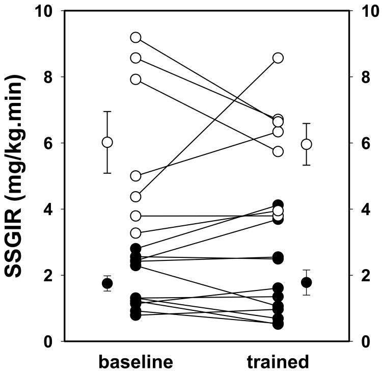
Seven control subjects and eleven metabolic syndrome subjects had euglycemic clamp studies performed before and after eight weeks of increasing duration and intensity stationary cycle training. In both groups some subjects improved and others did not change or their insulin responsiveness declined. The mean steady state glucose infusion rate (SSGIR) was unchanged in both groups. The individual subject data are shown in Figure 1. Interestingly, the control subjects who improved their insulin responsiveness were those who started at lower baseline responsiveness, whereas among the metabolic syndrome subjects, the lower baseline subjects showed little or no improvement.
Figure 1. Insulin responsiveness quantified by euglycemic insulin clamps was not increased by eight weeks of supervised stationary cycle training.
This graph plots the pre- and post-training steady state glucose infusion rates (SSGIR) for seven sedentary control subjects (open circles) and eleven subjects with the metabolic syndrome (black filled circles). The means and standard errors for the two groups before and after training are also plotted. The SSGIR of the metabolic syndrome group was 29% of the corresponding insulin responsiveness quantified in the control group both in the baseline study and after training.
Our subjects' data on insulin resistance can be expressed by many different calculated indicators as described in detail by Ferranini and coworkers from the European Group for the Study of Insulin Resistance (16). When the insulin clamp glucose infusion data were expressed as “M” (μmol/min/kg fat-free mass), differences between our groups before and after exercise training were amplified. Controls increased from 47.7±5.6 to 87.1±19.7 and metabolic syndrome subjects decreased from 17.4±2.3 to 12.9±2.7. Further, if the Insulin Sensitivity Index (ISI) were calculated as the ratio of “M” to fasting insulin (16), there was further separation of the group data. The controls averaged 17.5±3.9 pre-training and 34.7±12.1 post-training, with the metabolic syndrome group changing from 1.98±0.68 to 2.12±1.33 after training. The insulin clamp data expressed by these two alternative calculations suggest controls improved their insulin responsiveness but the metabolic syndrome group did not. When using these data to calculate HOMA IR (23), similar trends in training effects were present, but the changes were not statistically significant. The control subjects' HOMA IR decreased from 1.32±0.45 to 0.99±0.23 and metabolic syndrome subjects increased from 3.29±0.47 to 5.62±1.77.
Cycle training induces changes in muscle fiber composition and fiber diameters
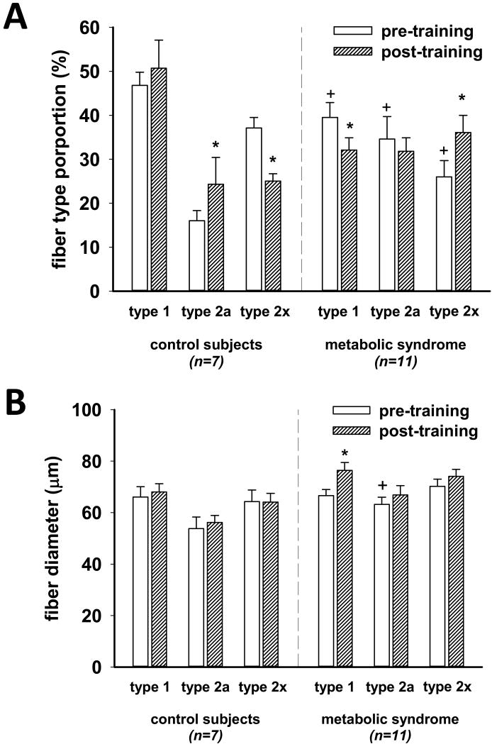
Muscle fiber composition was different in baseline assessments and changes induced by stationary cycle training was in opposite directions for the controls and metabolic syndrome subject groups as shown in Figure 2A. The metabolic syndrome subjects had lower type 1, higher type 2a, and lower 2x fiber proportions in vastus lateralis pre-training. After eight weeks of training, the control subject group muscle had a decrease in 2x fibers and an increase in 2a fiber content suggesting the training caused a shift away from purely strength, fast-twitch fibers. In contrast, metabolic syndrome trained muscle appeared to have shifted toward strength-power fibers, because there were lower type 1 and higher type 2x fiber proportions.
Figure 2. Change in muscle fiber composition and in fiber size after eight weeks of cycle training.
Panel A show the fiber composition of pre-training and post-training biopsies of vastus lateralis muscle in sedentary controls and in subjects with the metabolic syndrome. Shown here are the mean and SEM of the percent of each of the three principal fiber types as quantified by the slow-twitch/fast-twitch myosin antibody technique of Behan, et al (4). The asterix indicates significantly different post-training (p<0.05, paired t test), and the plus sign indicates a significant pre-training difference from the corresponding fiber type proportion in the control group (p<0.05, independent t test). Panel B displays the fiber diameter data derived as described in Methods above. Fresh biopsy material was immediately mounted on cork for transverse sectioning and frozen in a slurry of isopentane cooled in liquid nitrogen. The asterix indicates significant difference from baseline (p<0.05, paired t test). In the metabolic syndrome group, type 1 fiber type diameter increased by 15% after training.
Fiber size, shown in Figure 2B as fiber diameter, was not different between controls and metabolic syndrome subjects type 1 and 2x fibers in the baseline biopsies. Size did not change after training in the control subjects, but increased significantly in type 1 fibers in the metabolic syndrome subjects.
Eight weeks of cycle training of metabolic syndrome subjects increased insulin receptor and GLUT4 expression in vastus lateralis muscle
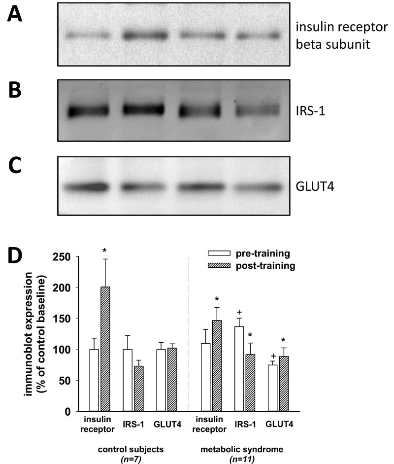
In order to evaluate the mechanisms underlying potential improvement in whole body insulin responsiveness in exercise trained subjects, expression of three key elements of the insulin pathway in muscle were quantified. Figure 3 displays changes in expression of insulin receptors, insulin receptor substrate-1 (IRS-1), and GLUT4 after supervised stationary cycle training for eight weeks. Both groups exhibited increases in insulin receptor expression after training. GLUT4 was increased in metabolic syndrome muscle. IRS-1 expression was higher in baseline metabolic syndrome muscle and significantly decreased after training.
Figure 3. The impact of exercise training on muscle expression of the insulin receptor, IRS-1, and GLUT4.
Panels A, B, and C display images of examples of immunoblots of the insulin receptor beta subunit, IRS-1, and GLUT4, respectively, expressed in muscle from biopsies of our subjects. Each lane of polyacrylamide gels contained a sample with 10 μg protein from muscle homogenate. Each sample was run in four separate experiments. The mean expression for each subject's sample quantified on an arbitrary scale relative to a reference muscle sample was then averaged with the mean expression of the other subjects in the group. These data were then expressed in the graph of Panel D in proportion to the control baseline data for each of the three factors that were quantified. The asterix indicates significantly different from baseline (p<0.05, paired t test) and the plus sign indicates baseline data that is different from the control subjects (p<0.05, independent t test).
Exercise training increased expression of activated AMPK and mitochondria in muscle fibers
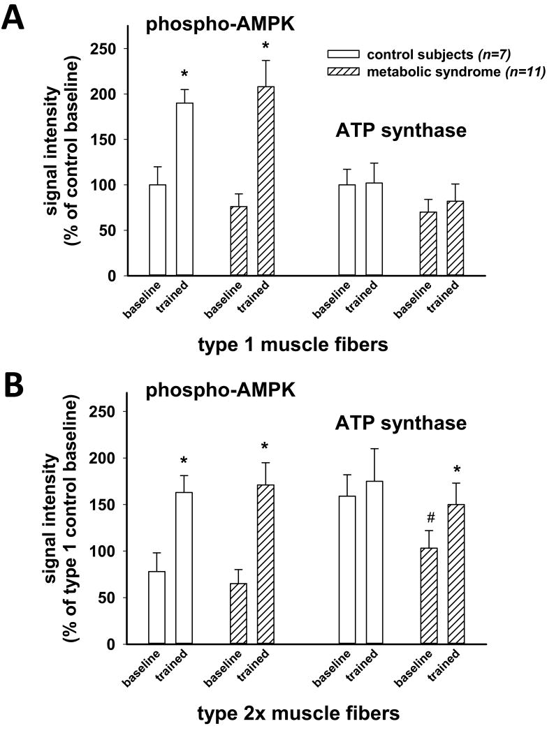
Post-exercise training improvement in maximal oxygen uptake seen in our subjects was likely due in part to increased mitochondria in muscle. Activation of AMPK is a necessary step in exercise-related activation of mitochondrial biogenesis and quantification of the expression of mitochondrial enzyme ATP synthase will reflect substantial changes in mitochondrial production. Eight weeks of stationary cycle training more than doubled the expression of phosphorylated AMPK in both type 1 and type 2x muscle fibers in both controls and metabolic syndrome subjects as shown in Figure 4. These data are from immunohistochemical studies as previously described for GLUT4 and activated mTOR (31). Unexpectedly, ATP synthase expression was significantly greater in metabolic syndrome 2x fibers than in type 1 fibers (p=0.039, paired t). This relative trend also appeared in the sedentary control muscle, but the difference did not achieve statistical significance. ATP synthase expression increased in metabolic syndrome type 2x muscle fibers by immunohistochemisty (Figure 4) and trended toward an increase in type 1 in metabolic syndrome and in type 1 and type 2 fibers in controls.
Figure 4. Fiber-specific changes in expression of activated AMPK and mitochondrial marker ATP synthase.
Since it was anticipated that exercise that was primarily endurance training would impact mitochondrial biogenesis, muscle AMPK activation and ATP synthase expression were quantified. Sections of muscle from each subject pre- and post-training were incubated with a monoclonal antibody against slow-twitch myosin heavy chain to identify the type 1 fibers on the section. A second rabbit polyclonal antibody against either phospho-AMPK or ATP synthase was added and incubated overnight at 4° C. Fluorescent tagged anti-rabbit IgG and anti-mouse IgG were added after the primary incubation and dual color images were obtained using a Leica confocal microscope. The signal intensity generated with either the phospho-AMPK or the ATP synthase antibody for the identified fiber type was quantified using digital imaging software (Quantity One from BioRad). At least thirty fibers had the signal intensity quantified for each antigen. Panel A displays the data for the control and metabolic syndrome subjects before and after training in the type 1 fibers. The expression of phospho-AMPK tended to be lower in the type 2x fibers for both controls and metabolic syndrome subjects, but ATP synthase expression appeared higher in 2x fibers. The activated AMPK was 2-fold increased after training for both groups in both fiber types. The ATP synthase was significantly increased by training in type 2x only in the metabolic syndrome group, indicating training-induced increased mitochondrial production, albeit not nearly to the level of increased activated AMPK. An asterix indicates significant post-training increase in expression compared to corresponding baseline (p<0.05, paired t test). The hatch mark designates a significant difference between type 2x fiber and type 1 fiber expression (p<0.05, paired t).
Immunoblots of muscle homogenates (data not shown) demonstrated a 16% lower pre-training expression of ATP synthase in metabolic syndrome subjects' muscle (p=0.040). There was a 9% increase post-training in controls (p=0.038) and a 12% increase post-training in metabolic syndrome subjects (p=0.055).
Muscle IRS-1 phosphorylation before and after exercise training
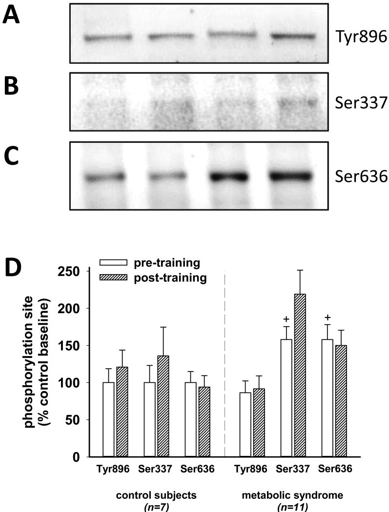
Phosphorylation of IRS-1 at tyrosine 896 is critical in the signal pathway activated by insulin interaction with its cell surface receptor. Previous studies have shown excess phosphorylation of IRS-1 at one of a few key serines can inhibit the ability of the insulin receptor tyrosine kinase to phosphorylate the tyrosine at 896. We hypothesized that beneficial exercise training might decrease the inhibitory serine phosphorylation and enhance the phosphorylation at Tyr896. Figure 5 summarizes the impact of cycle training for eight weeks on the phosphorylation of IRS-1 at Tyr896, Ser337, and Ser636. All muscle biopsies in these studies were performed after an overnight fast and thus circulating insulin concentrations were minimized in both groups (see Table 1). In spite of a higher expression of total IRS-1 (Figure 3), phosphorylation of IRS-1 at tyrosine 896 was not increased in metabolic syndrome muscle, either pre-training or post-training. In contrast, phosphorylation at serines 337 and 636 were increased compared to control subject phosphorylation at these sites. Training did not change the phosphorylation level at Ser636, but tended to increase the phosphorylation at Ser337, although this training-related difference was not significant in either group.
Figure 5. Cycle training impact on tyrosine and serine phosphorylation of muscle IRS-1.
Panels A, B, and C show typical immunoblot images of muscle homogenates probed with antibodies specific for phospho-Tyr896, phospho-Ser337, and phospho-Ser636, respectively. Immunoblot images like these were evaluated for signal strength using digital analysis software for each subject in four separate experiments. Panel D summarizes the results of these analyses. In the pre-training samples, Tyr896 phosphorylation was not different, but phosphorylation at Ser337 and Ser636 were about 50% higher in the metabolic syndrome muscle samples. The exercise training protocol did not decrease the amount of phosphorylation at Ser337 or Ser636, and there appears to be a trend toward increased phosphorylation at Ser337 in both controls and metabolic syndrome muscle. The plus sign on the graph indicates a difference from the corresponding control subject data (p<0.05).
Discussion
The study described in this report was designed to employ stationary cycle training as a predominantly endurance exercise training intervention to decrease insulin resistance in participants who were at high risk for type 2 diabetes because they met the criteria for the metabolic syndrome. This protocol, like its predecessor using only strength training (22), successfully adjusted caloric intake to prevent weight loss. Like the previous strength training of eight weeks duration, less than half of the subjects improved their insulin responsiveness, measured by euglycemic insulin clamps, but the others either showed no change or decreased their insulin responsiveness. These mixed individual results caused the group data to be unchanged. Thus, with this duration and intensity of closely supervised exercise training, either endurance or strength, no improvement in insulin resistance can be anticipated in such interventions in the absence of weight loss.
Several studies have shown that combined endurance and resistance training provided the best response in glycemic control and improved insulin sensitivity. Many of these studies did not keep the time and effort constant between the comparison groups, making it hard to be confident that it was the type of exercise rather than just total time spent or energy expended that was the critical variable. Sigal and coworkers evaluated the impact of exercise training (aerobic, resistance, or combined) on HbA1c in 251 adults with type 2 diabetes (28). Resistance training was approximately 120 minutes per week, aerobic training was 135 minutes per week, and combined training totaled about 250 minutes per week. All three training groups improved their HbA1c, but combined training did the best with a change of -0.97%, aerobic training improved by -0.51%, and the resistance training group improved by -0.38%. The starting weights averaged 101.5 kg, with resistance training losing an average of 1.1 kg, and the aerobic and the combined groups averaged a loss of 2.6 kg. Since time of exercise also correlated with change in HbA1c, the type of exercise alone cannot be concluded from their data to have an advantage of one over the other. Church and coworkers performed exercise training for nine months on 262 men and women with type 2 diabetes at the Pennington Biomedical Research Center in Baton Rouge (7). Volunteers were randomly assigned to no exercise, resistance exercise, aerobic exercise, or combined aerobic and resistance. All three exercise training groups lost weight, body fat, and waist circumference. All three exercise training groups decreased their HbA1c (-0.16%, -0.24%, and -0.34%, respectively), but only the combined exercise group achieved statistical significance. VO2max increased significantly only in the combined exercise group. Strength increased in the resistance and the combined groups, but not in the aerobic training only group. Cuff and coworkers at the University of British Columbia evaluated the impact of adding resistance training to aerobic training in older women with type 2 diabetes (9). Ten subjects with combined resistance and aerobic training for 16 weeks were compared to nine women with aerobic training only and nine women who did not train. Euglycemic clamp testing showed more improvement in the combined training group than in the aerobic only group (+1.82 vs +0.55 mg/kg.min). Both groups were demonstrated by CT to have less visceral fat coincident with weight loss (-2.9 and -1.2, compared to +2.0 kg in the untrained control group).
Davidson et al measured exercise training-related improvement in insulin responsiveness with euglycemic clamps in 136 sedentary older subjects who did not have diabetes (10). The glucose infusion rate data showed that the largest enhancement of insulin responsiveness occurred in the combined training group. Resistance training alone was not significantly different from non-trained controls, but substituting resistance training for a portion of the aerobic training time resulted in substantially higher enhancement of insulin responsiveness than the aerobic training that it replaced (10). These investigators concluded that the combined training was the optimal protocol.
Comparison of aerobic and resistance exercise training for subjects with the metabolic syndrome were carried out in the STRRIDE-AT/RT study (3, 29). There were 196 participants aged 18-70 participating. Resistance training was 45-60 minutes three days per week, aerobic training was about 120 minutes per week, equivalent to running 12 miles. The combined program added the two programs together, requiring twice the time and energy expenditure. An MS score was calculated from measures of HDL, triglycerides, fasting glucose concentration, waist circumference, and blood pressure. The scores improved with aerobic and combined training but did not improve with resistance training alone (3). VO2max increased in all three groups (3), but weight loss, decreased visceral fat, and improved insulin responsiveness by HOMA occurred only in the aerobic only and the aerobic plus resistance groups (29).
Stensvold, et al., also compared exercise modalities in treatment of men and women with the metabolic syndrome (30). They had eleven volunteers in each group where they trained for 12 weeks, three times per week at either aerobic, strength, combined exercise, or no intervention. With no weight loss, all three exercise groups had a decrease in waist circumference. Strength increased in the strength trained and the combined groups. VO2max increased in the aerobic and the combined groups, but not in the strength group. Insulin resistance, quantified by HOMA, did not change significantly (30).
Villareal and coworkers at Washington University evaluated diet-induced weight loss, exercise alone, and exercise with weight loss in older obese subjects in a program to reduce frailty (5, 35). The duration of the study was one year with the exercise being combined aerobic and strength training. Insulin sensitivity was quantified using an insulin sensitivity index calculated from insulin and glucose measurements from an oral glucose tolerance test. Weight loss and exercise showed the largest improvement in physical function in these subjects all of whom had some impairment at baseline (35). Weight loss occurred in the diet and diet plus exercise groups. Insulin sensitivity index improved 70% in the diet group and 86% in the diet plus exercise group, but did not change in the control or exercise only groups (5).
A randomized, controlled study of weight loss by diet or by aerobic exercise only was reported by Ross and coworkers from Kingston, Ontario (27). Fifty-two obese men were randomized to diet only, exercise without weight loss, exercise-induce weight loss, and no intervention controls for a duration of twelve weeks. Weight loss of 7 kg (∼8% of body weight) in both weight loss groups resulted in improved euglycemic clamp-quantified insulin responsiveness of 43% in diet only and 64% in weight loss by exercise (27). Aerobic exercise with no weight loss improved insulin responsiveness by 31%, but this change did not achieve statistical significance. MRI-quantified visceral fat improved significantly in the diet only, exercise-induced weight loss, and exercise without weight loss groups (-28%, -28%, -12%, respectively) (27).
The exercise intervention studies reviewed above are consistent with our finding that the impact on insulin resistance of exercise training without weight loss is minimal. Comparisons of types of exercise effectiveness suggest that with weight loss, a combination of aerobic and resistance training is usually associated with the most improvement in glycemic control in type 2 diabetes subjects or in insulin responsiveness in non-diabetic subjects with the metabolic syndrome. Typically, however, the time spent exercising is least with resistance and most with combined training, making time and/or volume of exercise an important variable. This often makes it difficult to determine whether it is the type of exercise or the time spent that is the factor determining the effectiveness of an exercise program.
Muscle fiber composition in metabolic syndrome subjects was different from sedentary controls with decreased slow-twitch (type 1) fibers and increased fast-twitch (type 2) fibers, particularly the mixed fibers (type 2a). Does this predominance of strength (type 2) fibers make metabolic syndrome subjects better suited to strength training than endurance training? The results from this study and our previous study (22), suggest that there was no advantage to strength training, in so far as improvement in insulin responsiveness is concerned. Studies from other investigators that allowed weight loss generally ranked combination training better than endurance and strength training the least effective at improving insulin responsiveness or diabetes glycemic control. Whether these comparisons of exercise protocols actually reflect the type of exercise or only the sum of time and effort is not clear, since workloads were generally not matched in the different protocols that were compared.
The data included in this report and our previous evaluation of strength training without weight loss (22) suggest that eight weeks of increasing intensity exercise training alone is largely futile at decreasing the risk for diabetes (decreasing insulin resistance). The current eight week cycle training intervention was designed to approximate the volume load and energy expenditure of the eight week resistance training protocol (22). Both cycle training and strength training can increase aerobic fitness (VO2max) and there were trends toward increasing fat oxidation and lowering blood pressure (Table 1). Strength training increased muscle mass and strength. If weight loss is the critical factor in improving insulin responsiveness, then regular exercise will be additive or even synergistic with a decrease in dietary calories, if a negative energy balance persists. The impact of regular exercise on weight loss, if not compensated during the remainder of the day, will at least be increased energy expenditure and may have other beneficial effects such as decreasing the leptin resistance that is nearly always present in human obesity (8). Unfortunately, exercise training alone at the volume we selected is largely futile in the metabolic syndrome.
Stationary cycle training in metabolic syndrome and controls increased muscle insulin receptor and activated AMPK substantially, but GLUT4 and mitochondria were only modestly affected by the training intervention. The question arises whether the duration of the training period was inadequate to overcome the individual subject's previous sedentary lifestyle or is weight loss necessary in order for exercise training to exert its full effect on the insulin-stimulated glucose uptake pathway in muscle. Further, is weight loss necessary to overcome obesity-related inhibition of exercise training modulation of muscle mitochondrial biogenesis or muscle fiber hypertrophy? Elite runners are substantially more insulin responsive than healthy control subjects (14). Long term training is likely part of this, but genetically-determined advantages may underlie the higher insulin sensitivity seen in advanced and elite athletes.
Eight weeks of moderate stationary cycle training without weight loss is not effective at decreasing the insulin resistance (a surrogate for diabetes risk) characteristic of the metabolic syndrome. Some key components of muscle remodeling increased robustly (insulin receptors, phospho-AMPK), but others did not, suggesting impairment downstream in the pathways normally involved in muscle adaptation.
Acknowledgments
The authors wish to thank the subjects who volunteered for these studies and the students who coached them through the training, without whose participation and motivation, these data would not be available. Research nurse Susie Cooper Whitaker was very important in recruitment, retention, and coordination of these studies.
Grants These studies were funded by a grant from the National Institutes of Health (DK080488) to Stuart, Stone, and Ramsey.
Funding: NIH DK 080488 (Stuart)
Footnotes
Disclosures. The authors have no conflicts of interest to disclose. The results of the present study do not constitute endorsement by the American College of Sports Medicine.
Disclosures: The authors have no disclosures
References
- 1.Alberti KG, Zimmet P, Shaw J. The metabolic syndrome--a new worldwide definition. Lancet. 2005;366:1059–1062. doi: 10.1016/S0140-6736(05)67402-8. [DOI] [PubMed] [Google Scholar]
- 2.Baar K. Training for endurance and strength: lessons from cell signaling. Med Sci Sports Exerc. 2006;38:1939–1944. doi: 10.1249/01.mss.0000233799.62153.19. [DOI] [PubMed] [Google Scholar]
- 3.Bateman LA, Slentz CA, Willis LH, Shields AT, Piner LW, Bales CW, Houmard JA, Kraus WE. Comparison of aerobic versus resistance exercise training effects on metabolic syndrome (from the Studies of a Targeted Risk Reduction Intervention Through Defined Exercise - STRRIDE-AT/RT) Am J Cardiol. 2011;108:838–844. doi: 10.1016/j.amjcard.2011.04.037. [DOI] [PMC free article] [PubMed] [Google Scholar]
- 4.Behan WM, Cossar DW, Madden HA, McKay IC. Validation of a simple, rapid, and economical technique for distinguishing type 1 and 2 fibres in fixed and frozen skeletal muscle. J Clin Pathol. 2002;55:375–380. doi: 10.1136/jcp.55.5.375. [DOI] [PMC free article] [PubMed] [Google Scholar]
- 5.Bouchonville MF, Shah K, Armamento-Villareal R, Sinacore DR, Qualls C, Villareal DT. Weight loss, excercise, or both and cardiometabolic risk factors in obese older adults: Results of a randomized controlled trial. 2012:S18–1. doi: 10.1038/ijo.2013.122. [DOI] [PMC free article] [PubMed] [Google Scholar]
- 6.Cauza E, Hanusch-Enserer U, Strasser B, Ludvik B, Metz-Schimmerl S, Pacini G, Wagner O, Georg P, Prager R, Kostner K, Dunky A, Haber P. The relative benefits of endurance and strength training on the metabolic factors and muscle function of people with type 2 diabetes mellitus. Arch Phys Med Rehabil. 2005;86:1527–1533. doi: 10.1016/j.apmr.2005.01.007. [DOI] [PubMed] [Google Scholar]
- 7.Church TS, Blair SN, Cocreham S, Johannsen N, Johnson W, Kramer K, Mikus CR, Myers V, Nauta M, Rodarte RQ, Sparks L, Thompson A, Earnest CP. Effects of aerobic and resistance training on hemoglobin A1c levels in patients with type 2 diabetes: a randomized controlled trial. JAMA. 2010;304:2253–2262. doi: 10.1001/jama.2010.1710. [DOI] [PMC free article] [PubMed] [Google Scholar]
- 8.Considine RV, Sinha MK, Heiman ML, Kriauciunas A, Stephens TW, Nyce MR, Ohannesian JP, Marco CC, McKee LJ, Bauer TL, Caro JF. Serum immunoreative-leptin concentrations in normal-weight and obese humans. N Engl J Med. 1996;334:292–295. doi: 10.1056/NEJM199602013340503. [DOI] [PubMed] [Google Scholar]
- 9.Cuff DJ, Meneilly GS, Martin A, Ignaszewski A, Tildesley HD, Frohlich JJ. Effective exercise modality to reduce insulin resistance in women with type 2 diabetes. Diabetes Care. 2003;26:2977–2982. doi: 10.2337/diacare.26.11.2977. [DOI] [PubMed] [Google Scholar]
- 10.Davidson LE, Hudson R, Kilpatrick K, Kuk JL, McMillan K, Janiszewski PM, Lee S, Lam M, Ross R. Effects of exercise modality on insulin resistance and functional limitation in older adults: a randomized controlled trial. Arch Intern Med. 2009;169:122–131. doi: 10.1001/archinternmed.2008.558. [DOI] [PubMed] [Google Scholar]
- 11.Dubowitz V, Sewry CA, Lane R. Normal muscle. In: Houston MJ, Cook L, editors. Muscle Biopsy: a Practical Approach. Philadelphia: Saunders Elsevier; 2007. pp. 41–74. [Google Scholar]
- 12.Dunstan DW, Daly RM, Owen N, Jolley D, de Court, Shaw J, Zimmet P. High-intensity resistance training improves glycemic control in older patients with type 2 diabetes. Diabetes Care. 2002;25:1729–1736. doi: 10.2337/diacare.25.10.1729. [DOI] [PubMed] [Google Scholar]
- 13.Dunstan DW, Puddey IB, Beilin LJ, Burke V, Morton AR, Stanton KG. Effects of a short-term circuit weight training program on glycaemic control in NIDDM. Diabetes Res Clin Pract. 1998;40:53–61. doi: 10.1016/s0168-8227(98)00027-8. [DOI] [PubMed] [Google Scholar]
- 14.Ebeling P, Bourey R, Koranyi L, Tuominen JA, Groop LC, Henriksson J, Mueckler M, Sovijarvi A, Koivisto VA. Mechanism of enhanced insulin sensitivity in athletes. Increased blood flow, muscle glucose transport protein (GLUT-4) concentration, and glycogen synthase activity. J Clin Invest. 1993;92:1623–1631. doi: 10.1172/JCI116747. [DOI] [PMC free article] [PubMed] [Google Scholar]
- 15.Eriksson JG. Exercise and the treatment of type 2 diabetes mellitus. An update. Sports Med. 1999;27:381–391. doi: 10.2165/00007256-199927060-00003. [DOI] [PubMed] [Google Scholar]
- 16.Ferrannini E, Natali A, Bell P, Cavallo-Perin P, Lalic N, Mingrone G. Insulin resistance and hypersecretion in obesity. European Group for the Study of Insulin Resistance (EGIR) J Clin Invest. 1997;100:1166–1173. doi: 10.1172/JCI119628. [DOI] [PMC free article] [PubMed] [Google Scholar]
- 17.Frayn KN. Calculation of substrate oxidation rates in vivo from gaseous exchange. J Appl Physiol. 1983;55:628–634. doi: 10.1152/jappl.1983.55.2.628. [DOI] [PubMed] [Google Scholar]
- 18.Grontved A, Rimm EB, Willett WC, Andersen LB, Hu FB. A Prospective Study of Weight Training and Risk of Type 2 Diabetes Mellitus in Men. Arch Intern Med. 2012:1–7. doi: 10.1001/archinternmed.2012.3138. [DOI] [PMC free article] [PubMed] [Google Scholar]
- 19.Grundy SM, Brewer HB, Jr, Cleeman JI, Smith SC, Jr, Lenfant C. Definition of metabolic syndrome: Report of the National Heart, Lung, and Blood Institute/American Heart Association conference on scientific issues related to definition. Circulation. 2004;109:433–438. doi: 10.1161/01.CIR.0000111245.75752.C6. [DOI] [PubMed] [Google Scholar]
- 20.Kahn R, Buse J, Ferrannini E, Stern M. The metabolic syndrome: time for a critical appraisal: joint statement from the American Diabetes Association and the European Association for the Study of Diabetes. Diabetes Care. 2005;28:2289–2304. doi: 10.2337/diacare.28.9.2289. [DOI] [PubMed] [Google Scholar]
- 21.Knowler WC, Barrett-Connor E, Fowler SE, Hamman RF, Lachin JM, Walker EA, Nathan DM. Reduction in the incidence of type 2 diabetes with lifestyle intervention or metformin. N Engl J Med. 2002;346:393–403. doi: 10.1056/NEJMoa012512. [DOI] [PMC free article] [PubMed] [Google Scholar]
- 22.Layne AS, Nasrallah S, South MA, Howell ME, McCurry MP, Ramsey MW, Stone MH, Stuart CA. Impaired Muscle AMPK Activation in the Metabolic Syndrome May Attenuate Improved Insulin Action after Exercise Training. J Clin Endocrinol Metab. 2011;96:1815–1826. doi: 10.1210/jc.2010-2532. [DOI] [PMC free article] [PubMed] [Google Scholar]
- 23.Mather KJ, Hunt AE, Steinberg HO, Paradisi G, Hook G, Katz A, Quon MJ, Baron AD. Repeatability characteristics of simple indices of insulin resistance: implications for research applications. J Clin Endocrinol Metab. 2001;86:5457–5464. doi: 10.1210/jcem.86.11.7880. [DOI] [PubMed] [Google Scholar]
- 24.Mokdad AH, Bowman BA, Ford ES, Vinicor F, Marks JS, Koplan JP. The continuing epidemics of obesity and diabetes in the United States. JAMA. 2001;286:1195–1200. doi: 10.1001/jama.286.10.1195. [DOI] [PubMed] [Google Scholar]
- 25.Mokdad AH, Serdula MK, Dietz WH, Bowman BA, Marks JS, Koplan JP. The spread of the obesity epidemic in the United States, 1991-1998. JAMA. 1999;282:1519–1522. doi: 10.1001/jama.282.16.1519. [DOI] [PubMed] [Google Scholar]
- 26.Reaven GM. Role of insulin resistance in human disease. Diabetes. 1988;37:1595–1607. doi: 10.2337/diab.37.12.1595. [DOI] [PubMed] [Google Scholar]
- 27.Ross R, Dagnone D, Jones PJ, Smith H, Paddags A, Hudson R, Janssen I. Reduction in obesity and related comorbid conditions after diet-induced weight loss or exercise-induced weight loss in men. A randomized, controlled trial. Ann Intern Med. 2000;133:92–103. doi: 10.7326/0003-4819-133-2-200007180-00008. [DOI] [PubMed] [Google Scholar]
- 28.Sigal RJ, Kenny GP, Boule NG, Wells GA, Prud'homme D, Fortier M, Reid RD, Tulloch H, Coyle D, Phillips P, Jennings A, Jaffey J. Effects of aerobic training, resistance training, or both on glycemic control in type 2 diabetes: a randomized trial. Ann Intern Med. 2007;147:357–369. doi: 10.7326/0003-4819-147-6-200709180-00005. [DOI] [PubMed] [Google Scholar]
- 29.Slentz CA, Bateman LA, Willis LH, Shields AT, Tanner CJ, Piner LW, Hawk VH, Muehlbauer MJ, Samsa GP, Nelson RC, Huffman KM, Bales CW, Houmard JA, Kraus WE. Effects of aerobic vs. resistance training on visceral and liver fat stores, liver enzymes, and insulin resistance by HOMA in overweight adults from STRRIDE AT/RT. Am J Physiol Endocrinol Metab. 2011;301:E1033–E1039. doi: 10.1152/ajpendo.00291.2011. [DOI] [PMC free article] [PubMed] [Google Scholar]
- 30.Stensvold D, Tjonna AE, Skaug EA, Aspenes S, Stolen T, Wisloff U, Slordahl SA. Strength training versus aerobic interval training to modify risk factors of metabolic syndrome. J Appl Physiol. 2010;108:804–810. doi: 10.1152/japplphysiol.00996.2009. [DOI] [PubMed] [Google Scholar]
- 31.Stuart CA, Howell ME, Baker JD, Dykes RJ, Duffourc MM, Ramsey MW, Stone MH. Cycle training increased GLUT4 and activation of mammalian target of rapamycin in fast twitch muscle fibers. Med Sci Sports Exerc. 2010;42:96–106. doi: 10.1249/MSS.0b013e3181ad7f36. [DOI] [PMC free article] [PubMed] [Google Scholar]
- 32.Stuart CA, Howell ME, Zhang Y, Yin D. Insulin-stimulated translocation of glucose transporter (GLUT) 12 parallels that of GLUT4 in normal muscle. J Clin Endocrinol Metab. 2009;94:3535–3542. doi: 10.1210/jc.2009-0162. [DOI] [PMC free article] [PubMed] [Google Scholar]
- 33.Stuart CA, Yin D, Howell MEA, Dykes RJ, Laffan JJ, Ferrando AA. Hexose transporter mRNAs for GLUT4, GLUT5, and GLUT12 predominate in human muscle. Am J Physiol Endocrinol Metab. 2006;291:E1067–E1073. doi: 10.1152/ajpendo.00250.2006. [DOI] [PubMed] [Google Scholar]
- 34.Tresierras MA, Balady GJ. Resistance Training in the Treatment of Diabetes and Obesity: MECHANISMS AND OUTCOMES. J Cardiopulm Rehabil Prev. 2009;29:67–75. doi: 10.1097/HCR.0b013e318199ff69. [DOI] [PubMed] [Google Scholar]
- 35.Villareal DT, Chode S, Parimi N, Sinacore DR, Hilton T, Armamento-Villareal R, Napoli N, Qualls C, Shah K. Weight loss, exercise, or both and physical function in obese older adults. N Engl J Med. 2011;364:1218–1229. doi: 10.1056/NEJMoa1008234. [DOI] [PMC free article] [PubMed] [Google Scholar]
- 36.Wilkinson SB, Phillips SM, Atherton PJ, Patel R, Yarasheski KE, Tarnopolsky MA, Rennie MJ. Differential effects of resistance and endurance exercise in the fed state on signalling molecule phosphorylation and protein synthesis in human muscle. J Physiol. 2008;586:3701–3717. doi: 10.1113/jphysiol.2008.153916. [DOI] [PMC free article] [PubMed] [Google Scholar]
- 37.Zimmet P, Alberti KG, Shaw J. Global and societal implications of the diabetes epidemic. Nature. 2001;414:782–787. doi: 10.1038/414782a. [DOI] [PubMed] [Google Scholar]


