Abstract
17β-estradiol (E2 or estrogen) is an endogenous steroid hormone that is well known to exert neuroprotection. Along these lines, one mechanism through which E2 protects the hippocampus from cerebral ischemia is by preventing the post-ischemic elevation of Dkk1, a neurodegenerative factor that serves as an antagonist of the canonical Wnt signaling pathway, and simultaneously inducing pro-survival Wnt/β-Catenin signaling in hippocampal neurons. Intriguingly, while expression of Dkk1 is required for proper neural development, overexpression of Dkk1 is characteristic of many neurodegenerative diseases, such as stroke, Alzheimer’s disease, Parkinson’s disease, and temporal lobe epilepsy. In this review, we will briefly summarize the canonical Wnt signaling pathway, highlight the current literature linking alterations of Dkk1 and Wnt/β-Catenin signaling with neurological disease, and discuss E2’s role in maintaining the delicate balance of Dkk1 and Wnt/β-Catenin signaling in the adult brain. Finally, we will consider the implications of long-term E2 deprivation and hormone therapy on this crucial neural pathway.
Keywords: Brain, Dkk1, Estrogen, Neurodegenerative Disease, Wnt Signaling
1. Introduction – Estrogen as a Neuroprotective Agent
17β-estradiol (E2 or estrogen) is an endogenous steroid hormone produced in the ovaries and in the brain. In addition to its well-known roles in reproduction, bone homeostasis, and metabolic functions, E2 also serves as a neuroprotective agent. In support of this, premenopausal women, who have high circulating E2 levels, are relatively protected from neurodegenerative diseases, such as stroke, when compared to men (Brann et al., 2007; Murphy et al., 2004; Niewada et al., 2005; Roquer et al., 2003; Scott et al., 2012). In contrast, this pattern is reversed for postmenopausal women, who demonstrate dramatically reduced circulating E2 levels and actually have worse morbidity and mortality following a stroke than age-matched men (Appelros et al., 2009; Niewada et al., 2005; Persky et al., 2010; Roquer et al., 2003). Importantly, animal models of stroke corroborate the phenomenon of E2 neuroprotection observed in humans. In fact, male rodents demonstrate larger cortical infarcts than their female counterparts after cerebral ischemia (Alkayed et al., 1998; Roof and Hall, 2000), and both estrogen receptor antagonists and aromatase inhibitors, which block endogenous production of E2, enhance stroke damage in rodent models of cerebral ischemia (McCullough et al., 2003; Sawada et al., 2000). Intriguingly, since exogenous estrogens also afford neuroprotection in the cerebral cortex and hippocampus in animal models of cerebral ischemia (Brann et al., 2007; Gibson et al., 2006; Scott et al., 2012; Simpkins et al., 2012a; Simpkins et al., 2012b), E2 has the potential to serve as an acute treatment for stroke and/or as a preventative therapeutic to preserve optimal neuronal functioning in postmenopausal women. Along these lines, it is no surprise that the mechanisms of E2 neuroprotection and the feasibility of postmenopausal hormone therapy are areas of intense study. One mechanism proposed to contribute to E2 neuroprotection is E2’s attenuation of ischemic elevation of the neurodegenerative Wnt antagonist dickkopf-1 (Dkk1) and simultaneous activation of the pro-survival Wnt/β-Catenin pathway (Zhang et al., 2008). Thus, below we review canonical Wnt/β-catenin signaling and its regulation by the Wnt antagonist Dkk1, as well as the role of Dkk1 in neurodegenerative disorders and its regulation by estrogen.
2. Canonical Wnt/β-Catenin Signaling
Wnt is a secreted glycoprotein whose gene was separately discovered in mouse mammary tumors (int-1) and in Drosophila melanogaster (wingless), but due to sequence homology, both int-1 and wingless were determined to encode the same proto-oncogene, which was dubbed Wnt (McMahon and Moon, 1989a; McMahon and Moon, 1989b; Thomas and Capecchi, 1990). The canonical Wnt signaling pathway has since been deemed critical for several embryonic events, including cell proliferation, cell polarity, and determination of cell fate (Logan and Nusse, 2004; MacDonald et al., 2009). Canonical Wnt/β-Catenin signaling has also been implicated in development of the limbs (Grotewold and Ruther, 2002; Mukhopadhyay et al., 2001), neural tube (De Marco et al., 2012; Roelink and Nusse, 1991), forebrain (Mukhopadhyay et al., 2001), midbrain and cerebellum (McMahon and Bradley, 1990; Thomas and Capecchi, 1990) and in the maintenance of neurotransmission and synaptic plasticity (Ataman et al., 2008; Avila et al., 2010; Budnik and Salinas, 2011; Jensen et al., 2012; Speese and Budnik, 2007). In light of this knowledge, it is not surprising that, in addition to tumorigenesis, mutations that enhance Wnt signaling in humans have been linked to neurological disorders, such as autism, schizophrenia, and bipolar disorder (De Ferrari and Moon, 2006).
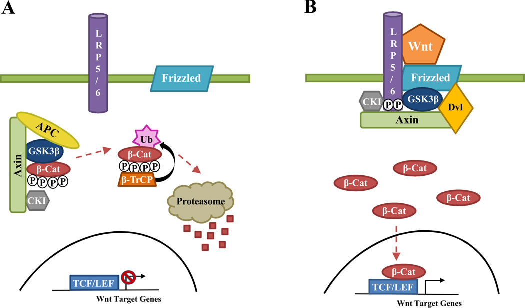
Wnt initiates an intracellular signaling cascade when it binds to its cognate membrane receptor, frizzled (Fz), and its co-receptor, low density lipoprotein-related protein 5/6 (LRP5/6) (Logan and Nusse, 2004; MacDonald et al., 2009) (See Figure 1 for Summary). The canonical Wnt signaling cascade, which ultimately determines intracellular levels of the transcriptional activator β-Catenin, begins with recruitment of disheveled (Dvl) to Fz and dual phosphorylation of LRP5/6 by glycogen synthase kinase 3β (GSK3β) and casein kinase I (CKI) (MacDonald et al., 2009). It is important to note that Wnt is also capable of signaling through non-canonical signaling pathways, which are independent of β-Catenin, but these are beyond the scope of this review. As such, the reader is referred to these excellent reviews on the subject (Clark et al., 2012; Komiya and Habas, 2008; Seifert and Mlodzik, 2007; Wang and Nathans, 2007). Intriguingly, GSK3β and CKI are both components of the Axin-adenomatous polyposis coli (APC) complex, which is devoted to the phosphorylation of the transcriptional activator β-Catenin (Logan and Nusse, 2004; MacDonald et al., 2009). As such, in the absence of Wnt ligand, the Axin-APC complex continually phosphorylates cytosolic β-Catenin, priming it for ubiquitination by the E3 ubiquitin ligase Beta-Transducing repeat-Containing Protein (β-TrCP) and subsequent proteasomal degradation in order to prevent expression of Wnt target genes. However, once Wnt signaling is initiated, GSK3β and CKI are recruited away from the Axin-APC complex to phosphorylate the co-receptor LRP5/6, and once doubly phosphorylated, LRP5/6 becomes a docking site for Axin (MacDonald et al., 2009). These events lead to the temporary disassembly of the Axin-APC complex and the subsequent stabilization of cytosolic β-Catenin, which remains untagged by either phosphate or ubiquitin (Logan and Nusse, 2004; MacDonald et al., 2009). Thus, the canonical Wnt signaling cascade ultimately ends in the elevation, stabilization, and nuclear translocation of cytosolic β-Catenin. Once inside the nucleus, β-Catenin, as a transcriptional activator, is able to convert the T-cell Factor/Lymphoid Enhancing Factor (TCF/LEF) transcriptional repression machinery into an active, mRNA transcription complex, which then promotes the expression of Wnt target genes, characterized by the presence of a consensus sequence called a Wnt response element (WRE) in their promoter regions (Logan and Nusse, 2004; MacDonald et al., 2009).
Figure 1. Summary of the Canonical Wnt Signaling Pathway.
This figure briefly summarizes canonical Wnt signaling. A, In the absence of Wnt ligand, cytosolic β-Catenin is phosphorylated, ubiquitinated, and degraded by the proteasome to prevent the expression of Wnt target genes. B, However, in the presence of Wnt ligand, cytosolic β-Catenin is stabilized and translocates to the nucleus, where it acts as a co-activator and facilitates the transcription of Wnt target genes. Dkk1 antagonizes canonical Wnt signaling by binding to the Wnt co-receptor LRP5/6 and preventing formation of the Wnt-Frizzled-LRP5/6 complex. See text for further details. APC, adenomatous polyposis coli, β-Cat, β-Catenin; β-TrCP, Beta-Transducing repeat-Containing Protein; Dvl, Disheveled; CKI, Casein Kinase I; Dkk1, Dickkopf-1; GSK3β, Glycogen Synthase Kinase 3β; LRP5/6, Low Density Lipoprotein-Related Protein 5/6; TCF/LEF, T Cell Factor/Lymphoid Enhancing Factor; Ub, Ubiquitin; Wnt, Wingless.
3. Dkk1
Several endogenous ligands, such as Shisa, Wnt inhibitory factor-1 (WIF-1), the secreted Frizzled related proteins (sFRPs), the WISE/SOST family, and the Dickkopf (Dkk) family, can serve as antagonists of canonical Wnt signaling (MacDonald et al., 2009). Arguably, the most important Wnt signaling antagonist is the prototypical Dkk family member Dkk1, which antagonizes Wnt signaling by binding to the LRP5/6 co-receptor and preventing Wnt from forming a signaling complex with Fz and LRP5/6 (Bafico et al., 2001; Fedi et al., 1999; Mao et al., 2001; Semenov et al., 2001; Wu et al., 2000). Similar to Wnt, Dkk1 expression is critical for neurodevelopment during the embryonic period. In fact, Dkk1 is known as a “head-inducer,” as its presence and antagonism of Wnt signaling is required for structures anterior of the midbrain to form (Glinka et al., 1998; Kazanskaya et al., 2000; Mukhopadhyay et al., 2001; Semenov et al., 2001). Importantly, Dkk1 is also responsible for orchestrating the apoptosis necessary for proper limb development (Grotewold and Ruther, 2002; Mukhopadhyay et al., 2001). Along these lines, Dkk1 null mice are not viable, with embryos lacking structures anterior of the midbrain and demonstrating limb polysyndactyly (Mukhopadhyay et al., 2001). Furthermore, doubleridge mice, which have hypomorphorphic expression of Dkk1, are viable, but they display hemivertebral fusions and polysyndactyly of the forelimbs, a phenotype that can be ameliorated by reducing the expression of LRP5/6 (MacDonald et al., 2004).
3.1. Dkk1 and Neurodegenerative Disease
While transgenic mouse models have overwhelmingly demonstrated that Dkk1 expression is crucial during neurodevelopment, elevation of Dkk1 later in life can be detrimental. In fact, many studies have linked elevated Dkk1 expression in the adult brain to neurodegenerative diseases, such as stroke, Alzheimer’s disease, Parkinson’s disease, and temporal lobe epilepsy (See Table 1 for Summary). Excitotoxicity has relevance to neurodegenerative disease because stroke leads to neuronal cell death, in part, due to excess release of the excitatory neurotransmitter glutamate, which subsequently leads to NMDA receptor activation and intracellular calcium overload (Choi, 1994a; Choi, 1994b; Zipfel et al., 2000). In vitro studies demonstrated that Dkk1 is induced in cultured cortical neurons following an excitotoxic pulse of NMDA and is also capable of potentiating NMDA neurotoxicity in a dose-dependent manner (Cappuccio et al., 2005). Further work confirmed that Dkk1 is, in fact, able to inhibit canonical Wnt signaling and initiate cell death in cultured cortical neurons, which was associated with loss of Bcl-2, induction of Bax, and hyperphosphorylation of the microtubule associated protein tau (Scali et al., 2006). Importantly, the same studies confirmed that these observations are relevant to ischemic insults in vivo, as hippocampal Dkk1 was induced following global cerebral ischemia in both gerbils and rats, and stereotaxic injection of recombinant Dkk1 into either the hippocampal CA1 region or nucleus basalis magnocellularis was sufficient to cause neuronal cell death (Cappuccio et al., 2005; Scali et al., 2006). Intriguingly, intracerebroventricular injection of Dkk1 anti-sense oligonucleotides attenuated the ischemia-induced cell death observed in gerbils, and intraperitoneal administration of lithium chloride, which rescues canonical Wnt signaling by inhibiting GSK3β, also attenuated the ischemia-induced cell death observed in rats (Cappuccio et al., 2005).
Table 1. Summary of Evidence Associating Dkk1 with Neurodegenerative Disease.
This table briefly summarizes the major research findings to date linking the Wnt antagonist Dkk1 to four neurodegenerative diseases: stroke, temporal lobe epilepsy, Alzheimer’s disease, and Parkinson’s disease. See text for further details. Aβ, β-Amyloid; Dkk1, Dickkopf-1; GCI, Global Cerebral Ischemia; ET-1, Endothelin-1; MCAO, Middle Cerebral Artery Occlusion; MPP+, 1-methyl-4-phenylpyridinium;NMDA, N-Methyl-D-Aspartate; 6-OHDA, 6-Hydroxydopamine.
| References | Finding | Model System | Neurodegenerative Disease |
|---|---|---|---|
| (Cappuccio et al., 2005) | Dkk1 ↑ after NMDA administration and potentiates NMDA excitotoxicity | Cultured Cortical Neurons | Stroke |
| (Scali et al., 2006) | Dkk1 causes cell death and tau hyperphosphorylation | Cultured Cortical Neurons | Stroke |
| (Cappuccio et al., 2005; Zhang et al., 2008) | Dkk1 ↑ in hippocampus after GCI | Gerbils and Rats | Stroke |
| (Mastroiacovo et al., 2009) | Dkk1 ↑ in cortex after ET-1 infusion and permanent MCAO | Rats | Stroke |
| (Scali et al., 2006) | Stereotaxic injection of Dkk1 into the brain causes cell death | Rats | Stroke |
| (Cappuccio et al., 2005) | ↓ of Dkk1 with anti-sense oligonucleotides attenuates cell death after GCI | Gerbils | Stroke |
| (Mastroiacovo et al., 2009) | Doubleridge mice with ↓ Dkk1 have smaller cortical infarcts after MCAO | Mice | Stroke |
| (Seifert-Held et al., 2011) | Serum Dkk1 ↑ after ischemic stroke | Humans | Stroke |
| (Busceti et al., 2007) | Dkk1 ↑ after kainate-induced seizures | Rats | Epilepsy |
| (Busceti et al., 2007) | ↓ of Dkk1 with anti-sense oligonucleotides protects against kainate damage | Rats | Epilepsy |
| (Busceti et al., 2007) | Dkk1 ↑ in temporal lobe epilepsy brain specimens | Humans | Epilepsy |
| (Caricasole et al., 2004; Purro et al., 2012) | Aβ ↑ Dkk1 | Cultured Cortical Neurons | Alzheimer’s Disease |
| (Caricasole et al., 2004) | ↓ of Dkk1 with anti-sense oligonucleotides protects against Aβ toxicity and prevents tau hyperphosphorylation | Cultured Cortical Neurons | Alzheimer’s Disease |
| (Caricasole et al., 2004) | Dkk1 ↑in post-mortem Alzheimer’s disease brain specimens and co-localizes with neurofibrillary tangles | Humans | Alzheimer’s Disease |
| (Rosi et al., 2010) | Dkk1 ↑ in the brain of transgenic models of Alzheimer’s disease, particularly surrounding amyloid deposits and in neurons with hyperphosphorylated tau | Mice | Alzheimer’s Disease |
| (Purro et al., 2012) | Dkk1 rapidly and reversibly ↓ synapse size and number | Rat Hippocampal Neurons | Alzheimer’s Disease |
| (Purro et al., 2012) | Dkk1 neutralizing antibodies suppress synapse loss related to acute Aβ exposure | Mouse Hippocampal Slices | Alzheimer’s Disease |
| (L’Episcopo et al., 2011) | Dkk1 reversed astrocyte-induced neuroprotection from MPP+ toxicity | Mouse Astrocyte-Neuron Co-Cultures | Parkinson’s Disease |
| (Dun et al., 2012) | Dkk1↑ after 6-OHDA administration and potentiates 6-OHDA neurotoxicity in substantia nigra | Rats | Parkinson’s Disease |
A later study demonstrated that neural Dkk1 is also induced in animal models of focal cerebral ischemia (local endothelin-1 infusion and permanent middle cerebral artery occlusion [MCAO]) and reiterated that administration of lithium ions was neuroprotective in rodents (Mastroiacovo et al., 2009). The same authors also performed MCAO in doubleridge mice, which have reduced expression of Dkk1, and noted a significant reduction in cortical infarct volume (Mastroiacovo et al., 2009). As such, these studies demonstrate the importance of the Wnt antagonist Dkk1 in the pathophysiology of cerebral ischemia and suggest that Dkk1 antagonists and/or Wnt agonists may be effective treatments for stroke. Finally, a recent study associated elevated circulating Dkk1 levels with acute ischemic stroke (<24 hours) in humans. While there was no relationship between Dkk1 and stroke severity or outcome, the authors found that plasma levels of Dkk1 were significantly higher in patients presenting with acute ischemic stroke versus healthy controls or patients with clinically stable cerebrovascular disease (Seifert-Held et al., 2011). Intriguingly, this study is in agreement with findings by Kim et al., which suggested that Dkk1 was elevated in the plasma of patients with coronary atherosclerotic plaques, even if they demonstrated low Agatston calcium scores (Kim et al., 2011). Thus, in addition to being a plausible therapeutic target for stroke, the Wnt antagonist Dkk1 may also be a promising biomarker for cardiovascular and/or cerebrovascular disease.
Several studies have also implicated dysregulation of Dkk1 and Wnt/β-Catenin signaling in Alzheimer’s disease (AD), both in familial/early-onset AD and in sporadic/late-onset AD [For review, see (De Ferrari and Moon, 2006) and (Boonen et al., 2009)]. In regard to Dkk1, Caricasole and colleagues noted that the beta-amyloid peptide induced expression of Dkk1, hyperphosphorylation of tau, and cell death in cultured cortical neurons (Caricasole et al., 2004). Furthermore, they showed that anti-sense knockdown of Dkk1 in vitro attenuated beta amyloid neurotoxicity and prevented the hyperphosphorylation of tau, which forms neurofibrillary tangles, one of the neuropathological hallmarks of AD (Caricasole et al., 2004). Along these lines, the same authors also observed enhanced Dkk1 expression in neurons from post-mortem human AD specimens, which consistently co-localized with neurofibrillary tangles of hyperphosphorylated tau (Caricasole et al., 2004), suggesting that Dkk1 may play an important role in human AD neuropathology. A subsequent study revealed that Dkk1 was also upregulated in transgenic mouse models of AD and fronto-temporal dementia (Rosi et al., 2010). In particular, Rosi et al. noted that Dkk1 was significantly increased in brain regions affected by the respective neurodegenerative disease and co-localized with neurons containing neurofibrillary tangles, similar to what is seen in humans (Rosi et al., 2010). Additionally, in the TgCRND8 mouse model of AD, Dkk1 was expressed in choline acetyltransferase-positive neurons of the basal forebrain, neurons thought to be primarily affected by AD, and in neurons adjacent to beta-amyloid deposits (Rosi et al., 2010). Recent work also demonstrated that acute treatment with oligomeric beta-amyloid enhanced Dkk1 expression and led to a loss of synapses, which occurs early in the pathophysiology of AD and may facilitate cognitive impairment (Purro et al., 2012). The authors further showed that brief exposure to Dkk1, through the inhibition of Wnt signaling, decreased the size of both presynaptic and postsynaptic terminals in mature neurons without affecting cell viability and disassembled synapses within hours by inducing the release of synaptic vesicles (Purro et al., 2012). Intriguingly, they also showed that antibodies capable of neutralizing Dkk1 suppressed the aforementioned synapse loss in mouse hippocampal slices (Purro et al., 2012). As such, these results support the idea that Dkk1 could be responsible for synaptic loss in the early stages of AD and further suggest that Dkk1 may serve a feasible target for the treatment of AD. It is important to mention that while no conclusive evidence has been provided, Dkk1 was identified in two screens for late-onset AD susceptibility genes (Morgan et al., 2007; Morgan et al., 2008). Furthermore, reduced Wnt/β-Catenin signaling has already been associated with genetic susceptibility to late-onset AD. Along these lines, a common variant of the LRP6 co-receptor (Val-1062), which has reduced β-Catenin signaling in vitro, was shown to interact with apolipoprotein E-epsilon4 (APOE-ε4) carrier status to form a risk haplotype for AD (De Ferrari et al., 2007). These results are in agreement with several studies implicating reduced Wnt signaling and activation of GSK3β, a kinase downstream of Dkk1 that is known to phosphorylate the microtubule associated protein tau, with various neurodegenerative diseases [See (Lei et al., 2011) for Review] and AD, in particular (Forlenza et al., 2011; Rockenstein et al., 2007).
Intriguingly, Dkk1 has also been implicated in two different animal models of Parkinson’s disease. L’Episcopo et al. demonstrated that reactive astrocytes may afford neuroprotection against 1-methyl-4-phenylpyridinium (MPP+) neurotoxicity by upregulating Wnt1 in the ventral midbrain and striatum in vivo (L'Episcopo et al., 2011). Importantly, the same authors also noted that blocking canonical Wnt signaling with Dkk1 prevented astrocyte-induced neuroprotection in vitro (L'Episcopo et al., 2011). Furthermore, Dkk1 was found to be upregulated in rats after stereotaxic administration of the selective dopaminergic neurotoxin 6-hydroxydopamine (6-OHDA), and administration of Dkk1 was found to potentiate the neurotoxicity of 6-OHDA in the substantia nigra in vivo (Dun et al., 2012). As such, these recent studies suggest that Dkk1 may play a role in the pathobiology of Parkinson’s disease and warrant further study in the human condition. Finally, a single study linked Dkk1 expression and subsequent Wnt inhibition to neurodegeneration caused by temporal lobe epilepsy. Busceti et al. demonstrated that Dkk1 was elevated in rat olfactory and hippocampal neurons following systemic administration of kainate, a compound known to induce seizure activity (Busceti et al., 2007). Intriguingly, Dkk1 was only induced in rats that were characterized as “high responders” to kainate, displayed reduced levels of nuclear β-Catenin, and experienced neuronal cell death following seizures. Furthermore, the researchers showed that either knockdown of Dkk1 or pre-treatment with lithium ions was sufficient to reduce kainate-induced cell death (Busceti et al., 2007). Importantly, Dkk1 was also strongly expressed in brain biopsies from patients with mesial temporal lobe epilepsy and hippocampal sclerosis, further implicating Dkk1 in the neurodegenerative processes associated with this disorder (Busceti et al., 2007). Thus, as a whole, these studies suggest that elevations in the Wnt antagonist Dkk1 in adulthood and subsequent reductions in canonical Wnt signaling are associated with neurodegenerative disease.
4. Estrogen Regulation of Dkk1 and Wnt/β-Catenin Signaling
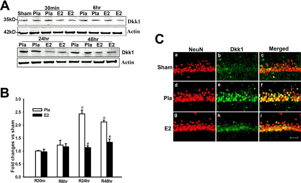
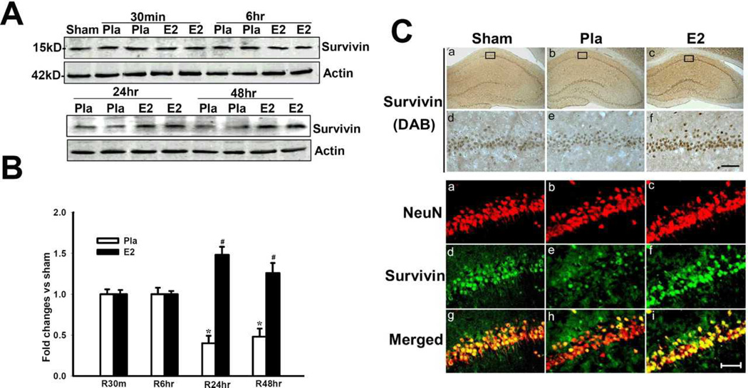
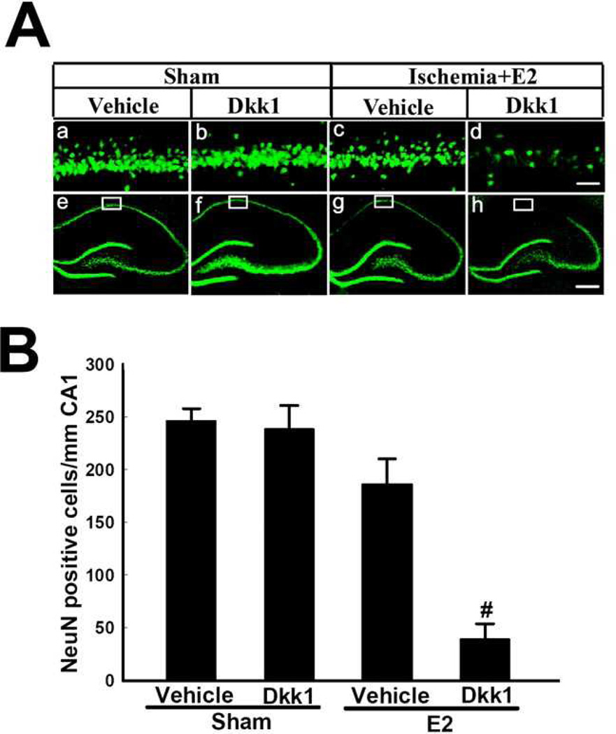
Intriguingly, E2, as an endogenous steroid hormone, is not only capable of preventing the neuronal damage associated with neurodegenerative diseases, but is also able to promote a favorable balance of Dkk1 and Wnt signaling in the brain. As mentioned earlier, our laboratory reported that one mechanism through which E2 prevents neuronal cell death from global cerebral ischemia (GCI) is by suppressing the post-ischemic elevation of Dkk1 and simultaneously facilitating pro-survival Wnt/β-Catenin signaling in the CA1 hippocampal region (Zhang et al., 2008). Using Western blotting, we showed that Dkk1 is significantly upregulated in ischemic animals at 24 and 48 hours following GCI, and we demonstrated that a low, Diestrus I dose of E2, given via subcutaneous osmotic mini-pump one week before induction of ischemia, was able to prevent this elevation at both of these post-reperfusion time points (Figure 2). Furthermore, we showed that Dkk1 expression co-localized with NeuN, a neuronal marker, suggesting that Dkk1 is primarily expressed in neurons 24 hours after GCI (Figure 2). In the same study, we also examined the status of canonical Wnt signaling in the CA1 hippocampal region after GCI and E2 treatment by measuring protein levels of Wnt3, phospho-β-Catenin, nuclear β-Catenin, and the canonical Wnt signaling product Survivin. In addition to elevations of Wnt3 and nuclear β-Catenin, we observed that E2 treatment not only prevented GCI-related loss but also enhanced the neuronal expression of Survivin, a Wnt target gene that facilitates survival through preventing the cleavage of pro-apoptotic caspases, at 24 and 48 hours after GCI (Figure 3). Since these are the same time points that we noted significant E2 suppression of the Wnt antagonist Dkk1 following GCI, we concluded that E2 is able to maintain a favorable balance of Dkk1-Wnt/β-Catenin signaling in the hippocampus following GCI. Finally, we demonstrated that E2’s post-ischemic suppression of Dkk1 elevation is required for its neuroprotective ability, as intracerebroventricular injection of Dkk1 peptides into E2-treated animals was sufficient to reverse E2 neuroprotection status (Figure 4).
Figure 2. A,B, Effect of 17β-estradiol on Dkk1 protein levels in hippocampus CA1 after global cerebral ischemia.
Values are mean ± SEM of determinations from five to six individual rats and expressed as fold change versus sham control. Pla, Placebo; R, reperfusion. *p < 0.05 versus sham control; #p < 0.05 versus placebo treatment group. Ca–Ci, Confocal analysis of NeuN and Dkk1 immunostaining in hippocampus CA1 at 24 h after global cerebral ischemia (magnification, 40X). Scale bar, 50µm. Reprinted, with permission, from Journal of Neuroscience (Zhang et al., 2008).
Figure 3. A,B, 17β-Estradiol enhances expression of the anti-apoptotic protein Survivin in hippocampus CA1 after global cerebral ischemia.
Values are mean ± SEM of determinations from five to six individual rats expressed as fold change versus sham control. Pla, Placebo; R, reperfusion. *p < 0.05 versus sham control; #p < 0.05 versus the Pla group at the same time point. C, DAB and confocal analysis shows that survivin is induced in NeuN-positive neurons by E2 in hippocampus CA1 at 24 h after global cerebral ischemia. Results are representative of staining observed in five individual animals per group (magnifications, 40X). Scale bars, 50 µm. Reprinted, with permission, from Journal of Neuroscience (Zhang et al., 2008).
Figure 4. Exogenous Dkk1 Administration Reverses 17β-Estradiol-Induced Neuroprotection after Global Cerebral Ischemia.
Dkk1 (5µg/5µl) was administered via intracerebroventricular injection into both lateral ventricles at 12 h after global cerebral ischemia. For a control, separate animals received vehicle into both lateral cerebral ventricles. Additionally, a non-ischemic sham control also received Dkk1 into the lateral ventricles. Aa–Ad, High-power magnification of NeuN staining in hippocampus CA1 at 7 d in sham animals and after 7 d reperfusion after global cerebral ischemia in E2-treated rats that received vehicle or exogenous Dkk1 in both lateral ventricles. Magnification is 40X. Scale bar, 50µm. Ae–Ah, Low-power magnifications of representative whole hippocampus sections showing NeuN staining in hippocampus CA1 at 7 d in sham animals and after 7 d reperfusion after global cerebral ischemia in E2-treated rats that received vehicle or exogenous Dkk1 in both lateral ventricles. Magnification is 5X. Scale bar, 200µm. B, CA1 cell counts of NeuN-positive neurons in all animals show that exogenous Dkk1 had no significant effect on CA1 neuronal cell survival in non-ischemic sham controls, although it significantly reversed E2 neuroprotection in ischemic animals. Values are mean ± SEM of determinations from five to six individual rats. #p < 0.01 versus vehicle. Figure adapted from (Zhang et al., 2008).
While our lab demonstrated that pre-treatment with low-dose E2 increased expression of Wnt3 in CA1 hippocampal neurons 24 and 48 hours following GCI and led to increased expression of Survivin, a Wnt target gene, E2 regulation of β-Catenin-dependent transcription may also occur independently of canonical Wnt signaling. However, regardless of the mechanism involved, the evidence overwhelmingly suggests that E2 stabilizes β-Catenin via inhibition of GSK3β [See (Varea et al., 2010) for review]. In fact, E2 effectively increases the amount of inactive GSK3β in neuronal cells in vitro (Cardona-Gomez et al., 2004; Varea et al., 2009), and E2 treatment protects hippocampal slice cultures from kainate-induced neurotoxicity via rapid, ER-mediated phosphorylation and inactivation of GSK3β at Serine 9 (Goodenough et al., 2005). Cardona-Gomez et al. further demonstrated that acute E2 treatment increased the amount of inactivated GSK3β in the rat hippocampus, which subsequently prevented the hyperphosphorylation of tau (Cardona-Gomez et al., 2004). This is consistent with our previous study, which showed that E2 increased the amount of inactive GSK3β at 24 and 48 hours following GCI and led to the stabilization of β-Catenin and the prevention of tau hyperphosphorylation at these same time points (Zhang et al., 2008). Varea et al. further demonstrated that estrogen receptor alpha (ERα) and estrogen receptor beta (ERβ) both directly interact with GSK3β and β-Catenin in vitro and that E2, PPT (an ERα agonist), and DPN (an ERβ agonist) were all capable of inducing β-Catenin-mediated transcription through the TCF/LEF-1 family of transcription factors in primary cortical neurons and neuroblastoma cells (Varea et al., 2009). Importantly, they observed that the ER antagonist ICI 182780 essentially blocked E2’s induction of TCF/LEF-1-mediated transcription, further suggesting that E2’s stabilization of β-Catenin requires the estrogen receptor, and they noted that PPT was more effective than DPN, suggesting that while both ERα and ERβ can mediate E2’s effect on β-Catenin-mediated transcription in the brain, E2 may be acting primarily through ERα to upregulate TCF/LEF-1-mediated transcription in vitro (Varea et al., 2009). Finally, two independent studies suggested that E2-induced β-Catenin-mediated transcription requires LEF-1 and activates a set of genes that is similar, but not identical to, that activated by canonical Wnt signaling (Varea et al., 2009; Wandosell et al., 2012). These observations further promote the idea that, in addition to regulating the canonical Wnt signaling pathway, E2 can act independently of the canonical Wnt signaling pathway to enhance β-Catenin-mediated transcription in neural cells.
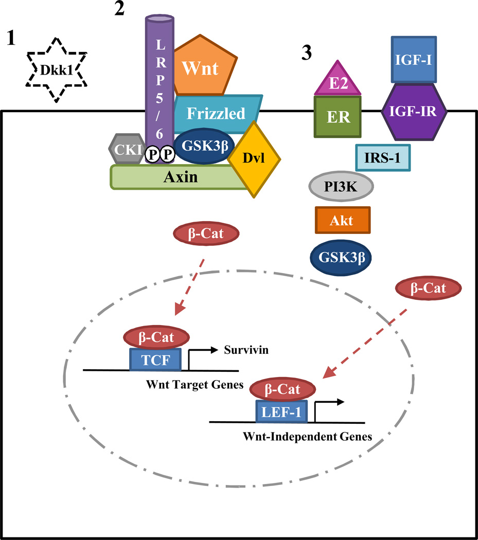
Recently, it has been determined that hypothalamic β-Catenin expression naturally fluctuates across the estrous cycle in rats, with elevations of β-Catenin observed during proestrus and estrus, two days characterized by high, circulating levels of E2 (Barrera-Ocampo et al., 2012). Intriguingly, the peaks in hypothalamic β-Catenin expression were concomitant with increases of activated Akt and inactivated GSK3β, suggesting that these two events are critical for E2-induced stabilization of β-Catenin in the brain (Barrera-Ocampo et al., 2012). E2 is well known to activate the neuroprotective phosphatidylinositol 3 kinase (PI3K)-Akt signaling pathway, either by itself or in concert with IGF-1 signaling, leading to the activation of the serine/threonine kinase Akt through phosphorylation at Serine 473 (Brann et al., 2007; Burgering and Coffer, 1995; Mendez et al., 2003; Singh, 2001; Varea et al., 2010). Importantly, activated Akt has also been shown to inhibit GSK3β via phosphorylation at Serine 9 (Cross et al., 1995), and Mendez et al. demonstrated that blockade of the PI3K-Akt pathway with wortmannin, a specific PI3K inhibitor, decreased the amount of cytosolic β-Catenin in neuronal cultures (Mendez and Garcia-Segura, 2006). In light of this knowledge, Varea et al. propose a novel, additional mechanism for E2’s regulation of β-Catenin-mediated transcription, where E2 binds to membrane-bound ERα and rapidly activates the neuroprotective PI3K-Akt pathway, possibly through interaction with IGF-1 signaling components. A multi-molecular complex is then formed consisting of ERα, GSK3β, β-Catenin, and others (Varea et al., 2009; Varea et al., 2010). Once activated via phosphorylation at Serine 473, Akt then inactivates GSK3β, which leads to the subsequent stabilization and nuclear retention of cytosolic β-Catenin. Inside the nucleus, β-Catenin then interacts with the TCF/LEF-1 transcription machinery to promote the expression of target genes that are independent of Wnt (Varea et al., 2009; Varea et al., 2010; Wandosell et al., 2012). Thus, it is plausible that E2 may maintain the delicate balance of Dkk1 and Wnt/β-Catenin signaling in the adult brain in three different ways: 1) by suppressing neuronal expression of the neurodegenerative Wnt antagonist Dkk1 2) by enhancing Wnt3 expression and subsequently facilitating canonical Wnt/β-Catenin signaling in neurons, and 3) by promoting Wnt-independent, β-Catenin-mediated transcription through a membrane ER-initiated intracellular signaling cascade involving PI3K/Akt/GSK3β (Figure 5).
Figure 5. Summary of Estrogen Regulation of Dkk1 and Wnt/β-Catenin Signaling.
This figure depicts three currently proposed mechanisms for 17β-estradiol’s regulation of Dkk1 and Wnt/β-Catenin signaling in the brain. 1) E2 can suppress expression of the neurodegenerative Wnt antagonist Dkk1 in the hippocampus, particularly following an ischemic insult. 2) E2 can enhance canonical Wnt signaling in the hippocampus, particularly following an ischemic insult, through induction of Wnt3, which leads to expression of pro-survival Wnt target genes, such as Survivin, through β-Catenin and TCF. 3) E2, either acting alone or in concert with IGF-1, can initiate a membrane receptor-mediated PI3K/Akt/GSK3β signaling cascade, which leads to the stabilization of cytosolic β-Catenin and Wnt-independent gene expression through β-Catenin and LEF-1. Akt, A Serine/Threonine Kinase (also known as Protein Kinase B); β-Cat, Beta-Catenin; CKI, Casein Kinase 1; Dkk1, Dickkopf-1; Dvl, Disheveled; E2, 17β-Estradiol; ER, Estrogen Receptor; GSK3β, Glycogen Synthase Kinase 3 Beta; IGF-I, Insulin-like Growth Factor-1; IGF-IR, Insulin-like Growth Factor-1 Receptor; IRS-1, Insulin Receptor Substrate 1; LEF-1, Lymphoid Enhancing Factor 1; LRP 5/6, Low-Density Lipoprotein 5/6; PI3K, Phosphatidylinositol 3 Kinase; TCF, T Cell Factor; Wnt, Wingless.
5. Future Directions – Long Term Estrogen Deprivation and Therapeutics
While it is apparent that elevations in Dkk1 and reductions in Wnt/β-Catenin signaling are associated with neurodegenerative disease and that E2 may prevent neurodegenerative disease through its favorable effect on neural Dkk1 and Wnt/β-Catenin signaling, many questions remain unanswered. Importantly, women who enter menopause prematurely (< 45 years of age) have increased risks for ischemic stroke, dementia, and mortality from neurological disorders, which can be ameliorated with timely replacement of E2 until the age of natural menopause (Rivera et al., 2009; Rocca et al., 2007; Rocca et al., 2011; Rocca et al., 2012; Shuster et al., 2010). This suggests that dramatic and prolonged loss of the neuroprotective ovarian hormone E2 is detrimental to the brain and highlights the importance of studying the effects of long-term estrogen deprivation (surgical menopause and reproductive senescence) on the brain. In fact, we have preliminary data suggesting that E2’s post-ischemic regulation of Dkk1 and the Wnt target gene Survivin is lost in long-term E2-deprived rats and that even basal levels of these two critical proteins are altered in the hippocampus of non-ischemic sham animals (Scott et al., 2012, Submitted). As such, these changes could explain why postmenopausal women, particularly those who enter menopause prematurely due to bilateral oophorectomy, are more susceptible to neurodegenerative disease.
Along these lines, another important, yet controversial, area of study is postmenopausal hormone therapy for the alleviation of menopausal symptoms, cardioprotection, and neuroprotection. Nearly a decade after the Women’s Health Initiative (WHI) determined that conjugated equine estrogens ± medroxyprogesterone acetate increased the risk of ischemic stroke (Wassertheil-Smoller et al., 2003) and dementia in women aged 65 and older (Shumaker et al., 2003), despite a wide breadth of basic research suggesting a neuroprotective effect of estrogen, researchers are still hard at work in an attempt to determine the most effective regimen, dose, and route of administration of hormone therapy. Intriguingly, the timing of hormone therapy initiation also seems to be critical for its effectiveness, at least in rodents. In fact, our lab and others have demonstrated that E2’s neuroprotective ability is lost in rodents if treatment is delayed 10 weeks following bilateral ovariectomy or natural aging (Suzuki et al., 2007; Zhang et al., 2009; Zhang et al., 2011). This is consistent with the “critical period hypothesis” of estrogen replacement and the “healthy cell bias” of E2 action, which state that hormone therapy must be initiated at the time of menopause to provide neurological benefit (Brinton, 2008; Harman et al., 2004; Maki, 2006a; Maki, 2006b) [See (Daniel and Bohacek, 2010; Scott et al., 2012) for review of animal studies and clinical studies addressing the critical period hypothesis]. In fact, our lab recently provided a potential mechanism to explain the critical period hypothesis, as unliganded ERα, the principal mediator of E2 neuroprotection, was specifically degraded in the hippocampus of rats that were deprived of ovarian E2 long-term (Zhang et al., 2011). Furthermore, our preliminary data suggest that delayed treatment with E2 following surgical menopause is ineffective in preventing the post-ischemic elevation of neurodegenerative Dkk1 or enhancing pro-survival Wnt/β-Catenin signaling (Scott et al., 2012, Submitted). Thus, since the brain appears to lose its sensitivity to the beneficial effects of E2 over time following menopause, this underscores the importance of immediate hormone therapy for optimal neurological benefit. As such, more well-designed randomized, controlled clinical trials of hormone therapy are necessary to effectively study this concept in postmenopausal women.
Finally, since elevation of Dkk1 plays a prominent role in neurodegenerative diseases, such as ischemic stroke, Alzheimer’s disease, Parkinson’s disease, and temporal lobe epilepsy, future studies are needed to explore the potential efficacy of Dkk1 inhibitors as effective therapeutics [See (Caraci et al., 2008) for review]. To date, there are no FDA-approved Dkk1 inhibitors available, and a single commercial Dkk1 inhibitor, WAY-262611, exists for laboratory study. Unfortunately, thus far, only one paper has been published using this inhibitor, which detailed the compound’s discovery and demonstrated that oral administration of WAY-262611 promoted trabecular bone growth in ovariectomized rats via inhibition of Dkk1 (Pelletier et al., 2009). Therefore, this compound should be tested in animal models of neurodegenerative disease in order to determine whether it would be a feasible treatment for humans. In addition, neutralizing Dkk1 antibodies have recently become available. In fact, similar to WAY-262611, fully human Dkk1 antibodies have been shown to regulate bone mass in vivo (Glantschnig et al., 2010). Furthermore, they have since been utilized to study the role of Dkk1 in glucocorticoid-mediated inhibition of human neural/stem cell progenitor proliferation and differentiation (Moors et al., 2012) and the role of Dkk1 in beta-amyloid-mediated synapse loss in Alzheimer’s disease (Purro et al., 2012). Hence, Dkk1 neutralizing antibodies could prove to be an asset to the field, and may also serve as a potential therapeutic treatment for neurodegenerative disease in humans.
In summary, Dkk1 is an antagonist of canonical Wnt signaling, and while it is absolutely critical for embryonic development of the limbs and forebrain, elevation of Dkk1 in the adult brain is associated with neurodegenerative disease. E2 is a neuroprotective ovarian steroid hormone capable of suppressing Dkk1 and activating pro-survival Wnt/β-Catenin signaling in vivo following global cerebral ischemia (Zhang et al., 2008), effects implicated to be critical for its neuroprotective actions. Furthermore, E2 is also able to enhance Wnt-independent β-Catenin-mediated transcription in vitro through rapid activation of the PI3K-Akt pathway and inactivation of GSK3β (Varea et al., 2009; Varea et al., 2010; Wandosell et al., 2012). While E2 modulation of Dkk1 and Wnt/β-catenin signaling has been well described, comparatively little is known regarding the effects of long-term E2 deprivation (surgical menopause and reproductive senescence) on this important signaling pathway in the brain. Studies on this issue are critically needed, as they may help explain why postmenopausal women have an increased risk of neurodegenerative disease and determine whether this increased risk can be reduced with timely administration of postmenopausal hormone therapy. It is also imperative that preclinical evaluation of Dkk1 inhibitors be fully explored, especially with regard to their neuroprotective efficacy in neurodegenerative disorders, as they may provide a broad-spectrum therapeutic agent for use in humans.
Acknowledgements
The authors’ research presented in this review was supported by a research grant to DWB from the NINDS (NS050730) and a pre-doctoral fellowship to ELS from the American Heart Association (12PRE11530009).
Abbreviations
- APC
adenomatous polyposis coli
- CKI
Casein Kinase I
- Dkk1
Dickkopf-1
- Dvl
Disheveled
- E2 or Estrogen
17β-Estradiol
- GCI
Global Cerebral Ischemia
- Fz
Frizzled
- GSK3β
Glycogen Synthase Kinase 3β
- HT
Hormone Therapy
- JNK
c-Jun N-terminal Kinase
- LRP5/6
Low Density Lipoprotein-Related Protein 5/6
- LTED
Long-Term Estrogen Deprivation or Long-Term Estrogen-Deprived
- MCAO
Middle Cerebral Artery Occlusion
- NMDA
N-Methyl-D-Aspartate
- PI3K
Phosphatidylinositol 3 Kinase
- sFRP
secreted Frizzled-Related Proteins
- TCF/LEF
T Cell Factor/Lymphoid Enhancing Factor
- Wnt
Wingless
- WHI
Women’s Health Initiative
- WIF-1
Wnt Inhibitory Factor-1
- WRE
Wnt Response Element
References
- Alkayed NJ, Harukuni I, Kimes AS, London ED, Traystman RJ, Hurn PD. Gender-linked brain injury in experimental stroke. Stroke; a journal of cerebral circulation. 1998;29:159–165. doi: 10.1161/01.str.29.1.159. discussion 166. [DOI] [PubMed] [Google Scholar]
- Appelros P, Stegmayr B, Terent A. Sex differences in stroke epidemiology: a systematic review. Stroke; a journal of cerebral circulation. 2009;40:1082–1090. doi: 10.1161/STROKEAHA.108.540781. [DOI] [PubMed] [Google Scholar]
- Ataman B, Ashley J, Gorczyca M, Ramachandran P, Fouquet W, Sigrist SJ, Budnik V. Rapid activity-dependent modifications in synaptic structure and function require bidirectional Wnt signaling. Neuron. 2008;57:705–718. doi: 10.1016/j.neuron.2008.01.026. [DOI] [PMC free article] [PubMed] [Google Scholar]
- Avila ME, Sepulveda FJ, Burgos CF, Moraga-Cid G, Parodi J, Moon RT, Aguayo LG, Opazo C, De Ferrari GV. Canonical Wnt3a modulates intracellular calcium and enhances excitatory neurotransmission in hippocampal neurons. The Journal of biological chemistry. 2010;285:18939–18947. doi: 10.1074/jbc.M110.103028. [DOI] [PMC free article] [PubMed] [Google Scholar]
- Bafico A, Liu G, Yaniv A, Gazit A, Aaronson SA. Novel mechanism of Wnt signalling inhibition mediated by Dickkopf-1 interaction with LRP6/Arrow. Nature cell biology. 2001;3:683–686. doi: 10.1038/35083081. [DOI] [PubMed] [Google Scholar]
- Barrera-Ocampo A, Gutierrez-Vargas J, Garcia-Segura LM, Cardona-Gomez GP. Glycogen synthase kinase-3beta/beta-catenin signaling in the rat hypothalamus during the estrous cycle. Journal of neuroscience research. 2012;90:1078–1084. doi: 10.1002/jnr.22816. [DOI] [PubMed] [Google Scholar]
- Boonen RA, van Tijn P, Zivkovic D. Wnt signaling in Alzheimer's disease: up or down, that is the question. Ageing research reviews. 2009;8:71–82. doi: 10.1016/j.arr.2008.11.003. [DOI] [PubMed] [Google Scholar]
- Brann DW, Dhandapani K, Wakade C, Mahesh VB, Khan MM. Neurotrophic and neuroprotective actions of estrogen: basic mechanisms and clinical implications. Steroids. 2007;72:381–405. doi: 10.1016/j.steroids.2007.02.003. [DOI] [PMC free article] [PubMed] [Google Scholar]
- Brinton RD. The healthy cell bias of estrogen action: mitochondrial bioenergetics and neurological implications. Trends in neurosciences. 2008;31:529–537. doi: 10.1016/j.tins.2008.07.003. [DOI] [PMC free article] [PubMed] [Google Scholar]
- Budnik V, Salinas PC. Wnt signaling during synaptic development and plasticity. Current opinion in neurobiology. 2011;21:151–159. doi: 10.1016/j.conb.2010.12.002. [DOI] [PMC free article] [PubMed] [Google Scholar]
- Burgering BM, Coffer PJ. Protein kinase B (c-Akt) in phosphatidylinositol-3-OH kinase signal transduction. Nature. 1995;376:599–602. doi: 10.1038/376599a0. [DOI] [PubMed] [Google Scholar]
- Busceti CL, Biagioni F, Aronica E, Riozzi B, Storto M, Battaglia G, Giorgi FS, Gradini R, Fornai F, Caricasole A, Nicoletti F, Bruno V. Induction of the Wnt inhibitor, Dickkopf-1, is associated with neurodegeneration related to temporal lobe epilepsy. Epilepsia. 2007;48:694–705. doi: 10.1111/j.1528-1167.2007.01055.x. [DOI] [PubMed] [Google Scholar]
- Cappuccio I, Calderone A, Busceti CL, Biagioni F, Pontarelli F, Bruno V, Storto M, Terstappen GT, Gaviraghi G, Fornai F, Battaglia G, Melchiorri D, Zukin RS, Nicoletti F, Caricasole A. Induction of Dickkopf-1, a negative modulator of the Wnt pathway, is required for the development of ischemic neuronal death. The Journal of neuroscience : the official journal of the Society for Neuroscience. 2005;25:2647–2657. doi: 10.1523/JNEUROSCI.5230-04.2005. [DOI] [PMC free article] [PubMed] [Google Scholar]
- Caraci F, Busceti C, Biagioni F, Aronica E, Mastroiacovo F, Cappuccio I, Battaglia G, Bruno V, Caricasole A, Copani A, Nicoletti F. The Wnt antagonist, Dickkopf-1, as a target for the treatment of neurodegenerative disorders. Neurochemical research. 2008;33:2401–2406. doi: 10.1007/s11064-008-9710-0. [DOI] [PubMed] [Google Scholar]
- Cardona-Gomez P, Perez M, Avila J, Garcia-Segura LM, Wandosell F. Estradiol inhibits GSK3 and regulates interaction of estrogen receptors, GSK3, and beta-catenin in the hippocampus. Molecular and cellular neurosciences. 2004;25:363–373. doi: 10.1016/j.mcn.2003.10.008. [DOI] [PubMed] [Google Scholar]
- Caricasole A, Copani A, Caraci F, Aronica E, Rozemuller AJ, Caruso A, Storto M, Gaviraghi G, Terstappen GC, Nicoletti F. Induction of Dickkopf-1, a negative modulator of the Wnt pathway, is associated with neuronal degeneration in Alzheimer's brain. The Journal of neuroscience : the official journal of the Society for Neuroscience. 2004;24:6021–6027. doi: 10.1523/JNEUROSCI.1381-04.2004. [DOI] [PMC free article] [PubMed] [Google Scholar]
- Choi DW. Calcium and excitotoxic neuronal injury. Annals of the New York Academy of Sciences. 1994a;747:162–171. doi: 10.1111/j.1749-6632.1994.tb44407.x. [DOI] [PubMed] [Google Scholar]
- Choi DW. Glutamate receptors and the induction of excitotoxic neuronal death. Progress in brain research. 1994b;100:47–51. doi: 10.1016/s0079-6123(08)60767-0. [DOI] [PubMed] [Google Scholar]
- Clark CE, Nourse CC, Cooper HM. The tangled web of non-canonical Wnt signalling in neural migration. Neuro-Signals. 2012;20:202–220. doi: 10.1159/000332153. [DOI] [PubMed] [Google Scholar]
- Cross DA, Alessi DR, Cohen P, Andjelkovich M, Hemmings BA. Inhibition of glycogen synthase kinase-3 by insulin mediated by protein kinase B. Nature. 1995;378:785–789. doi: 10.1038/378785a0. [DOI] [PubMed] [Google Scholar]
- Daniel JM, Bohacek J. The critical period hypothesis of estrogen effects on cognition: Insights from basic research. Biochimica et biophysica acta. 2010;1800:1068–1076. doi: 10.1016/j.bbagen.2010.01.007. [DOI] [PubMed] [Google Scholar]
- De Ferrari GV, Moon RT. The ups and downs of Wnt signaling in prevalent neurological disorders. Oncogene. 2006;25:7545–7553. doi: 10.1038/sj.onc.1210064. [DOI] [PubMed] [Google Scholar]
- De Ferrari GV, Papassotiropoulos A, Biechele T, Wavrant De-Vrieze F, Avila ME, Major MB, Myers A, Saez K, Henriquez JP, Zhao A, Wollmer MA, Nitsch RM, Hock C, Morris CM, Hardy J, Moon RT. Common genetic variation within the low-density lipoprotein receptor-related protein 6 and late-onset Alzheimer's disease. Proceedings of the National Academy of Sciences of the United States of America. 2007;104:9434–9439. doi: 10.1073/pnas.0603523104. [DOI] [PMC free article] [PubMed] [Google Scholar]
- De Marco P, Merello E, Consales A, Piatelli G, Cama A, Kibar Z, Capra V. Genetic Analysis of Disheveled 2 and Disheveled 3 in Human Neural Tube Defects. Journal of molecular neuroscience : MN. 2012 doi: 10.1007/s12031-012-9871-9. [DOI] [PMC free article] [PubMed] [Google Scholar]
- Dun Y, Li G, Yang Y, Xiong Z, Feng M, Wang M, Zhang Y, Xiang J, Ma R. Inhibition of the canonical Wnt pathway by Dickkopf-1 contributes to the neurodegeneration in 6-OHDA-lesioned rats. Neuroscience letters. 2012;525:83–88. doi: 10.1016/j.neulet.2012.07.030. [DOI] [PubMed] [Google Scholar]
- Fedi P, Bafico A, Nieto Soria A, Burgess WH, Miki T, Bottaro DP, Kraus MH, Aaronson SA. Isolation and biochemical characterization of the human Dkk-1 homologue, a novel inhibitor of mammalian Wnt signaling. The Journal of biological chemistry. 1999;274:19465–19472. doi: 10.1074/jbc.274.27.19465. [DOI] [PubMed] [Google Scholar]
- Forlenza OV, Torres CA, Talib LL, de Paula VJ, Joaquim HP, Diniz BS, Gattaz WF. Increased platelet GSK3B activity in patients with mild cognitive impairment and Alzheimer's disease. Journal of psychiatric research. 2011;45:220–224. doi: 10.1016/j.jpsychires.2010.06.002. [DOI] [PubMed] [Google Scholar]
- Gibson CL, Gray LJ, Murphy SP, Bath PM. Estrogens and experimental ischemic stroke: a systematic review. Journal of cerebral blood flow and metabolism : official journal of the International Society of Cerebral Blood Flow and Metabolism. 2006;26:1103–1113. doi: 10.1038/sj.jcbfm.9600270. [DOI] [PubMed] [Google Scholar]
- Glantschnig H, Hampton RA, Lu P, Zhao JZ, Vitelli S, Huang L, Haytko P, Cusick T, Ireland C, Jarantow SW, Ernst R, Wei N, Nantermet P, Scott KR, Fisher JE, Talamo F, Orsatti L, Reszka AA, Sandhu P, Kimmel D, Flores O, Strohl W, An Z, Wang F. Generation and selection of novel fully human monoclonal antibodies that neutralize Dickkopf-1 (DKK1) inhibitory function in vitro and increase bone mass in vivo. The Journal of biological chemistry. 2010;285:40135–40147. doi: 10.1074/jbc.M110.166892. [DOI] [PMC free article] [PubMed] [Google Scholar]
- Glinka A, Wu W, Delius H, Monaghan AP, Blumenstock C, Niehrs C. Dickkopf-1 is a member of a new family of secreted proteins and functions in head induction. Nature. 1998;391:357–362. doi: 10.1038/34848. [DOI] [PubMed] [Google Scholar]
- Goodenough S, Schleusner D, Pietrzik C, Skutella T, Behl C. Glycogen synthase kinase 3beta links neuroprotection by 17beta-estradiol to key Alzheimer processes. Neuroscience. 2005;132:581–589. doi: 10.1016/j.neuroscience.2004.12.029. [DOI] [PubMed] [Google Scholar]
- Grotewold L, Ruther U. The Wnt antagonist Dickkopf-1 is regulated by Bmp signaling and c-Jun and modulates programmed cell death. The EMBO journal. 2002;21:966–975. doi: 10.1093/emboj/21.5.966. [DOI] [PMC free article] [PubMed] [Google Scholar]
- Harman SM, Brinton EA, Clarkson T, Heward CB, Hecht HS, Karas RH, Judelson DR, Naftolin F. Is the WHI relevant to HRT started in the perimenopause? Endocrine. 2004;24:195–202. doi: 10.1385/ENDO:24:3:195. [DOI] [PubMed] [Google Scholar]
- Jensen M, Hoerndli FJ, Brockie PJ, Wang R, Johnson E, Maxfield D, Francis MM, Madsen DM, Maricq AV. Wnt signaling regulates acetylcholine receptor translocation and synaptic plasticity in the adult nervous system. Cell. 2012;149:173–187. doi: 10.1016/j.cell.2011.12.038. [DOI] [PMC free article] [PubMed] [Google Scholar]
- Kazanskaya O, Glinka A, Niehrs C. The role of Xenopus dickkopf1 in prechordal plate specification and neural patterning. Development. 2000;127:4981–4992. doi: 10.1242/dev.127.22.4981. [DOI] [PubMed] [Google Scholar]
- Kim KI, Park KU, Chun EJ, Choi SI, Cho YS, Youn TJ, Cho GY, Chae IH, Song J, Choi DJ, Kim CH. A novel biomarker of coronary atherosclerosis: serum DKK1 concentration correlates with coronary artery calcification and atherosclerotic plaques. Journal of Korean medical science. 2011;26:1178–1184. doi: 10.3346/jkms.2011.26.9.1178. [DOI] [PMC free article] [PubMed] [Google Scholar]
- Komiya Y, Habas R. Wnt signal transduction pathways. Organogenesis. 2008;4:68–75. doi: 10.4161/org.4.2.5851. [DOI] [PMC free article] [PubMed] [Google Scholar]
- L'Episcopo F, Tirolo C, Testa N, Caniglia S, Morale MC, Cossetti C, D'Adamo P, Zardini E, Andreoni L, Ihekwaba AE, Serra PA, Franciotta D, Martino G, Pluchino S, Marchetti B. Reactive astrocytes and Wnt/beta-catenin signaling link nigrostriatal injury to repair in 1-methyl-4-phenyl-1,2,3,6-tetrahydropyridine model of Parkinson's disease. Neurobiology of disease. 2011;41:508–527. doi: 10.1016/j.nbd.2010.10.023. [DOI] [PMC free article] [PubMed] [Google Scholar]
- Lei P, Ayton S, Bush AI, Adlard PA. GSK-3 in Neurodegenerative Diseases. International journal of Alzheimer's disease. 2011;2011:189246. doi: 10.4061/2011/189246. [DOI] [PMC free article] [PubMed] [Google Scholar]
- Logan CY, Nusse R. The Wnt signaling pathway in development and disease. Annual review of cell and developmental biology. 2004;20:781–810. doi: 10.1146/annurev.cellbio.20.010403.113126. [DOI] [PubMed] [Google Scholar]
- MacDonald BT, Adamska M, Meisler MH. Hypomorphic expression of Dkk1 in the doubleridge mouse: dose dependence and compensatory interactions with Lrp6. Development. 2004;131:2543–2552. doi: 10.1242/dev.01126. [DOI] [PubMed] [Google Scholar]
- MacDonald BT, Tamai K, He X. Wnt/beta-catenin signaling: components, mechanisms, and diseases. Developmental cell. 2009;17:9–26. doi: 10.1016/j.devcel.2009.06.016. [DOI] [PMC free article] [PubMed] [Google Scholar]
- Maki PM. Potential importance of early initiation of hormone therapy for cognitive benefit. Menopause. 2006a;13:6–7. doi: 10.1097/01.gme.0000194822.76774.30. [DOI] [PubMed] [Google Scholar]
- Maki PM. Hormone therapy and cognitive function: is there a critical period for benefit? Neuroscience. 2006b;138:1027–1030. doi: 10.1016/j.neuroscience.2006.01.001. [DOI] [PubMed] [Google Scholar]
- Mao B, Wu W, Li Y, Hoppe D, Stannek P, Glinka A, Niehrs C. LDL-receptor-related protein 6 is a receptor for Dickkopf proteins. Nature. 2001;411:321–325. doi: 10.1038/35077108. [DOI] [PubMed] [Google Scholar]
- Mastroiacovo F, Busceti CL, Biagioni F, Moyanova SG, Meisler MH, Battaglia G, Caricasole A, Bruno V, Nicoletti F. Induction of the Wnt antagonist, Dickkopf-1, contributes to the development of neuronal death in models of brain focal ischemia. Journal of cerebral blood flow and metabolism : official journal of the International Society of Cerebral Blood Flow and Metabolism. 2009;29:264–276. doi: 10.1038/jcbfm.2008.111. [DOI] [PubMed] [Google Scholar]
- McCullough LD, Blizzard K, Simpson ER, Oz OK, Hurn PD. Aromatase cytochrome P450 and extragonadal estrogen play a role in ischemic neuroprotection. The Journal of neuroscience : the official journal of the Society for Neuroscience. 2003;23:8701–8705. doi: 10.1523/JNEUROSCI.23-25-08701.2003. [DOI] [PMC free article] [PubMed] [Google Scholar]
- McMahon AP, Moon RT. int-1--a proto-oncogene involved in cell signalling. Development. 1989a;107(Suppl):161–167. doi: 10.1242/dev.107.Supplement.161. [DOI] [PubMed] [Google Scholar]
- McMahon AP, Moon RT. Ectopic expression of the proto-oncogene int-1 in Xenopus embryos leads to duplication of the embryonic axis. Cell. 1989b;58:1075–1084. doi: 10.1016/0092-8674(89)90506-0. [DOI] [PubMed] [Google Scholar]
- McMahon AP, Bradley A. The Wnt-1 (int-1) proto-oncogene is required for development of a large region of the mouse brain. Cell. 1990;62:1073–1085. doi: 10.1016/0092-8674(90)90385-r. [DOI] [PubMed] [Google Scholar]
- Mendez P, Azcoitia I, Garcia-Segura LM. Estrogen receptor alpha forms estrogen-dependent multimolecular complexes with insulin-like growth factor receptor and phosphatidylinositol 3-kinase in the adult rat brain. Brain research. Molecular brain research. 2003;112:170–176. doi: 10.1016/s0169-328x(03)00088-3. [DOI] [PubMed] [Google Scholar]
- Mendez P, Garcia-Segura LM. Phosphatidylinositol 3-kinase and glycogen synthase kinase 3 regulate estrogen receptor-mediated transcription in neuronal cells. Endocrinology. 2006;147:3027–3039. doi: 10.1210/en.2005-1224. [DOI] [PubMed] [Google Scholar]
- Moors M, Bose R, Johansson-Haque K, Edoff K, Okret S, Ceccatelli S. Dickkopf 1 mediates glucocorticoid-induced changes in human neural progenitor cell proliferation and differentiation. Toxicological sciences : an official journal of the Society of Toxicology. 2012;125:488–495. doi: 10.1093/toxsci/kfr304. [DOI] [PubMed] [Google Scholar]
- Morgan AR, Turic D, Jehu L, Hamilton G, Hollingworth P, Moskvina V, Jones L, Lovestone S, Brayne C, Rubinsztein DC, Lawlor B, Gill M, O'Donovan MC, Owen MJ, Williams J. Association studies of 23 positional/functional candidate genes on chromosome 10 in late-onset Alzheimer's disease. American journal of medical genetics. Part B, Neuropsychiatric genetics : the official publication of the International Society of Psychiatric Genetics. 2007;144B:762–770. doi: 10.1002/ajmg.b.30509. [DOI] [PubMed] [Google Scholar]
- Morgan AR, Hamilton G, Turic D, Jehu L, Harold D, Abraham R, Hollingworth P, Moskvina V, Brayne C, Rubinsztein DC, Lynch A, Lawlor B, Gill M, O'Donovan M, Powell J, Lovestone S, Williams J, Owen MJ. Association analysis of 528 intra-genic SNPs in a region of chromosome 10 linked to late onset Alzheimer's disease. American journal of medical genetics. Part B, Neuropsychiatric genetics : the official publication of the International Society of Psychiatric Genetics. 2008;147B:727–731. doi: 10.1002/ajmg.b.30670. [DOI] [PubMed] [Google Scholar]
- Mukhopadhyay M, Shtrom S, Rodriguez-Esteban C, Chen L, Tsukui T, Gomer L, Dorward DW, Glinka A, Grinberg A, Huang SP, Niehrs C, Izpisua Belmonte JC, Westphal H. Dickkopf1 is required for embryonic head induction and limb morphogenesis in the mouse. Developmental cell. 2001;1:423–434. doi: 10.1016/s1534-5807(01)00041-7. [DOI] [PubMed] [Google Scholar]
- Murphy SJ, McCullough LD, Smith JM. Stroke in the female: role of biological sex and estrogen. ILAR journal / National Research Council, Institute of Laboratory Animal Resources. 2004;45:147–159. doi: 10.1093/ilar.45.2.147. [DOI] [PubMed] [Google Scholar]
- Niewada M, Kobayashi A, Sandercock PA, Kaminski B, Czlonkowska A. Influence of gender on baseline features and clinical outcomes among 17,370 patients with confirmed ischaemic stroke in the international stroke trial. Neuroepidemiology. 2005;24:123–128. doi: 10.1159/000082999. [DOI] [PubMed] [Google Scholar]
- Pelletier JC, Lundquist JTt, Gilbert AM, Alon N, Bex FJ, Bhat BM, Bursavich MG, Coleburn VE, Felix LA, Green DM, Green P, Hauze DB, Kharode YP, Lam HS, Lockhead SR, Magolda RL, Matteo JJ, Mehlmann JF, Milligan C, Murrills RJ, Pirrello J, Selim S, Sharp MC, Unwalla RJ, Vera MD, Wrobel JE, Yaworsky P, Bodine PV. (1-(4-(Naphthalen-2-yl)pyrimidin-2-yl)piperidin-4-yl)methanamine: a wingless beta-catenin agonist that increases bone formation rate. Journal of medicinal chemistry. 2009;52:6962–6965. doi: 10.1021/jm9014197. [DOI] [PubMed] [Google Scholar]
- Persky RW, Turtzo LC, McCullough LD. Stroke in women: disparities and outcomes. Current cardiology reports. 2010;12:6–13. doi: 10.1007/s11886-009-0080-2. [DOI] [PMC free article] [PubMed] [Google Scholar]
- Purro SA, Dickins EM, Salinas PC. The secreted Wnt antagonist Dickkopf-1 is required for amyloid beta-mediated synaptic loss. The Journal of neuroscience : the official journal of the Society for Neuroscience. 2012;32:3492–3498. doi: 10.1523/JNEUROSCI.4562-11.2012. [DOI] [PMC free article] [PubMed] [Google Scholar]
- Rivera CM, Grossardt BR, Rhodes DJ, Rocca WA. Increased mortality for neurological and mental diseases following early bilateral oophorectomy. Neuroepidemiology. 2009;33:32–40. doi: 10.1159/000211951. [DOI] [PMC free article] [PubMed] [Google Scholar]
- Rocca WA, Bower JH, Maraganore DM, Ahlskog JE, Grossardt BR, de Andrade M, Melton LJ., 3rd Increased risk of cognitive impairment or dementia in women who underwent oophorectomy before menopause. Neurology. 2007;69:1074–1083. doi: 10.1212/01.wnl.0000276984.19542.e6. [DOI] [PubMed] [Google Scholar]
- Rocca WA, Grossardt BR, Shuster LT. Oophorectomy, menopause, estrogen treatment, and cognitive aging: clinical evidence for a window of opportunity. Brain research. 2011;1379:188–198. doi: 10.1016/j.brainres.2010.10.031. [DOI] [PMC free article] [PubMed] [Google Scholar]
- Rocca WA, Grossardt BR, Miller VM, Shuster LT, Brown RD., Jr Premature menopause or early menopause and risk of ischemic stroke. Menopause. 2012;19:272–277. doi: 10.1097/gme.0b013e31822a9937. [DOI] [PMC free article] [PubMed] [Google Scholar]
- Rockenstein E, Torrance M, Adame A, Mante M, Bar-on P, Rose JB, Crews L, Masliah E. Neuroprotective effects of regulators of the glycogen synthase kinase-3beta signaling pathway in a transgenic model of Alzheimer's disease are associated with reduced amyloid precursor protein phosphorylation. The Journal of neuroscience : the official journal of the Society for Neuroscience. 2007;27:1981–1991. doi: 10.1523/JNEUROSCI.4321-06.2007. [DOI] [PMC free article] [PubMed] [Google Scholar]
- Roelink H, Nusse R. Expression of two members of the Wnt family during mouse development--restricted temporal and spatial patterns in the developing neural tube. Genes & development. 1991;5:381–388. doi: 10.1101/gad.5.3.381. [DOI] [PubMed] [Google Scholar]
- Roof RL, Hall ED. Gender differences in acute CNS trauma and stroke: neuroprotective effects of estrogen and progesterone. Journal of neurotrauma. 2000;17:367–388. doi: 10.1089/neu.2000.17.367. [DOI] [PubMed] [Google Scholar]
- Roquer J, Campello AR, Gomis M. Sex differences in first-ever acute stroke. Stroke; a journal of cerebral circulation. 2003;34:1581–1585. doi: 10.1161/01.STR.0000078562.82918.F6. [DOI] [PubMed] [Google Scholar]
- Rosi MC, Luccarini I, Grossi C, Fiorentini A, Spillantini MG, Prisco A, Scali C, Gianfriddo M, Caricasole A, Terstappen GC, Casamenti F. Increased Dickkopf-1 expression in transgenic mouse models of neurodegenerative disease. Journal of neurochemistry. 2010;112:1539–1551. doi: 10.1111/j.1471-4159.2009.06566.x. [DOI] [PubMed] [Google Scholar]
- Sawada M, Alkayed NJ, Goto S, Crain BJ, Traystman RJ, Shaivitz A, Nelson RJ, Hurn PD. Estrogen receptor antagonist ICI182,780 exacerbates ischemic injury in female mouse. Journal of cerebral blood flow and metabolism : official journal of the International Society of Cerebral Blood Flow and Metabolism. 2000;20:112–118. doi: 10.1097/00004647-200001000-00015. [DOI] [PubMed] [Google Scholar]
- Scali C, Caraci F, Gianfriddo M, Diodato E, Roncarati R, Pollio G, Gaviraghi G, Copani A, Nicoletti F, Terstappen GC, Caricasole A. Inhibition of Wnt signaling, modulation of Tau phosphorylation and induction of neuronal cell death by DKK1. Neurobiology of disease. 2006;24:254–265. doi: 10.1016/j.nbd.2006.06.016. [DOI] [PubMed] [Google Scholar]
- Scott E, Zhang QG, Wang R, Vadlamudi R, Brann D. Estrogen neuroprotection and the critical period hypothesis. Frontiers in neuroendocrinology. 2012;33:85–104. doi: 10.1016/j.yfrne.2011.10.001. [DOI] [PMC free article] [PubMed] [Google Scholar]
- Seifert-Held T, Pekar T, Gattringer T, Simmet NE, Scharnagl H, Stojakovic T, Fazekas F, Storch MK. Circulating Dickkopf-1 in acute ischemic stroke and clinically stable cerebrovascular disease. Atherosclerosis. 2011;218:233–237. doi: 10.1016/j.atherosclerosis.2011.05.015. [DOI] [PubMed] [Google Scholar]
- Seifert JR, Mlodzik M. Frizzled/PCP signalling: a conserved mechanism regulating cell polarity and directed motility. Nature reviews. Genetics. 2007;8:126–138. doi: 10.1038/nrg2042. [DOI] [PubMed] [Google Scholar]
- Semenov MV, Tamai K, Brott BK, Kuhl M, Sokol S, He X. Head inducer Dickkopf-1 is a ligand for Wnt coreceptor LRP6. Current biology : CB. 2001;11:951–961. doi: 10.1016/s0960-9822(01)00290-1. [DOI] [PubMed] [Google Scholar]
- Shumaker SA, Legault C, Rapp SR, Thal L, Wallace RB, Ockene JK, Hendrix SL, Jones BN, 3rd, Assaf AR, Jackson RD, Kotchen JM, Wassertheil-Smoller S, Wactawski-Wende J. Estrogen plus progestin and the incidence of dementia and mild cognitive impairment in postmenopausal women: the Women's Health Initiative Memory Study: a randomized controlled trial. JAMA : the journal of the American Medical Association. 2003;289:2651–2662. doi: 10.1001/jama.289.20.2651. [DOI] [PubMed] [Google Scholar]
- Shuster LT, Rhodes DJ, Gostout BS, Grossardt BR, Rocca WA. Premature menopause or early menopause: long-term health consequences. Maturitas. 2010;65:161–166. doi: 10.1016/j.maturitas.2009.08.003. [DOI] [PMC free article] [PubMed] [Google Scholar]
- Simpkins JW, Richardson TE, Yi KD, Perez E, Covey DF. Neuroprotection with non-feminizing estrogen analogues: An overlooked possible therapeutic strategy. Hormones and behavior. 2012a doi: 10.1016/j.yhbeh.2012.03.013. [DOI] [PMC free article] [PubMed] [Google Scholar]
- Simpkins JW, Singh M, Brock C, Etgen AM. Neuroprotection and estrogen receptors. Neuroendocrinology. 2012b;96:119–130. doi: 10.1159/000338409. [DOI] [PMC free article] [PubMed] [Google Scholar]
- Singh M. Ovarian hormones elicit phosphorylation of Akt and extracellular-signal regulated kinase in explants of the cerebral cortex. Endocrine. 2001;14:407–415. doi: 10.1385/ENDO:14:3:407. [DOI] [PubMed] [Google Scholar]
- Speese SD, Budnik V. Wnts: up-and-coming at the synapse. Trends in neurosciences. 2007;30:268–275. doi: 10.1016/j.tins.2007.04.003. [DOI] [PMC free article] [PubMed] [Google Scholar]
- Suzuki S, Brown CM, Dela Cruz CD, Yang E, Bridwell DA, Wise PM. Timing of estrogen therapy after ovariectomy dictates the efficacy of its neuroprotective and antiinflammatory actions. Proceedings of the National Academy of Sciences of the United States of America. 2007;104:6013–6018. doi: 10.1073/pnas.0610394104. [DOI] [PMC free article] [PubMed] [Google Scholar]
- Thomas KR, Capecchi MR. Targeted disruption of the murine int-1 proto-oncogene resulting in severe abnormalities in midbrain and cerebellar development. Nature. 1990;346:847–850. doi: 10.1038/346847a0. [DOI] [PubMed] [Google Scholar]
- Varea O, Garrido JJ, Dopazo A, Mendez P, Garcia-Segura LM, Wandosell F. Estradiol activates beta-catenin dependent transcription in neurons. PloS one. 2009;4:e5153. doi: 10.1371/journal.pone.0005153. [DOI] [PMC free article] [PubMed] [Google Scholar]
- Varea O, Arevalo MA, Garrido JJ, Garcia-Segura LM, Wandosell F, Mendez P. Interaction of estrogen receptors with insulin-like growth factor-I and Wnt signaling in the nervous system. Steroids. 2010;75:565–569. doi: 10.1016/j.steroids.2009.09.006. [DOI] [PubMed] [Google Scholar]
- Wandosell F, Varea O, Arevalo MA, Garcia-Segura LM. Oestradiol regulates beta-catenin-mediated transcription in neurones. Journal of neuroendocrinology. 2012;24:191–194. doi: 10.1111/j.1365-2826.2011.02186.x. [DOI] [PubMed] [Google Scholar]
- Wang Y, Nathans J. Tissue/planar cell polarity in vertebrates: new insights and new questions. Development. 2007;134:647–658. doi: 10.1242/dev.02772. [DOI] [PubMed] [Google Scholar]
- Wassertheil-Smoller S, Hendrix SL, Limacher M, Heiss G, Kooperberg C, Baird A, Kotchen T, Curb JD, Black H, Rossouw JE, Aragaki A, Safford M, Stein E, Laowattana S, Mysiw WJ. Effect of estrogen plus progestin on stroke in postmenopausal women: the Women's Health Initiative: a randomized trial. JAMA : the journal of the American Medical Association. 2003;289:2673–2684. doi: 10.1001/jama.289.20.2673. [DOI] [PubMed] [Google Scholar]
- Wu W, Glinka A, Delius H, Niehrs C. Mutual antagonism between dickkopf1 and dickkopf2 regulates Wnt/beta-catenin signalling. Current biology : CB. 2000;10:1611–1614. doi: 10.1016/s0960-9822(00)00868-x. [DOI] [PubMed] [Google Scholar]
- Zhang QG, Wang R, Khan M, Mahesh V, Brann DW. Role of Dickkopf-1, an antagonist of the Wnt/beta-catenin signaling pathway, in estrogen-induced neuroprotection and attenuation of tau phosphorylation. The Journal of neuroscience : the official journal of the Society for Neuroscience. 2008;28:8430–8441. doi: 10.1523/JNEUROSCI.2752-08.2008. [DOI] [PMC free article] [PubMed] [Google Scholar]
- Zhang QG, Raz L, Wang R, Han D, De Sevilla L, Yang F, Vadlamudi RK, Brann DW. Estrogen attenuates ischemic oxidative damage via an estrogen receptor alpha-mediated inhibition of NADPH oxidase activation. The Journal of neuroscience : the official journal of the Society for Neuroscience. 2009;29:13823–13836. doi: 10.1523/JNEUROSCI.3574-09.2009. [DOI] [PMC free article] [PubMed] [Google Scholar]
- Zhang QG, Han D, Wang RM, Dong Y, Yang F, Vadlamudi RK, Brann DW. From the Cover: C terminus of Hsc70-interacting protein (CHIP)-mediated degradation of hippocampal estrogen receptor-{alpha} and the critical period hypothesis of estrogen neuroprotection. PNAS. 2011;108:E617–E624. doi: 10.1073/pnas.1104391108. [DOI] [PMC free article] [PubMed] [Google Scholar]
- Zipfel GJ, Babcock DJ, Lee JM, Choi DW. Neuronal apoptosis after CNS injury: the roles of glutamate and calcium. Journal of neurotrauma. 2000;17:857–869. doi: 10.1089/neu.2000.17.857. [DOI] [PubMed] [Google Scholar]


