

Abstract
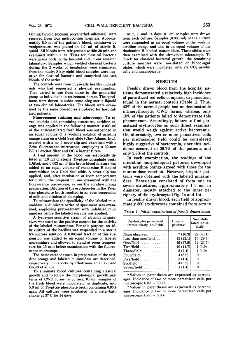
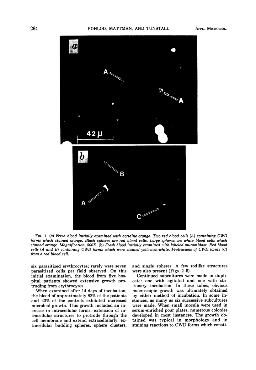
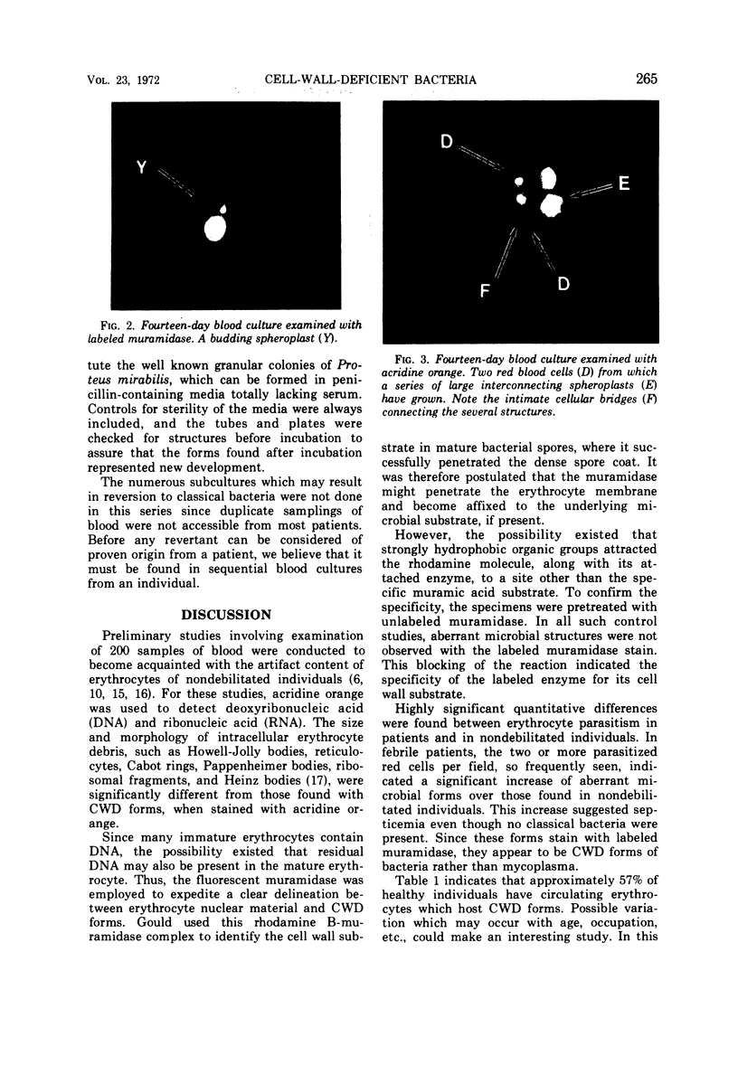
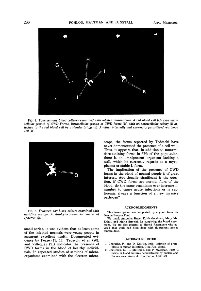
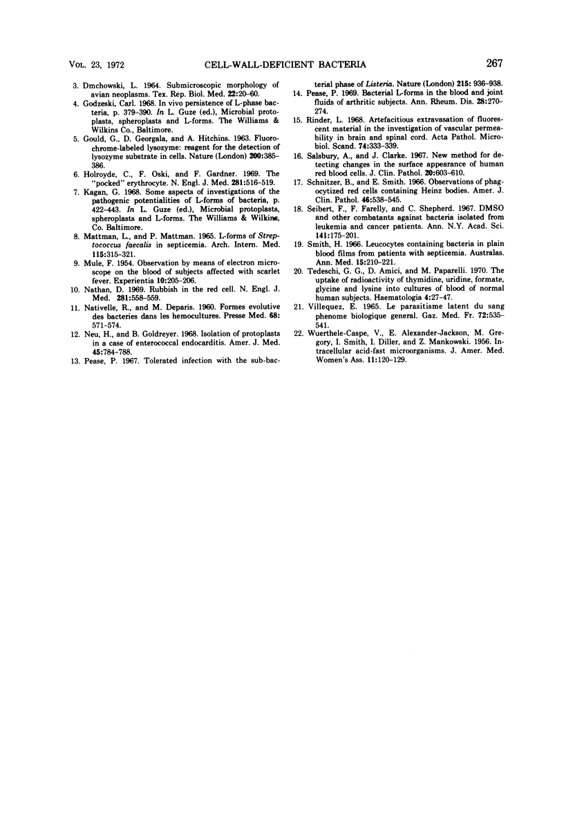
Cell-wall-deficient (CWD) forms of bacteria are associated with certain cases of idiopathic septicemia. In this preliminary study of blood examined immediately after venipuncture, structures with a morphology characteristic of CWD forms were seen parasitizing the erythrocytes. These inclusions were usually circumferential, but in some cases they protruded from the red cells. The CWD forms were detected by staining with Gould's rhodamine-labeled muramidase, which reacted similarly to acridine orange but with greater specificity. A blocking test, employing unlabeled muramidase, indicated the specificity of the reaction between muramidase and the microbial substrate. Reaction of the forms with muramidase indicates their bacterial, rather than mycoplasmal, nature. Thus in vivo CWD forms have a detectable component of muramic acid, at least in certain cases. Sixty-eight individuals with a diagnosis of fever of unknown origin were tested, with 51 nondebilitated individuals serving as controls. More intraerythrocytic forms reacting with muramidase were found in the patients than in the controls. Nearly 40% of the cases had a relatively high incidence of erythrocyte parasitism. In some instances when freshly drawn blood was examined, the structures, which appear to be microbial, extended in rhizoid filaments from the erythrocytes.
Full text
PDF
Images in this article
Selected References
These references are in PubMed. This may not be the complete list of references from this article.
- Chattman M. S., Mattman L. H., Mattman P. E. L forms in blood cultures demonstrated by nucleic acid fluorescence. Am J Clin Pathol. 1969 Jan;51(1):41–50. doi: 10.1093/ajcp/51.1.41. [DOI] [PubMed] [Google Scholar]
- DMOCHOWSKI L., GREY C. E., PADGETT F., LANGFORD P. L., BURMESTER B. R. SUBMICROSCOPIC MORPHOLOGY OF AVIAN NEOPLASMS. VI. COMPARATIVE STUDIES ON ROUS SARCOMA, VISCERAL LYMPHOMATOSIS, ERYTHROBLASTOSIS, MYELOBLASTOSIS, AND NEPHROBLASTOMA. Tex Rep Biol Med. 1964;22:20–60. [PubMed] [Google Scholar]
- GOULD G. W., GEORGALA D. L., HITCHINS A. D. FLUOROCHROME-LABELLED LYSOZYME: REAGENT FOR THE DETECTION OF LYSOZYME SUBSTRATE IN CELLS. Nature. 1963 Oct 26;200:385–386. doi: 10.1038/200385b0. [DOI] [PubMed] [Google Scholar]
- Holroyde C. P., Oski F. A., Gardner F. H. The "pocked" erythrocyte. Red-cell surface alterations in reticuloendothelial immaturity of the neonate. N Engl J Med. 1969 Sep 4;281(10):516–520. doi: 10.1056/NEJM196909042811002. [DOI] [PubMed] [Google Scholar]
- MATTMAN L. H., MATTMAN P. E. L FORMS OF STREPTOCOCCUS FECALIS IN SEPTICEMIA. Arch Intern Med. 1965 Mar;115:315–321. doi: 10.1001/archinte.1965.03860150059011. [DOI] [PubMed] [Google Scholar]
- MULE F. Observation by means of electron microscope on the blood of subjects affected with scarlet fever. Experientia. 1954 May 15;10(5):205–206. doi: 10.1007/BF02159270. [DOI] [PubMed] [Google Scholar]
- Nathan D. G. Rubbish in the red cell. N Engl J Med. 1969 Sep 4;281(10):558–559. doi: 10.1056/NEJM196909042811013. [DOI] [PubMed] [Google Scholar]
- Neu H. C., Goldreyer B. Isolation of protoplasts in a case of enterococcal endocarditis. Am J Med. 1968 Nov;45(5):784–788. doi: 10.1016/0002-9343(68)90211-8. [DOI] [PubMed] [Google Scholar]
- Pease P. E. Tolerated infection with the sub-bacterial phase of Listeria. Nature. 1967 Aug 26;215(5104):936–938. doi: 10.1038/215936a0. [DOI] [PubMed] [Google Scholar]
- Pease P. Bacterial L-forms in the blood and joint fluids of arthritic subjects. Ann Rheum Dis. 1969 May;28(3):270–274. doi: 10.1136/ard.28.3.270. [DOI] [PMC free article] [PubMed] [Google Scholar]
- Rinder L. Artefactitious extravasation of fluorescent indicators in the investigation of vascular permeability in brain and spinal cord. Acta Pathol Microbiol Scand. 1968;74(3):333–339. doi: 10.1111/j.1699-0463.1968.tb03486.x. [DOI] [PubMed] [Google Scholar]
- Salsbury A. J., Clarke J. A. New method for detecting changes in the surface appearance of human red blood cells. J Clin Pathol. 1967 Jul;20(4):603–610. doi: 10.1136/jcp.20.4.603. [DOI] [PMC free article] [PubMed] [Google Scholar]
- Schnitzer B., Smith E. B. Observations of phagocytized red cells containing Heinz bodies. A light and electron microscopic study. Am J Clin Pathol. 1966 Nov;46(5):538–545. doi: 10.1093/ajcp/46.5.538. [DOI] [PubMed] [Google Scholar]
- Seibert F. B., Farrelly F. K., Shepherd C. C. DMSO and other combatants against bacteria isolated from leukemia and cancer patients. Ann N Y Acad Sci. 1967 Mar 15;141(1):175–201. doi: 10.1111/j.1749-6632.1967.tb34879.x. [DOI] [PubMed] [Google Scholar]
- Tedeschi G. G., Amici D., Paparelli M. The uptake of radioactivity of thymidine, uridine, formate, glycine and lysine into cultures of blood of normal human subjects. Relationships with mycoplasma infection. Haematologia (Budap) 1970;4(1):27–47. [PubMed] [Google Scholar]
- VILLEQUEZ E. LE PARASITISME LATENT DU SANG PH'ENOM'ENE BIOLOGIQUE G'EN'ERAL. Gaz Med Fr. 1965 Feb 10;72:535–541. [PubMed] [Google Scholar]
- WUERTHELE-CASPE V., ALEXANDER-JACKSON E., GREGORY M., SMITH L. W., DILLER I. C., MANKOWSKI Z. Intracellular acid-fast microorganism; isolated from two cases of hepatolenticular degeneration. J Am Med Womens Assoc. 1956 Apr;11(4):120–129. [PubMed] [Google Scholar]