

Abstract
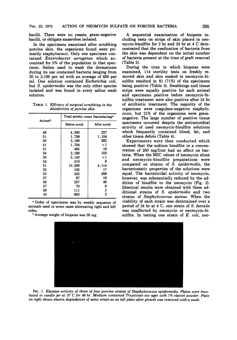
Homogenized 4-mm punch biopsies were taken from pigs and bacteriologically evaluated to determine the efficacy of surgical scrub procedures and the subsequent treatment of tissue with 0.5% neomycin sulfate-sodium bisulfite (neomycin-bisulfite) as a decontaminating agent. The majority of the lots of porcine skin taken directly from animals for xenografts in the treatment of burns contained viable bacteria at the time of grafting although scrubbing procedures substantially reduced the skin bacteria. The porcine bacteria consisted primarily of coagulase-negative staphylococci with most strains exhibiting caseinolytic and elastase activity. Staphylococci were the only abundant bacteria found in postscrub biopsies and in saline solutions used to wash the dermatome during its use. After an overnight exposure of grafting tissue soaked in neomycin-bisulfite, the spent neomycin-bisulfite solutions were tested for bacteriostatic and bactericidal activity by comparison to unused neomycin. All solutions tested were equal in bacteriostatic strength, but the bactericidal action of some spent solutions was decreased. Neomycin alone exerted a more lethal effect on sensitive bacteria than the neomycin-bisulfite solution. The desirability of having viable porcine skin for a xenograft necessitated using or discarding the tissue after storage in neomycin-bisulfite at 4 C for a maximum of 72 hr. Certain contaminating microorganisms were unaffected by antibiotic treatment, and the prolonged use of neomycin without bisulfite would have primarily eradicated only the porcine coagulase-negative staphylococci. Neither the presence of this group in grafting tissue nor their proteolytic activity had any observed adverse effect on xenografting success.
Full text
PDF
Images in this article
Selected References
These references are in PubMed. This may not be the complete list of references from this article.
- BAIRD-PARKER A. C. A classification of micrococci and staphylococci based on physiological and biochemical tests. J Gen Microbiol. 1963 Mar;30:409–427. doi: 10.1099/00221287-30-3-409. [DOI] [PubMed] [Google Scholar]
- Branson D. Identification of Micrococcaceae in clinical bacteriology. Appl Microbiol. 1968 Jun;16(6):906–911. doi: 10.1128/am.16.6.906-911.1968. [DOI] [PMC free article] [PubMed] [Google Scholar]
- Larson D. L., Abston S. Acutely burned patient. Initial care and closure of burn wound. N Y State J Med. 1970 Jun 15;70(12):1626–1633. [PubMed] [Google Scholar]
- Martley F. G., Jayashankar S. R., Lawrence R. C. An improved agar medium for the detection of proteolytic organisms in total bacterial counts. J Appl Bacteriol. 1970 Jun;33(2):363–370. doi: 10.1111/j.1365-2672.1970.tb02208.x. [DOI] [PubMed] [Google Scholar]
- Montes L. F., Wilborn W. H. Anatomical location of normal skin flora. Arch Dermatol. 1970 Feb;101(2):145–159. [PubMed] [Google Scholar]
- Smith R. F. Comparative enumeration of lipophilic and nonlipophilic cutaneous diphtheroids and cocci. Appl Microbiol. 1970 Feb;19(2):254–258. doi: 10.1128/am.19.2.254-258.1970. [DOI] [PMC free article] [PubMed] [Google Scholar]
- Varadi D. P., Saqueton A. C. Elastase from Staphylococcus epidermidis. Nature. 1968 May 4;218(5140):468–470. doi: 10.1038/218468a0. [DOI] [PubMed] [Google Scholar]