Abstract
[Purpose] Rheobase and chronaxie are used to confirm muscle degeneration. For stroke patients, however, the uses of rheobase and chronaxie in determining paretic side muscle degeneration is not yet fully understood. Thus, in this study, we examined the electrical properties of the quadriceps muscles of stroke patients’ paretic side and compared them with their respective values on the non-paretic side. [Method] The subjects were six stroke patients (three females, three males). The pad of an electrical stimulator was applied to the vastus lateralis and vastus medialis regions to measure rheobase and chronaxie until the contractive muscle response to electrical stimulation became visible. [Result] Rheobase was significantly increased on the paretic side compared to that of the non-paretic side of hemiplegic stroke patients. Furthermore, chronaxie was significantly increased on the paretic side compared to the non-paretic side of hemiplegic stroke patients. [Conclusion] These results suggest that stroke affects the sensitivity of skeletal muscle contraction. Therefore, this data may contribute to our understanding of the muscle status of stroke patients.
Key words: Rheobase, Chronaxie, Hemiplegic stroke patients
INTRODUCTION
Upper motor neuron injury, as occurs in stroke, is believed not to result in muscular atrophy, and only a few studies have examined muscle abnormalities after stroke and their relationship with fitness and function1). However, a recent study showed that stroke patients suffer from disproportionate muscle atrophy and other detrimental tissue composition changes on the paretic side2). This is the reason that many stroke survivors move using abnormal movement patterns3,4,5,6). Muscle weakness develops rapidly after stroke, adversely affecting motor performance, and contributing to reduced functional ability7). Especially, lower-extremity muscle weakness, particularly in the quadriceps muscle, has profound functional consequences8). Post-stroke rehabilitation therapy aims to restore partially lost functions9). Electric therapy in particular, such as functional electrical stimulation (FES) of the muscle, is known to be an effective method of improving the motor function of stroke patients10). Thus, electrical properties such as rheobase and chronaxie are important factors in electrotherapy for rehabilitation. Because electrical properties affect electric current, the appropriate adjustment of electric current increases the effectiveness of rehabilitation. Despite the importance of this for stroke patients, the paretic side is not fully understood in terms of rheobase and chronaxie, which are commonly used to confirm the electrical characteristics of the muscle. Thus, in this study, we examined the muscle properties of the quadriceps (especially, the vastus lateralis and vastus medialis) of stroke patients’ paretic side and compared them with their respective values on the non-paretic side.
SUBJECTS AND METHODS
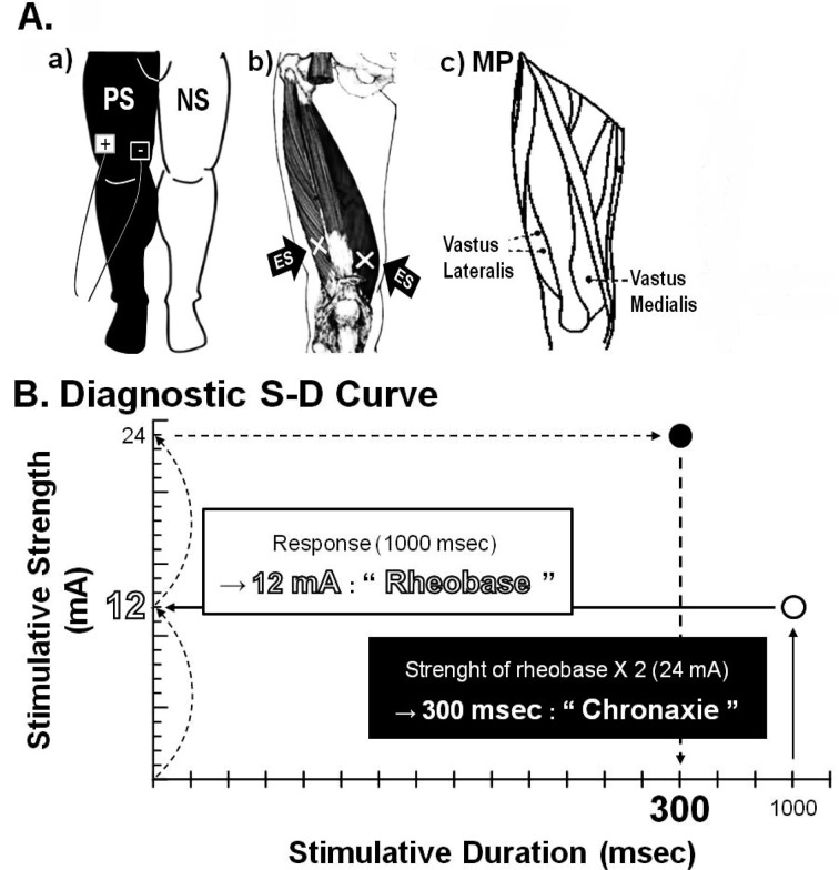
For the study, six participants (three females, three males) were recruited from B hospital located in Yong-in, Korea (weight: 68.2±4.7 kg; height: 164.8±2.8 cm). Participants had previously been diagnosed with cerebral infarction, or cerebral hemorrhage, according to established criteria (Table 1). Rheobase and chronaxie were measured at the regions of the vastus lateralis and vastus medialis muscles using an electrical stimulator (Duo 500, Gymnauniphy Co., Belgium) (Fig. 1). A rheobase measurement pad was applied to the regions of the vastus lateralis and vastus medialis muscles of subjects, who were in the sitting position, until the muscle contraction response became visible. A chronaxie measurement pad was applied to the same regions until muscle contraction responses became visible using double the rheobase current intensity (Fig. 1). After one side had been measured, subjects rested for 5 minutes; then, the process was repeated on the other side. Data were expressed as mean ± standard error (SE). A p value of < 0.05 was considered statistically significant. SPSS Version 18.0 (International Business Machines, Armonk, USA) for Microsoft Windows was used for data analysis in this study. The protocol for the study was approved by the Committee of Ethics in Research of the University of Yongin, in accordance with the terms of Resolution 5-1-20, December 2006.
Table 1. Clinical characteristics of the hemiplegic stroke patients.
| No | Age (year) | Gender | BMI (kg/m2) | Time post stroke (month) | K-MMSE (Score) | PS | Lesion site |
| 1 | 32 | Male | 27.9 | 42 | 30/30 | L | Left thalamic ICH |
| 2 | 57 | Male | 28.7 | 68 | 30/30 | L | Middle cerebral artery |
| 3 | 49 | Female | 23.5 | 40 | 30/30 | R | Middle cerebral artery |
| 4 | 64 | Female | 18.4 | 42 | 30/30 | R | Left thalamic ICH |
| 5 | 67 | Female | 27.9 | 61 | 28/30 | L | Basal ganglia ICH |
| 6 | 63 | Male | 24.1 | 18 | 30/30 | L | Basal ganglia ICH |
BMI, body mass index; K-MMSE, Korean version of mini mental status examination; PS, paretic side;
L, left side; R, right side; ICH, intra-cerebral hemorrhage
Fig. 1.
Schematic representation of the measurements of rheobase and chronaxie
In the analysis of rheobase and chronaxie, the thigh (parts of the vastus lateralis and vastus medialis) was stimulated using an electrical stimulator and two surface electrodes of the same size for bipolar stimulation. Rheobase and chronaxie were measured using monofilaments, as described in the Materials and Methods. +, anode; -, cathode; →, region of stimulation of the anterior parts of thigh; PS, paretic side of stroke patients; NS, non-paretic side of stroke patients; ES, electrical stimulation; MP, motor point; S-D Curve, strength-duration curve.
RESULTS
To confirm whether stroke state affects the sensitivity of skeletal muscle contraction, the rheobase and chronaxie properties of stroke patients were compared between the paretic and non-paretic sides. Rheobase was significantly higher on the paretic side than on the non-paretic side of hemiplegic stroke patients (Table 2). Furthermore, chronaxie was significantly higher on the paretic side than on the non-paretic side of hemiplegic stroke patients (Table 2).
Table 2. Differences in rheobase and chronaxie between the paretic and non-paretic sides of hemiplegic stroke patients.
| Non-paretic side | Paretic side | ||
| Rheobase (mA) | Chronaxie (msec) | Rheobase (mA) | Chronaxie (msec) |
| 12.3 ± 0.3 | 0.1 ± 0.0 | 15.1 ± 0.4* | 0.2 ± 0.0* |
Mean±SE. *, Significantly different from the non-paretic side p < 0.05
DISCUSSION
Stroke is a leading cause of neurological disability among adults and often leads to functional deficits in motor control4, 5, 11, 12). Stroke involves muscle weakness, spasms, disturbed muscle timing, and a reduced ability to selectively activate muscles5, 6, 9). Quadriceps weakness in particular is a common finding in stroke patients13, 14). Quadriceps muscle weakness is associated with decreased gait speed, balance, stair-climbing ability, and ability to rise from a seated position, as well as with an increased risk of falls15). Thus, quadriceps muscle function improvement is of utmost importance in stroke patients’ rehabilitation. Electrotherapy is known to be an effective method of improving motor function and is currently used in many forms to facilitate changes in muscle action and performance16). For example, FES has been widely used to treat patients with lesions in the central nervous system arising from stroke or spinal cord injury in order to improve motor control17). Thus, we measured rheobase and chronaxie to gain a better understanding of their implications in electrical therapy for stroke patients, because they are commonly accepted as parameters predicting the efficacy of electrical stimulation18). Rheobase is measured as the threshold stimulus current for an active response with a long-duration pulse and chronaxie is the pulse width at twice the rheobase threshold current19). There have been few studies that have focused on physical therapy using rheobase and chronaxie, and whether it should be applied differently based on these differences. Thus, we investigated this issue. All patients showed lower rheobase and chronaxie on their non-paretic sides than on their paretic sides. Based on the results of our study, we can carefully provide an important basis for electric current and duration when performing electrotherapy for stroke patients. In other words, the paretic sides of stroke patients require a higher current and longer pulse duration for quadriceps muscle reaction than the non-paretic sides. Therefore, it is necessary to consider the proper intensities for electrotherapy. A major limitation of this study is the lack of measurements of the other muscles. However, since few studies have been performed on differences in muscle electrical characteristics, we consider the present results are meaningful for physical therapy. Further studies including investigation of diverses muscle of stroke patients would contribute to the development of clinical physical therapy and to electrotherapy. In summary, there were significant differences in rheobase and chronaxie between the paretic and non-paretic sides of hemiplegic stroke patients. Rheobase and chronaxie of the paretic side had greater values than the non-paretic side. In this study, we found that there was a difference between the electrical properties of the paretic and non-paretic sides of hemiplegic stroke patients. Therefore, when performing physical therapy, the electrical properties of muscle of each stroke patient need to be carefully considered.
REFERENCES
- 1.Hafer-Macko CE, Ryan AS, Ivey FM, et al. : Skeletal muscle changes after hemiparetic stroke and potential beneficial effects of exercise intervention strategies. J Rehabil Res Dev, 2008, 45: 261–272 [DOI] [PMC free article] [PubMed] [Google Scholar]
- 2.Ryan AS, Ivey FM, Prior S, et al. : Skeletal muscle hypertrophy and muscle myostatin reduction after resistive training in stroke survivors. Stroke, 2011, 42: 416–420 [DOI] [PMC free article] [PubMed] [Google Scholar]
- 3.Twitchell TE: The restoration of motor function following hemiplegia in man. Brain, 1951, 74: 443–480 [DOI] [PubMed] [Google Scholar]
- 4.Kim MY, Kim JH, Lee JU, et al. : The effects of functional electrical stimulation on balance of stroke patients in the standing posture. J Phys Ther Sci, 2012, 24: 77–81 [Google Scholar]
- 5.Kim MY, Kim JH, Lee JU, et al. : The effect of low frequency repetitive transcranial magnetic stimulation combined with range of motion exercise on paretic hand function in female patients after stroke. Neurosci Med, 2012, (in press). [Google Scholar]
- 6.Jeon HJ, Kim JH, Hwang BY, et al. : Analysis of the sensory threshold between paretic and nonparetic sides for healthy rehabilitation in hemiplegic patients after stroke. Health, 2012, 4: 1241–1246 [Google Scholar]
- 7.Madhavan S, Krishnan C, Jayaraman A, et al. : Corticospinal tract integrity correlates with knee extensor weakness in chronic stroke survivors. Clin Neurophysiol, 2011, 122: 1588–1594 [DOI] [PMC free article] [PubMed] [Google Scholar]
- 8.Stevens-Lapsley JE, Balter JE, Wolfe P, et al. : Early neuromuscular electrical stimulation to improve quadriceps muscle strength after total knee arthroplasty: a randomized controlled trial. Phys Ther, 2012, 92: 210–226 [DOI] [PMC free article] [PubMed] [Google Scholar]
- 9.Krabben T, Molier BI, Houwink A, et al. : Circle drawing as evaluative movement task in stroke rehabilitation: an explorative study. J Neuroeng Rehabil, 2011, 8: 15 [DOI] [PMC free article] [PubMed] [Google Scholar]
- 10.Langhorne P, Coupar F, Pollock A: Motor recovery after stroke: a systematic review. Lancet Neurol, 2009, 8: 741–754 [DOI] [PubMed] [Google Scholar]
- 11.Takahashi M, Takeda K, Otaka Y, et al. : Event related desynchronization-modulated functional electrical stimulation system for stroke rehabilitation: a feasibility study. J Neuroeng Rehabil, 2012, 9: 56 [DOI] [PMC free article] [PubMed] [Google Scholar]
- 12.Kim JH, Kim MY, Lee JU: The effects of symmetrical self-performed facial muscle exercises on the neuromuscular facilitation of patients with facial palsy. J Phys Ther Sci, 2011, 23: 543–547 [Google Scholar]
- 13.Bohannon RW: Muscle strength and muscle training after stroke. J Rehabil Med, 2007, 39: 14–20 [DOI] [PubMed] [Google Scholar]
- 14.Patten C, Lexell J, Brown HE: Weakness and strength training in persons with poststroke hemiplegia: rationale, method, and efficacy. J Rehabil Res Dev, 2004, 41: 293–312 [DOI] [PubMed] [Google Scholar]
- 15.Rantanen T, Guralnik JM, Izmirlian G, et al. : Association of muscle strength with maximum walking speed in disabled older women. Am J Phys Med Rehabil, 1998, 77: 299–305 [DOI] [PubMed] [Google Scholar]
- 16.Doucet BM, Lam A, Griffin L: Neuromuscular electrical stimulation for skeletal muscle function. Yale J Biol Med, 2012, 85: 201–215 [PMC free article] [PubMed] [Google Scholar]
- 17.Rushton DN: Functional electrical stimulation and rehabilitation-an hypothesis. Med Eng Phys, 2003, 25: 75–78 [DOI] [PubMed] [Google Scholar]
- 18.Efimov IR: Chronaxie of defibrillation: a pathway toward further optimization of defibrillation waveform? J Cardiovasc Electrophysiol, 2009, 20: 315–317 [DOI] [PMC free article] [PubMed] [Google Scholar]
- 19.Leal-Cardoso JH, Matos-Brito BG, Lopes-Junior JE, et al. : Effects of estragole on the compound action potential of the rat sciatic nerve. Braz J Med Biol Res, 2004, 37: 1193–1198 [DOI] [PubMed] [Google Scholar]


