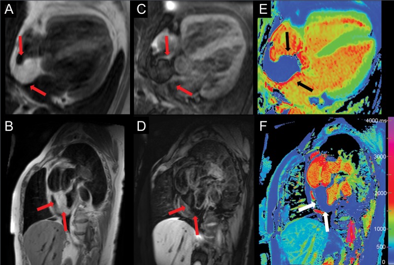
A 67-year-old woman initially presented with dyspnoea. Echocardiography demonstrated an inter-atrial mass. Cardiovascular magnetic resonance (CMR) showed a well-marginated mass (35 mm × 45 mm) in the inter-atrial septum (IAS) sparing the fossa ovalis, extending to the posterior right atrial wall. The mass had high signal intensity on T1-weighted images (Panels A and B, arrows) consistent with fat, which attenuated on fat-suppression imaging (Panels C and D, arrows). Native T1-maps (Panels E and F, arrows) showed homogenous and significantly lower T1 values (230–350 ms) compared with the normal myocardium (green) and blood (yellow/red), but similar to subcutaneous and epicardial fat (blue). The mass had no late gadolinium enhancement and was stable in the size compared with 1-year prior. Findings were consistent with lipomatous hypertrophy of the IAS.
This is a first demonstration of using native T1-mapping for immediate visual and quantitative tissue characterization of an intra-cardiac mass to help confirm its fat content. T1-maps provide pixel-wise estimates of the T1 relaxation times of the underlying tissue, and each tissue type exhibits a characteristic normal range of T1 values. This allows the visual differentiation of tissues using colour scales as shown. Lipomatous hypertrophy of the IAS is an unencapsulated proliferation of adipose tissue ≥20 mm within the atrial septum that is in continuity with epicardial fat. It is associated with older age, increased fat elsewhere in the body, atrial arrhythmias and in some cases, obstruction of the superior vena cava. The correct differentiation of this benign cardiac mass from other atrial tumours using multi-parametric tissue characterization techniques on CMR can prevent unnecessary invasive interventions.

Funding
Funding to pay the Open Access publication charges for this article was provided by the Division of Cardiovascular Medicine, Radcliffe Department of Medicine, University of Oxford.


