Abstract
Paramyxoviruses, including the emerging lethal human Nipah virus (NiV) and the avian Newcastle disease virus (NDV), enter host cells through fusion of the viral and target cell membranes. For paramyxoviruses, membrane fusion is the result of the concerted action of two viral envelope glycoproteins: a receptor binding protein and a fusion protein (F). The NiV receptor binding protein (G) attaches to ephrin B2 or B3 on host cells, whereas the corresponding hemagglutinin-neuraminidase (HN) attachment protein of NDV interacts with sialic acid moieties on target cells through two regions of its globular domain. Receptor-bound G or HN via its stalk domain triggers F to undergo the conformational changes that render it competent to mediate fusion of the viral and cellular membranes. We show that chimeric proteins containing the NDV HN receptor binding regions and the NiV G stalk domain require a specific sequence at the connection between the head and the stalk to activate NiV F for fusion. Our findings are consistent with a general mechanism of paramyxovirus fusion activation in which the stalk domain of the receptor binding protein is responsible for F activation and a specific connecting region between the receptor binding globular head and the fusion-activating stalk domain is required for transmitting the fusion signal.
INTRODUCTION
The entry of enveloped viruses into host cells requires fusion of the viral and cell membranes. Viral fusion is driven by specialized fusion proteins that bring the viral and host membranes in close apposition to form a fusion pore (reviewed previously [1–6]). The trigger that initiates a series of conformational changes in F leading to membrane fusion differs depending on the pathway that the virus uses to enter the cell, i.e., whether fusion occurs at neutral pH at the surface or at low pH in the endosome. For paramyxoviruses, the F protein is activated when the adjacent receptor binding protein binds to its receptor on host cell and initiates the fusion process (7). Once activation occurs, the fusion protein undergoes a coordinated series of conformational changes that progress toward the most stable form of the protein and promote membrane fusion (reviewed in references 8 and 9). The role of the receptor binding protein in this process is critical (10–15).
Paramyxoviruses possess envelope proteins that provide a receptor binding function and, depending on the specific paramyxovirus family member, a receptor cleaving (neuraminidase) activity. A recently identified function of the receptor binding protein of human parainfluenza virus 3 (HPIV3), which may apply to other paramyxoviruses (16), is to stabilize the fusion protein and prevent its activation until the virus engages receptor (17). Most paramyxovirus receptor binding proteins studied to date also serve the critical function of activating the fusion protein (F) upon receptor engagement. The receptor binding proteins possess a membrane distal globular head domain that engages the receptor and a membrane proximal stalk that confers specificity toward the homologous F protein.
For Newcastle disease virus (NDV), the envelope protein hemagglutinin-neuraminidase (HN) contains both receptor binding and neuraminidase activities. When bound to receptor, HN triggers F to undergo conformational changes that lead to membrane fusion (7, 18–20). HN is a type II membrane protein with a cytoplasmic domain, a membrane-spanning region, a stalk region, and a globular head that interacts with sialic acid receptors. Structural analysis of the HNs from avian NDV (21, 22), HPIV3 (23), and simian virus 5 (or parainfluenza virus type 5 [PIV5]) (24) has identified the locations of the primary binding/neuraminidase active-site residues on the globular head of the molecule, as well as several key structural elements that are required for the fusion-triggering function of HN (7, 18–20). The analyses of NDV revealed two sialic acid binding regions, sites I and II, on HN. We previously reported that site II can be activated for receptor binding by small molecules (e.g., zanamivir) that occupy site I (25), and this finding was supported by recent analysis of a series of NDV HN mutants (25–27).
We recently described a chimeric protein consisting of the globular head of NDV HN and the stalk domain of NiV G that activates NiV F, meaning that the head of a heterotypic paramyxovirus can signal F through a homotypic stalk. Activation of site II of the receptor binding protein is a determinant for fusion activation (27, 28). We now explore the hypothesis that the connecting region between the stalk domain and the globular head of the receptor binding protein plays a pivotal role in fusion promotion, whether the fusion protein is homotypic or heterotypic with respect to the globular head. Specific residues between the stalk and globular domains of the receptor binding protein are required for efficient triggering of NiV F, and alterations in this connecting region prevent the globular head from activating the stalk domain. Our results are consistent with a unified mechanism of fusion activation for paramyxoviruses, in which the globular domain of the receptor binding protein transmits the fusion signal to the F protein through the stalk domain of the binding protein.
MATERIALS AND METHODS
Cell cultures.
293T (human kidney epithelial cells) were grown in Dulbecco modified Eagle medium (Gibco) supplemented with 10% fetal bovine serum and antibiotics in a humidified incubator supplemented with 5% CO2.
Chemicals.
Zanamivir was prepared from Relenza Rotadisks (5 mg of zanamivir with lactose). A 50 mM stock solution was prepared by dissolving each 5-mg blister capsule in 285 μl of Opti-MEM (Gibco). Stock solutions were stored at −20°C.
Plasmids.
The NiV wild-type (wt) G and F genes were codon optimized and synthesized by GeneArt (Germany) and subcloned into the mammalian expression vector pCAGGS using the EcoRI or the XhoI and BglII restriction enzyme sites. The chimeric cDNAs were codon optimized and synthesized by Epoch Biolabs and subcloned into the mammalian expression vector pCAGGS between EcoRI and BglII. The pCAGGS NDV AV (Australia-Victoria) HN and F constructs were generously provided by Ronald Iorio (University of Massachusetts, Worcester, MA).
Transient expression of NDV HN/F, NiV G/F, and chimeric cDNA genes.
Transfections were performed according to the Lipofectamine 2000 manufacturer's protocol (Invitrogen).
Pseudotyped virus infection assay.
Generation and recovery of recombinant VSV-F(NiV) were carried out as previously described (29). Briefly, for the recovery of VSV-F(NiV) complemented with the indicated receptor binding proteins, BHK-21 cells transiently expressing the indicated receptor binding proteins were infected at 24 h posttransfection with VSV-F(NiV) complemented with VSV-G. Medium containing the virus was collected after 48 h. Virus stocks were stored at −80°C and titered in 293T cells transfected with NiV G.
Plaque assays and plaque size assessment.
Supernatant fluids containing recombinant pseudotyped viruses were serially diluted in medium with reduced serum (Opti-MEM) and added to confluent 293T cell monolayers transiently expressing NiV G. Cells were incubated at 37°C. After 90 min, minimum essential medium containing 0.5% agarose (for plaque size assessment) or 0.4% Avicel RC-591 (FMC BioPolymer) (for plaque counting) was added to the dishes, and the dishes were incubated for 24 h at 37°C. After the overlay was removed, the cells were fixed with 4% formaldehyde in phosphate-buffered saline (PBS) for 15 min and stained for plaque detection using polyclonal antibodies against NiV F.
Detection of protein expression and processing by immunoprecipitation.
293T cell monolayers were transiently transfected with NiV F and receptor binding protein constructs and then incubated overnight in medium containing 10 μM HPIV3-derived E459V fusion inhibitory peptide to prevent fusion (30). The transfected cells were washed with starvation medium (lacking glutamine, cysteine, and methionine), followed by incubation for an additional 2 h at 37°C in the same medium. The starvation medium was then replaced with fresh medium supplemented with 2 mM glutamine and 55 μCi of Expre35S[35S] cysteine-methionine labeling mix (Perkin-Elmer, Boston, MA) for 2 h at 37°C. After the incubation, 100 ng of cycloheximide/ml was added to prevent de novo protein synthesis (31). The cells were then washed with cold PBS and lysed in lysis buffer A (Invitrogen, catalog no. 143.21D). HN, G, and F and the chimeric glycoproteins were immunoprecipitated from postnuclear lysates with protein G-conjugated Sepharose beads that were preincubated either with anti-NDV HN, -NiV G, or -NiV F antibodies. After overnight incubation at 4°C, the beads were washed twice in lysis buffer, and the last wash was completely removed. The protein-bead complexes were suspended in Laemmli's sodium dodecyl sulfate (SDS) sample buffer, heated to 99°C for 5 min, and then subjected to SDS-PAGE, membrane transfer, and autoradiography.
Hemadsorption assay.
A hemadsorption assay was performed and quantified as previously described (32). Briefly, growth medium from 293T cell monolayers cotransfected with HN and F in 24- or 48-well Biocoat plates (Becton Dickinson Labware) was aspirated, replaced with 150 μl of CO2-independent medium (pH 7.3; Gibco) with or without the indicated concentrations of zanamivir and 1% red blood cells (RBCs) in serum-free, CO2-independent medium, and kept at 4°C for 30 min. The wells were then washed three times with 150 μl of cold CO2-independent medium. The bound RBCs were lysed with 200 μl of RBC lysis solution (0.145 M NH4Cl and 17 mM Tris-HCl), and the absorbance was read at 405 nm using a Spectramax M5 (Molecular Devices) microplate reader.
Cell surface expression and ephrin B2 binding assays.
Monolayers of 293T cells transiently transfected with the indicated constructs were washed twice in PBS and then incubated with a pool of mouse anti-NDV HN monoclonal antibodies (sc53561, sc53562, and sc53563 from Santa Cruz Biotechnology) or with a pool of rat anti-NiV G monoclonal antibodies (33) in 3% bovine serum albumin and 0.1% sodium azide in PBS for 1 h. Samples were then washed twice with PBS, followed by incubation with 1:100 of an anti-mouse IgG(H+L)-fluorescein isothiocyanate (FITC) conjugate (BD Pharmingen) or with an anti-rat-FITC conjugate (Jackson ImmunoResearch). To quantify the cell surface protein in each sample, indirect immunofluorescence was measured by fluorescence-activated cell sorting (FACS; FACScan; Becton Dickinson). For ephrin binding, cells expressing indicated construct were washed with PBS and then incubated with either ephrin B1-Fc or ephrin B2-Fc mouse chimera (Sigma-Aldrich) for 1 h. Samples were then washed twice with PBS and incubated with 1:100 of an anti-human IgG(H+L)-Alexa 488 conjugate (Invitrogen).
Measurement of neuraminidase activity.
Assays were performed with transiently transfected 293T cell monolayers as previously described (20, 32). Briefly, 293T cells expressing viral glycoproteins were added to 96-well plates in CO2-independent medium (Gibco) at pH 6.5 and incubated at 37°C for 1 h in the presence of reaction mixtures containing 20 mM substrate [2′-(4-methylumbelliferyl)-α-d-N-acetylneuraminic acid; Toronto Research Chemicals, Inc.] with or without zanamivir. Throughout this period, fluorescence resulting from the hydrolysis of the substrate was read at 365-nm excitation wavelength and 450-nm emission wavelength using a Spectramax M5 microplate reader.
β-Gal complementation-based fusion assay.
We previously adapted a fusion assay based on alpha complementation of β-galactosidase (β-Gal) (34, 35). In this assay, receptor-bearing cells expressing the omega peptide of β-Gal are mixed with cells coexpressing envelope glycoproteins and the alpha peptide of β-Gal, and cell fusion leads to complementation. Fusion is stopped by lysing the cells and, after addition of the substrate, fusion is quantified on a Spectramax M5 microplate reader.
Measurement of RBC fusion with envelope glycoprotein-expressing cells.
Monolayers of 293T cells in 24-well plates transiently coexpressing viral glycoproteins were washed and incubated with 1% RBC suspensions at pH 7.4 for 30 min at 4°C with or without zanamivir (2 mM). After a rinsing step to remove unbound RBCs, the cells were placed at 37°C for the indicated time with or without 2 mM zanamivir. The plates were then rocked, and the liquid phase was collected in V-bottom 96-well plates for the measurement of released RBCs. The monolayers were then incubated at 4°C with RBC lysis solution to remove RBCs that have not fused with cells coexpressing envelope glycoprotein. The liquid phase was collected in V-bottom 96-well plates for measurement of reversibly bound RBCs. The cells were then lysed in 200 μl of 0.2% Triton X-100–PBS and transferred to flat-bottom 96-well plates for quantification of the pool of fused RBCs. The percentage of RBCs in each of the above three compartments was determined by measurement of the absorption at 405 nm.
Assessment of F protein insertion into RBC membranes.
Monolayers of 293T cells in 24-well plates transiently coexpressing viral glycoproteins were washed and incubated with 1% RBC suspensions (pH 7.5) for 30 min at 4°C with zanamivir (2 mM). After a rinsing step to remove unbound RBCs, the cells were placed at 37°C for 90 min with 2 mM zanamivir. The cells were then washed and incubated at 37°C for 75 min in fresh medium at pH 6.5 without zanamivir. The plates were rocked, and the liquid phase was collected in V-bottom 96-well plates for the measurement of released RBCs. The cells were then incubated at 4°C with 200 ml of RBC lysis solution, wherein the lysis of unfused RBCs with NH4Cl removes RBCs that have not fused with cells coexpressing envelope glycoproteins. The liquid phase was collected in V-bottom 96-well plates for measurement of bound RBCs. The cells were then lysed in 200 μl of 0.2% Triton X-100–PBS and transferred to flat-bottom 96-well plates for quantification of the fused RBCs. The amount of RBCs in each of the above three compartments was determined by measuring the absorption at 405 nm.
RESULTS
Chimeric viral glycoproteins containing the NiV G stalk domain and NDV HN globular head (G-HN) are expressed on the cell surface and promote F-mediated fusion.
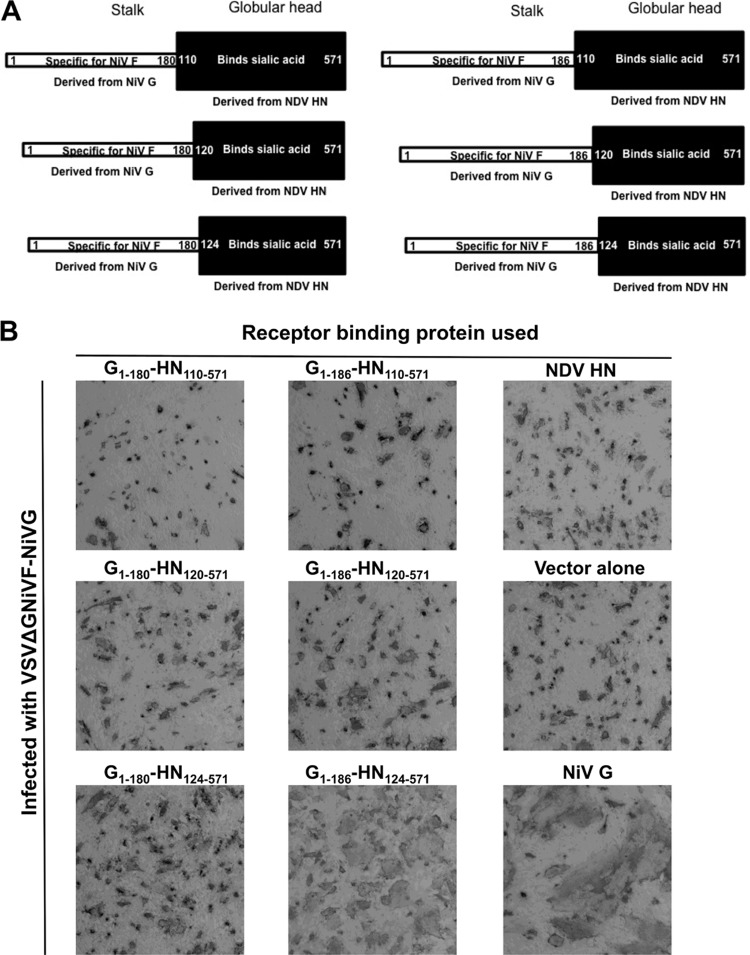
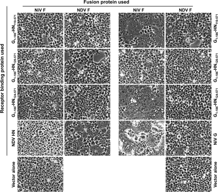
We have shown that two chimeric proteins with the NiV G stalk domain and NDV AV (Australia-Victoria) HN globular head domain, G1-186-HN124-571 and G1-188-HN124-571, activate NiV F to mediate fusion (27, 28) and that fusion activation requires a functional NDV globular domain site II (27, 36). A series of chimeric proteins were designed with NiV G stalk domains of different lengths, as well as NDV HN globular domains with or without segments from the NDV HN stalk domain (Fig. 1A), to investigate the role of the region connecting the globular head domain to the stalk domain. The activity of these chimeric proteins in viral plaque enlargement was evaluated by measuring plaque sizes 24 h postinfection (Fig. 1B). Cells transfected with the indicated receptor binding proteins (NiV G, NDV HN, or chimeric proteins, as indicated in Fig. 1A) were infected with a recombinant vesicular stomatitis virus (VSV) that encodes NiV F but not VSV-G [VSV-F(NiV)], complemented with VSV-G externally and is thus replication deficient. This replication-deficient virus, which carries VSVG and NiV F on its surface but only NiV F in its genome, has been shown to efficiently promote fusion and plaque formation when complemented with NiV G (29). We use this recombinant virus to evaluate the activity of the chimeric receptor proteins in viral plaque enlargement, which has been an accurate marker for the fusion activation properties of several HPIV3 HN proteins (32). Only the chimeric proteins that can promote fusion would be able to promote plaque enlargement. 293T cells transfected with G1-180-HN110-571, G1-180-HN120-571, or G1-180-HN124-571 failed to show any plaque enlargement when infected with the recombinant virus (Fig. 1B). 293T cells transfected with G1-186-HN124-571 showed plaque enlargement, comparable to that observed in the NiV G-transfected cells. Similarly, cotransfecting 293T cells with NiV F and the receptor binding proteins shown in Fig. 1A (Fig. 2) confirmed that the region in NiV G between 181 and 186 is required for cell-to-cell fusion mediated by NiV F. 293T cells transfected with either G1-186-HN110-571 or G1-186-HN124-571 showed cell-to-cell fusion comparable to that observed in the NiV G transfected cells. However, the chimeric protein G1-186-HN120-571 (which included the NDV HN domain present in the chimeric protein G1-186-HN124-571) failed to promote plaque enlargement or fusion.
Fig 1.
Chimeric proteins containing the NiV G stalk domain and the NDV globular can activate NiV F-mediated fusion. (A) Schematic diagram of chimera NiV-NDV. The stalk region was derived from residues 1 to 180 or from residues 1 to 186 of NiV G, and the globular head was derived from either residues 110 to 571, 120 to 571, or 124 to 571 of NDV HN. (B) Plaque size was evaluated by staining with anti-F antibodies.
Fig 2.
Chimeric proteins containing the NiV G stalk domain and the NDV globular can activate NiV/NDV F-mediated cell-cell fusion. Cell-to-cell fusion promoted by the chimeric proteins (shown in Fig. 1A) coexpressed with NiV F was assessed by syncytium formation.
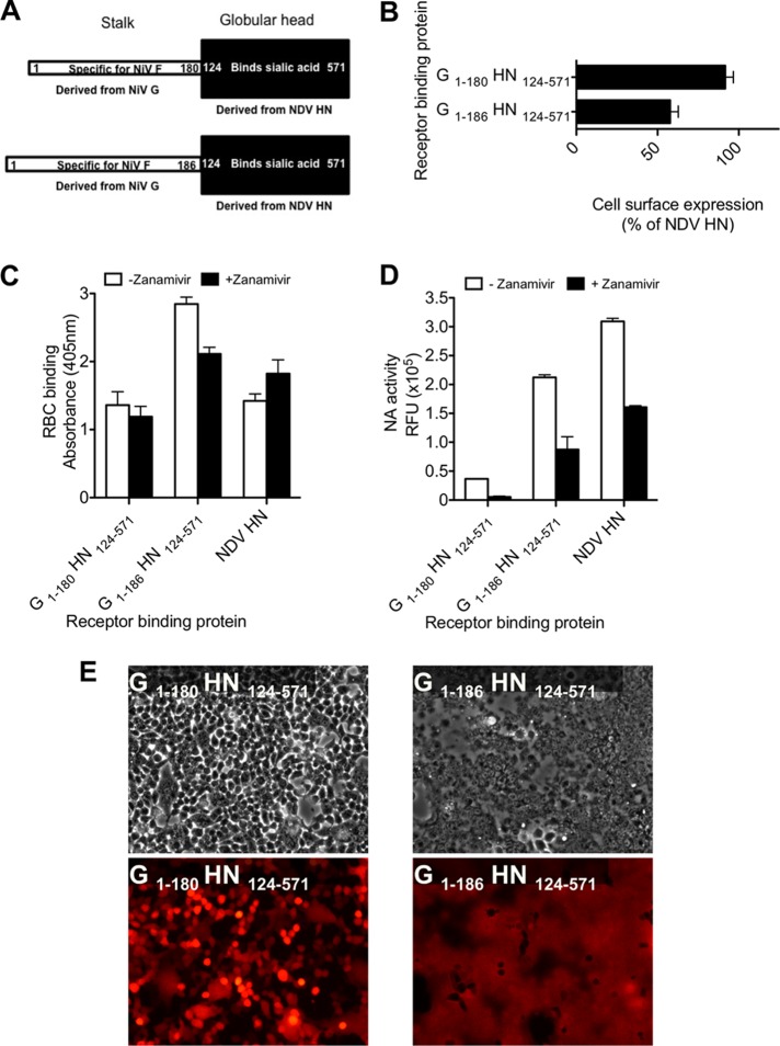
To address the role of the region between amino acids 181 and 186 of NiV G in fusion promotion, we focused on the properties of two chimeric proteins, G1-180-HN124-571 and G1-186-HN124-571 (Fig. 3A). Cell surface expression of the chimeric proteins, measured by FACS analysis (Fig. 3B) using pooled anti-NDV AV HN monoclonal antibodies, was similar to NDV AV HN (27, 36). Sialic acid receptor binding by the chimeric proteins was assessed by a quantitative hemadsorption assay (27, 35). The receptor binding avidities of the expressed chimeric proteins were similar to those of NDV AV HN (Fig. 3C) (27). As shown previously, zanamivir occupies site I of NDV HN and sialic acid binding switches to site II (27) without a significant effect on overall receptor binding.
Fig 3.
Chimeric proteins containing the NiV G stalk domain and the NDV globular head are efficiently expressed and can activate NiV F-mediated fusion. (A) Schematic diagram of chimera NiV-NDV. The stalk region is derived from residues 1 to 180 or residues 1 to 186 of NiV G, and the globular head is derived from residues 124 to 571 of NDV HN. (B) FACS analysis of cell surface expression from cells transfected with the chimeric proteins shown in Fig. 1A. The results are presented as percentages of NDV HN cell surface expression. (C) Receptor binding in the absence (□) or presence (■) of 2 mM zanamivir. (D) Neuraminidase activity of the receptor binding proteins, expressed in relative fluorescence intensity units (RFU) in the absence (□) or presence (■) of 2 mM zanamivir. The values in panels B, C, and D are means ± the standard deviations (SD) of results from samples assessed in triplicate and are representative of the experiment repeated at least three times. (E) Cell-to-cell fusion promoted by the chimeric proteins coexpressed with NiV F is observed in the top panel by syncytium formation using visible microscopy and in the bottom panel by redistribution of RFP (red fluorescent protein) using fluorescence microscopy.
Neuraminidase activity was assessed in the presence or absence of zanamivir, which inhibits NDV's neuraminidase activity (25). For chimeric proteins with mutated stalk domains, neuraminidase activity was decreased compared to NDV AV HN wt, especially for G1-180-HN124-571 (Fig. 3D); this finding is in accord with previous observations that a single amino acid change in the stalk domain significantly altered the neuraminidase activity (7, 19, 37). The neuraminidase activity was reduced in the presence of zanamivir for all of the expressed viral chimeric proteins, indicating that zanamivir effectively interacts with catalytic site I. Figure 3C and 3D show that receptor binding activity is maintained despite zanamivir's blockade of site I (as evidenced by the inhibition of neuraminidase), indicating that the NDV site II was activated for receptor binding in the chimeric proteins as described for NDV AV HN wt and other G-HN and HN-HN chimeric proteins (25–27).
To assess the fusion promotion function of the chimeric proteins with NiV F, cells were cotransfected with the indicated chimeric glycoproteins and NiV F, along with red fluorescent protein (RFP) (38), and syncytium formation was monitored. The chimeric protein G1-180-HN124-571 did not promote significant cell-to-cell fusion, i.e., no diffusion of the red fluorescence could be observed, when coexpressed with NiV F despite proper expression and binding (Fig. 3E). The chimeric protein G1-186-HN124-571 efficiently promoted cell-to-cell fusion in the presence of NiV F, as shown previously (27). These results suggest that the connecting region between the globular head and the stalk does not affect binding activity of the chimeric proteins, but it has a role in fusion activation.
Activation of NiV F by the G1-186-HN124-571 chimeric protein is determined by amino acids 181 and 182.
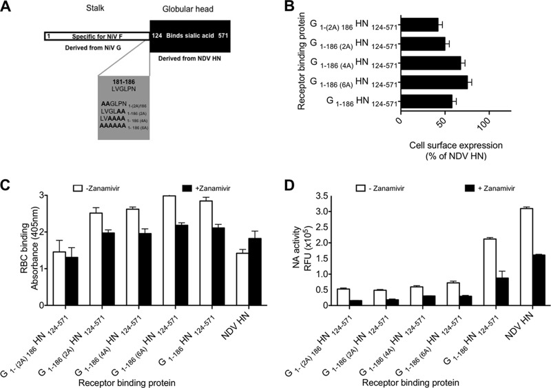
The chimeric protein G1-180-HN124-571 is impaired in fusion promotion (Fig. 3E), implicating that the region between amino acids 181 and 186 is crucial in proper NiV F activation. To assess the role of individual residues in this region, a new set of chimeric proteins was designed. Each of the amino acids 181 and 186 were replaced with alanine in different combinations (Fig. 4A). The four new chimeric proteins were expressed on the cell surface (Fig. 4B), retained RBC binding (Fig. 4C), and retained receptor cleaving activities (Fig. 4D). Similarly to NDV HN, the chimeric proteins' neuraminidase activities were sensitive to zanamivir inhibition but their binding activity was resistant to zanamivir inhibition.
Fig 4.
Requirement for specific residues at residues 181 to 186 of the NiV G stalk for fusion activity of chimeric binding proteins. (A) Schematic diagram of alanine scanning mutagenesis of chimeric protein G1-186-HN124-571 constructs. (B) FACS analysis of cell surface expression from cells transfected with the chimeric proteins shown in panel A. The results are presented as percentages of NDV HN cell surface expression. (C) Receptor binding in the absence (□) or presence (■) of 2 mM zanamivir. (D) Neuraminidase activity of the receptor binding proteins, expressed in relative fluorescence intensity units (RFU) in the absence (□) or presence (■) of 2 mM zanamivir. The values in panels B, C, and D are means ± the SD of results from samples assessed in triplicate and are representative of the experiment repeated at least three times.
Fusion properties of the chimeric proteins were evaluated (Fig. 5) using a β-galactosidase complementation assay. Cells coexpressing the indicated receptor binding proteins with either NDV F (Fig. 5A) or NiV F (Fig. 5B) together with the alpha-peptide of β-galactosidase were overlaid with cells expressing the omega peptide. Upon cell-to-cell fusion, the alpha and omega peptides reconstitute β-galactosidase activity (34) in proportion to the extent of fusion (20, 35, 39, 40). In Fig. 5A, as expected, none of the chimeric proteins promoted fusion mediated by NDV F. In Fig. 5B, while G1-186-HN124-571 achieved ca. 50% of the fusion achieved by NiV G/F (27, 36), the G1-180-HN124-571 chimeric protein showed significant reduction in NiV F activation compared to NiV G. Fusion was promoted to various degrees by other chimeric proteins in which amino acids between 181 and 186 were mutated to alanines. The G1-186(2A)-HN124-571 and G1-186(4A)-HN124-571 chimeric proteins (with alanines at the last two and four positions in the intervening regions) promoted ca. 70% of the fusion promoted by G1-186-HN124-571 (the chimera with all original residues in the intervening region). However, the fusion promoted by G1-(2A)186-HN124-571 and G1-186(6A)-HN124-571 (with alanines at the first two and all six positions in the intervening region) was almost as low as for G1-180-HN124-571.
Fig 5.
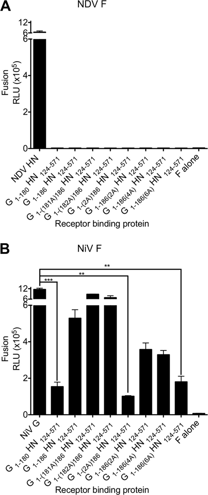
NiV G stalk residues 181 to 182 are required for activation of NiV F. Cell-to-cell fusion mediated by the chimeric NiV-NDV proteins with NDV F (A) or with NiV F (B) compared to the NDV HN/F (A) or NiV G/F (B) proteins. Fusion is measured by a β-Gal complementation assay. The values are means (± the standard errors) of results from four experiments. **, P < 0.05; ***, P < 0.005 (one-way analysis of variance, Dunn's multiple-comparison test).
The NiV receptor binding protein G does not have receptor cleaving activity and remains receptor bound during F activation, raising the possibility that receptor disengagement could account for the failure of some chimeric proteins to promote fusion (27). We considered the possibility that the failure of the chimeric proteins G1-(2A)186-HN124-571, G1-186(6A)-HN124-571, and G1-180-HN124-571 to activate NiV F may be due to a curtailed engagement of the receptor. To prevent detachment of the NDV globular head from its receptor and thereby assess the effect of constant receptor interaction on fusion promotion by chimeric proteins, we used zanamivir to inhibit the neuraminidase activity of NDV (Fig. 6). The fusion assay in Fig. 6 shows that in the absence of zanamivir to prevent receptor detachment, none of the chimeric proteins promoted fusion in our RBC fusion assay. Most of the receptor-bound RBCs were released (Fig. 6A), and no fusion was observed (no black bars). However, in the presence of zanamivir to inhibit NDV's neuraminidase activity, there was no release of receptor-bound RBCs (27, 28), allowing assessment of the chimeric proteins' potential to activate NiV F (Fig. 6B). The chimeric protein G1-180-HN124-571 (missing its connecting region) exhibited minimal NiV F fusion activation despite constant receptor interaction, with only 10 to 15% of the total bound RBCs fused after incubation at 37°C for 120 min. In contrast, in the presence of the G1-186-HN124-571 chimeric protein with an intact connector, 100% of the bound RBCs fused (Fig. 6B), as previously described (27). The chimeric proteins with two and four alanines substituted at amino acids 185 to 186 and amino acids 183 to 186 [G1-186(2A)-HN124-571 and G1-186(4A)-HN124-571, respectively] promoted fusion with 85 to 90% of the total bound RBCs. On the other hand, the chimeric proteins G1-(2A)186-HN124-571 and G1-186(6A)-HN124-571 in which amino acids 181 and 182 or amino acids 181 to 186 were substituted with alanines, promoted fusion with only 10 to 15% of the total bound RBCs, similar to the G1-180-HN124-571 (no connector sequence) chimeric protein. These data indicate that the specific residues at positions 181 and 182, but not at positions 183 to 186, are required for fusion activation.
Fig 6.
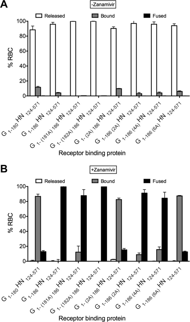
Engagement of site II of the receptor binding protein is required for activation of NiV F. 293T cells coexpressing NiV F with the NiV-NDV chimeric binding protein were allowed to bind to receptor-bearing RBCs at 4°C in the absence (A) or presence (B) of zanamivir. Zanamivir was added to activate NDV HN site II. Unbound RBCs were then washed, and standard medium without (A) or with (B) zanamivir, for the activation of binding site II, was added at 37°C for 120 min. The values on the y axis reflect quantification of RBCs that were (i) released (□), (ii) bound (▩), or (iii) fused (■). Note that there is no fusion (■) in panel A and that there are no released cells (□) in panel B. The values are means (± the standard errors) of results from three experiments.
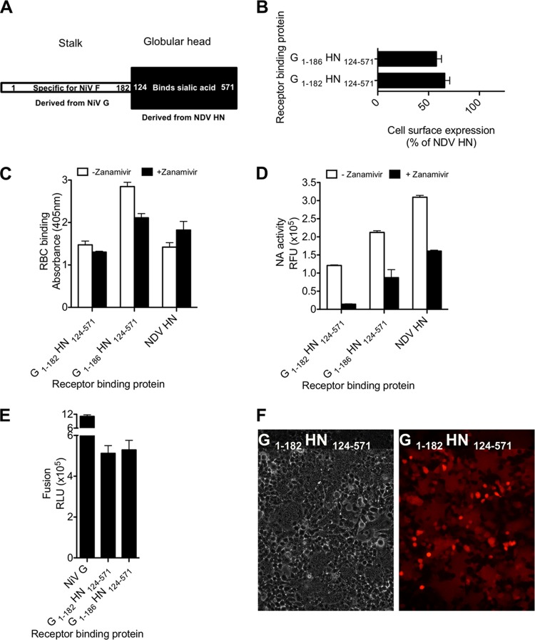
The finding that positions 181 and 182 are important for fusion promotion by the chimeric protein G1-186-HN124-571 led to the design of chimeric protein G1-182-HN124-571 (Fig. 7A) in which only residues 181 and 182 remain of the original NiV intervening region. This chimeric protein was expressed at 65% of NDV HN (Fig. 7B). The expressed chimeric proteins bound RBCs (Fig. 7C), possessed neuraminidase activity that was inhibited in the presence of zanamivir (Fig. 7D), and promoted fusion at a level comparable to G1-186-HN124-571 (Fig. 7E and F) with ∼50% efficiency of the wt NiV G/F fusion machinery. These data confirm the importance of the two amino acids at positions 181 and 182 for proper fusion activation and indicate that the minimal stalk region for maintaining the NiV G fusion activation is from position 1 to position 182.
Fig 7.
NiV G stalk residues 181 to 182 are sufficient for the activation of NiV F. (A) Schematic diagram of the G1-182-HN124-571 chimeric protein. (B) FACS analysis of cell surface expression from cells transfected with the chimeric proteins shown in Fig. 6A. The results are presented as percentages of NDV HN cell surface expression. (C) Receptor binding in the absence (□) or presence (■) of 2 mM zanamivir. (D) Neuraminidase activity of the receptor binding proteins, expressed in relative fluorescence intensity units (RFU) in the absence (□) or presence (■) of 2 mM zanamivir. The values are means ± the SD of results from samples assessed in triplicate and are representative of the experiment repeated at least three times. (E) Cell-to-cell fusion of the chimeric protein coexpressed with NiV F. Cell-to-cell fusion was measured by a β-Gal complementation assay. The values in panels B, C, D, and E are means (± the standard errors) of results from three experiments. (F) Cell-to-cell fusion promoted by the chimeric protein expressed with NiV F is observed in the left panel by syncytium formation using visible microscopy and in the right panel by redistribution of RFP (red fluorescent protein) using fluorescence microscopy.
To assess whether the important function of the NiV G stalk connector region in the chimeric protein can be substituted by the connector region from the NDV HN, we assessed chimeric proteins with NiV stalk lengths from positions 1 to 180 or from positions 1 to 186 and NDV regions spanning either positions 110 to 571 or positions 120 to 571; the NDV sequence starting at position 110 includes the head-stalk connecting region for NDV (Fig. 8). Chimeric proteins with a NiV G stalk length of amino acids 1 to 180 do not promote fusion in the RBC fusion assay, even with the NDV HN region positions 110 to 571 (or 120 to 571), indicating that the connecting region between the head and the stalk in the chimeric proteins cannot be substituted by the analogous NDV HN sequences.
Fig 8.
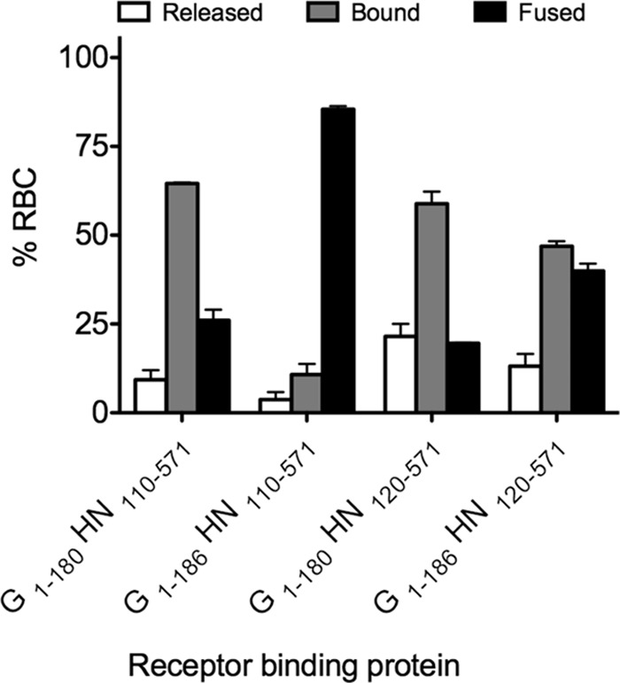
The connecting region from NiV G stalk cannot be substituted by the NDV HN 110-124 or 120-124 stalk region. 293T cells coexpressing NiV F with the NiV-NDV chimeric binding protein were allowed to bind to receptor-bearing RBCs at 4°C in the presence of zanamivir. Zanamivir was added to activate NDV HN site II. Unbound RBCs were then washed, and standard medium with zanamivir, for the activation of binding site II, was added at 37°C for 120 min. The values on the y axis reflect quantification of RBCs that were (i) released (□), (ii) bound (▩), or (iii) fused (■). The values are means (± the standard errors) of results from two representative experiments.
Amino acids at positions 181 and 182 in the stalk domain of the receptor binding protein regulate an early stage in F activation.
We have recently shown that receptor engagement by the receptor binding protein is required to promote fusion beyond the stage of F insertion (28). To investigate whether the chimeric proteins G1-180-HN124-571 and G1-(2A)186-HN124-571 are defective at initial F triggering or at this later stage in the fusion process, we determined whether the defect is before or after insertion of the fusion protein into the target cell. Insertion of F's fusion peptide into the target cell indicates that the prehairpin intermediate has formed (18, 28, 41–43); at this stage, F mediates attachment to the target cells, and disengagement of the receptor binding protein does not lead to release from the target cell (13, 18, 28, 40, 41, 44). To determine whether the G1-180-HN124-571 and G1-(2A)186-HN124-571 chimeric proteins activate NiV F up to the stage of the prehairpin intermediate with fusion peptide inserted but then fail to complete the fusion process, we modified the assay described in Fig. 6. The receptor-bound RBCs were eliminated from the bound pool of RBCs by a zanamivir-free incubation step at pH 6.5 to allow NDV neuraminidase to release receptor-bound HN (28). The bound RBC pool consisted only of RBCs retained via F's insertion into the target cell, forming a bridge between the glycoprotein-expressing cell and the target RBCs (Fig. 9) (40, 41). If the chimeric glycoproteins G1-180-HN124-571 and G1-(2A)186-HN124-571 trigger F to the prehairpin intermediate, then fusion peptide insertion will retain RBCs attached to the cell monolayer, but if they are defective in the initial stage of F activation (i.e., before fusion peptide insertion), then the RBCs will be released (18, 28, 41).
Fig 9.
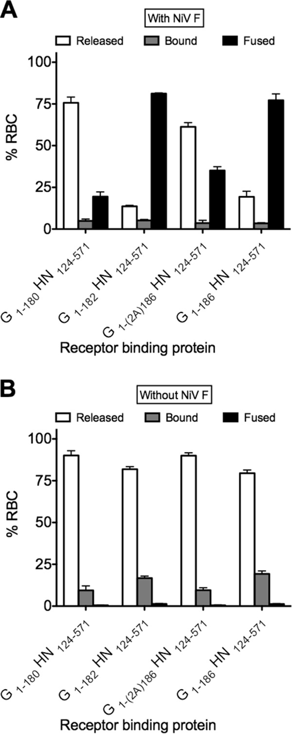
Amino acids 181 and 182 regulate an early stage in F activation. 293T cells expressing the NiV-NDV chimeric binding protein alone (B) or coexpressing with NiV F (A) were allowed to bind to receptor-bearing RBCs at 4°C in the presence of zanamivir. Unbound RBCs were then removed, and medium with zanamivir, to activate binding site II, was added, followed by incubation at 37°C for 90 min. After incubation, the cells were washed, and medium at pH 6.5 was added. The cells were incubated at 37°C for 75 min. Values on the y axis reflect quantification of RBCs that were (i) released (□), (ii) bound (▩), or (iii) fused (■). Note that there is no fusion (■) in panel B, and there are no bound cells (▩) in panel A. The values are means (± the standard errors) of results from three experiments.
For cells coexpressing the chimeric receptor binding proteins G1-180-HN124-571 and G1-(2A)186-HN124-571 with NiV F (Fig. 9A), most of the bound RBCs were released into the medium after zanamivir was removed, indicating the failure of F insertion. In the presence of G1-182-HN124-571 and G1-186-HN124-571, however, most of the RBCs were fused. Without NiV F (Fig. 9B), all RBCs were released by the neuraminidase activity of NDV HN globular domain, as expected. The defect in fusion activation by the chimeric proteins G1-180-HN124-571 and G1-(2A)186-HN124-571—chimeric proteins that lack either the whole connecting region or the correct residues at 181 and 182—occurs at an early step in the F triggering process, prior to insertion of the fusion peptide into the target cell membrane.
NiV G stalk domains containing the 181 and 182 mutations are altered in receptor binding and fusion activation.
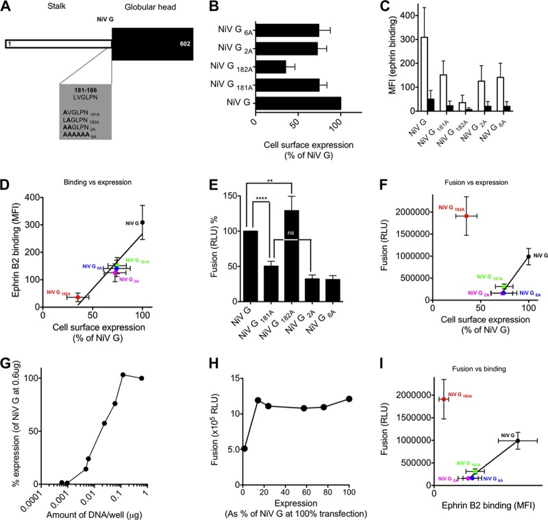
A series of NiV G constructs were designed to determine the role of the amino acids 181 and 182 in the context of the NiV G (Fig. 10A). The mutated NiV Gs had a lower level of cell surface expression than wt NiV G, assessed by FACS analysis using an anti-NiV G monoclonal antibody (Fig. 10B). Of the six mutants shown in Fig. 10A, three (NiV G181A, NiV G2A, and NiV G6A) showed 70 to 80% expression compared to wt NiV G. Only NiV G182A had a significantly lower expression (35% of wt NiV G). The receptor binding capacity of the mutated NiV G proteins was evaluated by quantification of binding to soluble ephrin B2 since ephrin B2 (but not ephrin B1) acts as a cellular receptor for NiV (45–51). Cells expressing the indicated proteins were allowed to bind ephrin at 4°C for 60 min, and bound ephrin was quantified by FACS. Receptor binding for ephrin B2 by the mutated proteins was 20 to 60% of that of NiV G (Fig. 10C). All of the NiV G mutants, as well as wt NiV G, showed negligible ephrin B1 binding, as expected. The effect of stalk domain alterations on binding activity could be attributed to altered expression levels. Indeed, when we normalized for expression level, ephrin B2 binding was similar to ephrin B2 binding and was directly proportional to the expression level of G protein (Fig. 10D).
Fig 10.
Requirement for specific residues at residues 181 and 182 of the NiV G stalk for binding and fusion promoting activity of NiV G. (A) Schematic diagram of alanine scanning mutagenesis of NiV G constructs. (B) FACS analysis of cell surface expression from cells transfected with the chimeric proteins shown in panel A. The results are presented as percentages of NiV G cell surface expression. (C) Receptor binding activity of the NiV G proteins to ephrin B2 (□) or ephrin B1 (■). (D) Ephrin B2 binding versus cell surface expression. (E) Cell-to-cell fusion mediated by NiV F coexpressed with the NiV G proteins in panel A. Fusion is measured by a β-Gal complementation assay. The values in panels B, C and D are means (± the standard errors) of results from four experiments. (F) Fusion measured by β-Gal complementation assay versus cell surface expression. (G) FACS analysis of cell surface expression from cells transfected with different levels of wt NiV G cDNA. The results are presented as percentages of NiV G cell surface expression at the highest cDNA concentration. (H) Fusion measured by β-Gal complementation under the same cell surface expression. (I) Ephrin B2 binding versus fusion measured by β-Gal complementation assay. **, P < 0.05; ****, P < 0.001 (one-way analysis of variance, Dunn's multiple comparison test).
The fusion-promoting capacity of the mutated NiV G proteins was assessed (Fig. 10E). Fusion promoted by the mutants NiV G2A and NiV G6A was much lower than that promoted by NiV G. The NiV G181A has relatively low fusion-promoting activity, causing 50% less fusion than NiV G. Surprisingly, the NiV G182A was more efficient than NiV G in promoting fusion in the presence of NiV F. In the analysis of fusion promotion versus cell surface expression, all of the mutants showed a direct correlation between cell surface expression and fusion except for NiV G182A (Fig. 10F). When we compared fusion promotion efficiencies of the wt NiV G proteins at expression levels similar to those of mutant NiV G, wt NiV G promoted 100% fusion at the same levels as mutant NiV Gs (Fig. 10G and H). The enhanced F-promotion activity of NiV G182A is even more apparent when plotted in this fashion. Fusion mediated by the mutated NiV G proteins was proportional to the binding activity of the mutants, with NiV G182A being the only outlier (Fig. 10I).
Substitution of cysteine for tryptophan at position 123, a potential site for an intersubunit disulfide bridge in the NDV HN globular head, prevents efficient NiV F activation.
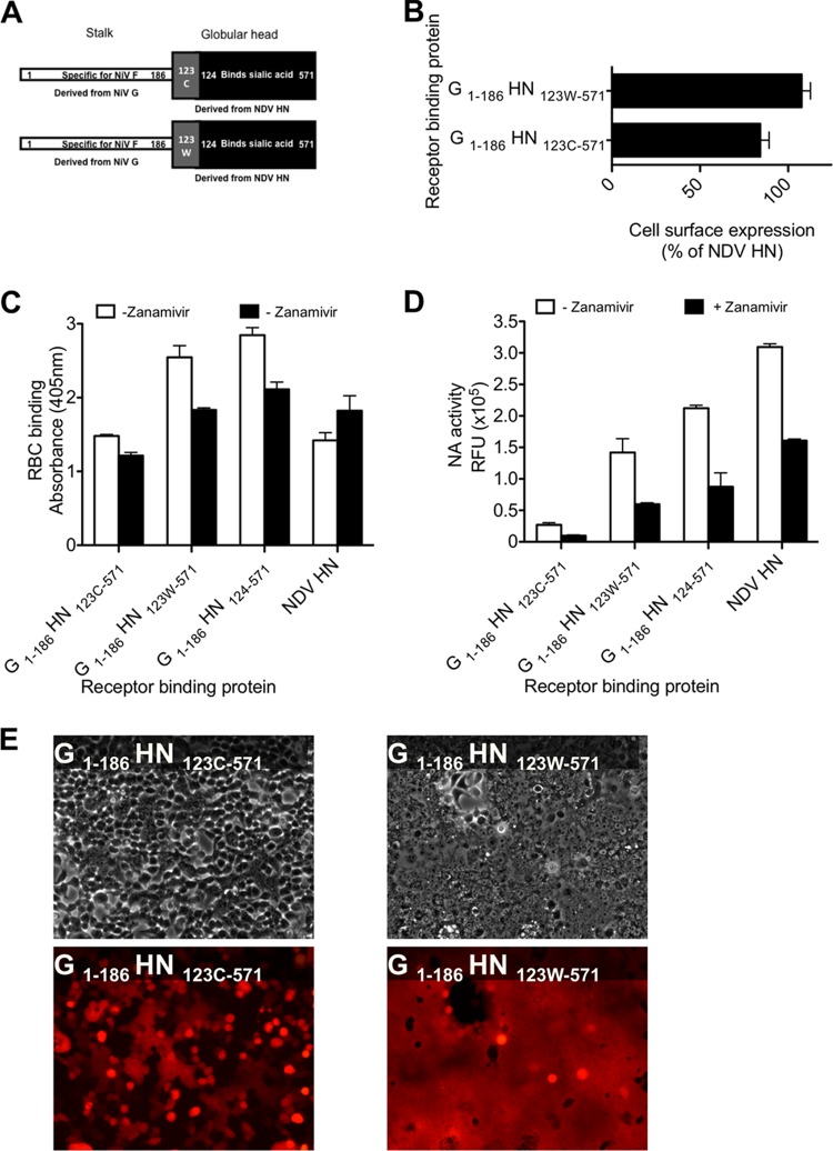
We then investigated the role of the globular head region of the head-stalk interface of our chimeric constructs to determine its effect on the propagation of the fusion signal. In Fig. 2, the cells transfected with G1-186-HN120-571, a chimeric protein containing the full connector region provided by NiV G but containing additional residues 120 to 123 from NDV HN, did not show cell-to-cell fusion. The other two chimeric proteins with the same NiV G stalk length but different contributions from NDV HN, G1-186-HN110-571 (with additional NDV HN residues 110 to 123) and G1-186-HN124-571, were functional with respect to fusion promotion, leading us to hypothesize that residues in the NDV HN region between residues 120 and 123 may interfere with the proper NiV G stalk activation. The NDV AV HN globular head contains a cysteine at position 123 that (in the NDV AV HN) forms an intersubunit disulfide bridge (52, 53), which could limit the flexibility of the heads with respect to the stalks. Other NDV strains have either a tryptophan or tyrosine at the same position (53), indicating that the disulfide linkage in HN can be removed by substitution of the cysteine with tryptophan at this position without inactivating the protein. We hypothesized that the degree of flexibility between the NDV HN globular head and the NiV G stalk could affect the fusion signal transmitted to F. Based on naturally occurring NDV HN variations, two new chimeric proteins were constructed with the NiV G stalk region (residues 1 to 186) used for the previous chimeric proteins, but with the NDV HN globular head starting at residue 123 instead of residue 124 (Fig. 11A). The G1-186-HN123C-571 (with a cysteine at position 123) and G1-186-HN123w-571 (with a tryptophan at position 123) chimeras showed cell surface expression (Fig. 11B) and RBC binding (Fig. 11C) (both in the absence and in the presence of zanamivir) similar to NDV HN. The neuraminidase activity of the G1-186-HN123C-571 was decreased compared to the G1-186-HN123W-571 (Fig. 11D). The presence of the cysteine (compared to tryptophan) at position 123 decreased the ability of the chimeric protein to promote fusion by NiV F (Fig. 11E). The lack of fusion promotion in the G1-186-HN123C-571 chimeric protein indicates that the presence of cysteine at position 123 in the NDV HN head is detrimental for transmitting the fusion signal.
Fig 11.
Cysteine 123 in the NDV HN globular head alters neuraminidase and fusion promotion. (A) Schematic diagram of chimeric protein G1-186-HN123-571 constructs. (B) FACS analysis of cell surface expression from cells transfected with the chimeric proteins shown in panel A. The results are presented as percentages of NDV HN cell surface expression. (C) Receptor binding in the absence (□) or presence (■) of 2 mM zanamivir. (D) Neuraminidase activity of the receptor binding proteins, expressed in relative fluorescence intensity units (RFU) in the absence (□) or presence (■) of 2 mM zanamivir. The values in panels B, C, and D are means ± the SD of results from samples assessed in triplicate and are representative of the experiment repeated at least three times. (E) Cell-to-cell fusion promoted by the chimeric proteins expressed with NiV F was observed in the top panel by syncytium formation using visible microscopy and in the bottom panel by redistribution of RFP (red fluorescent protein) using fluorescence microscopy.
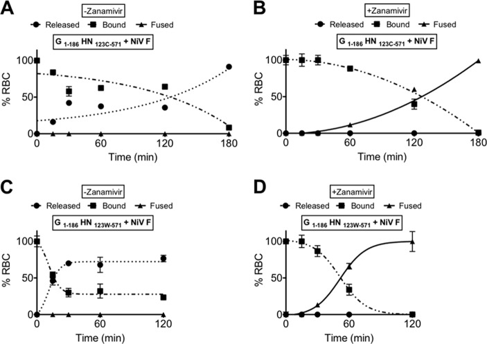
We next determined whether constant receptor engagement by G/HN could compensate for the defect of the G1-186-HN123C-571 chimera in fusion activation, using the fusion assay described in Fig. 6. In the absence of zanamivir, receptor binding site II in NDV's globular head is not activated (25), so that there is no fusion (Fig. 12A and C) and the RBCs are released. However, in the presence of zanamivir (Fig. 12B and D) to activate NDV HN′s site II (25), G1-186-HN123W-571 activated NiV F for fusion (Fig. 12D). Under the same conditions, G1-186-HN123C-571 also activated NiV F, but it took longer (Fig. 12B). The difference between panels B and D shows that, with a cysteine at position 123, fusion activation is less efficient, indicating that transmission of the fusion signal is impaired but not abolished.
Fig 12.
Cysteine 123 in the NDV HN globular head decreases the rate of F activation. 293T cells coexpressing NiV F and chimeric glycoproteins G1-186-HN123C-571 (A and B) or G1-186-HN123W-571 (C and D) were allowed to bind to RBCs at 4°C with (B and D) or without (A and C) zanamivir. Unbound RBCs were then washed, and standard medium with (B and D) or without (A and C) zanamivir was added at 37°C for up to 120 min. The values on the y axis reflect quantification of RBCs that were (i) released (●, dotted line), (ii) bound (■, dashed line), or (iii) fused (▲, solid line). The values are means (± the SD) of results from triplicate samples and are representative of the experiment repeated at least three times.
Oligomeric state of envelope chimeric proteins and NiV F processing.
Paramyxovirus fusion proteins are expressed as a precursor, F0, which requires proteolytic cleavage to assume an activation-ready form composed of disulfide-linked F1 and F2 (54). For most paramyxoviruses, the F protein arrives at the cell membrane already cleaved. However, NiV F processing requires an initial step in which uncleaved F is transported to the membrane to be reinternalized, cleaved by cathepsin L, and finally returned to the surface as a cleaved protein ready for activation (55, 56). We considered that the fusion phenotypes of the chimeric proteins might be attributable to altered processing of NiV F when coexpressed with these chimeric proteins. Alternatively, differences in oligomerization could account for altered function.
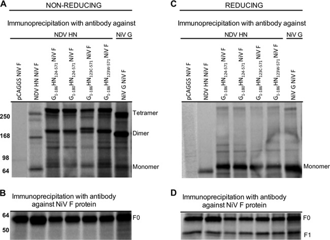
To assess NiV processing, cells were cotransfected with NiV F alone or in combination with chimeric glycoproteins, NDV HN, or NiV G (Fig. 13). Radiolabeled lysates were immunoprecipitated using antibodies against the receptor binding proteins (Fig. 13A and C) or NiV F (Fig. 13B and D). The migration pattern of the envelope glycoproteins on SDS-PAGE under nonreducing conditions revealed the oligomeric form of NiV G, and all of the chimeric proteins showed a similar level of oligomerization, with ca. 50% dimeric, 50% tetrameric, and very little monomeric protein (Fig. 13A). Under reducing conditions, a similar level of chimeric proteins and NDV AV HN was immunoprecipitated (Fig. 13C), but their expression levels were lower than NiV G. The differences in fusion-triggering activity between the G1-186-HN123C-571 and the G1-186-HN123W-571 chimeras were not due to altered oligomeric states or differential expression but rather to the cysteine at position 123 in the globular region, which may generate an extra disulfide bond at this position. The processing of F protein was not altered by coexpression of the chimeric envelope glycoproteins; Western blot analysis of immunoprecipitated NiV F showed similar quantities of NiV F0, F1, and F2 (Fig. 13B and D). Thus, the failure of specific chimeric receptor glycoproteins to activate NiV F was not due to altered F processing, oligomeric state, or expression.
Fig 13.
NiV G stalk domain determines the oligomerization state of the chimeric binding proteins. Monolayers of cells coexpressing NiV F and either the indicated chimeric glycoproteins, NDV HN, or NiV G were incubated in a medium supplemented with 35S-labeled amino acids. The cells were lysed, and the envelope glycoproteins were immunoprecipitated and subjected to SDS-PAGE under nonreducing (A and B) or reducing (C and D) conditions. Representative autoradiography shows the oligomeric state (A) and the level of protein expression (C) of the receptor binding glycoproteins, immunoprecipitated with anti-NDV HN antibodies. Note that monoclonal antibodies against NiV G were used in the lane marked NiV G. In panels B and D, the same samples were immunoprecipitated with anti-NiV F antibodies showing similar F expression (B) and processing (D) regardless of the coexpressed viral glycoproteins.
Chimeric viral glycoproteins containing the NiV G stalk domain and the NDV globular head can mediate viral infection.
To assess the function of each chimeric protein in a way that more closely mimics authentic infection, we used the virion-based infection assay (see Fig. 1). The envelope glycoproteins were pseudotyped onto a recombinant vesicular stomatitis virus (VSV) that expresses NiV F but lacks VSV G to generate the pseudotyped virus VSV-ΔG-NiVF/(G-HN) bearing the chimeric binding proteins and NiV F. Entry of the pseudotyped virus into target cells was quantified by plaque assay. Only the chimeric proteins that effectively promoted fusion generated infectious particles (Fig. 14A). The two chimeric proteins (G1-180-HN124-571 and G1-186-HN123C-571) that did not promote fusion failed to generate infective particles, whereas the G1-186-HN124-571 and G1-186-HN123W-571 chimeras complemented NiV F in the context of a live virus.
Fig 14.
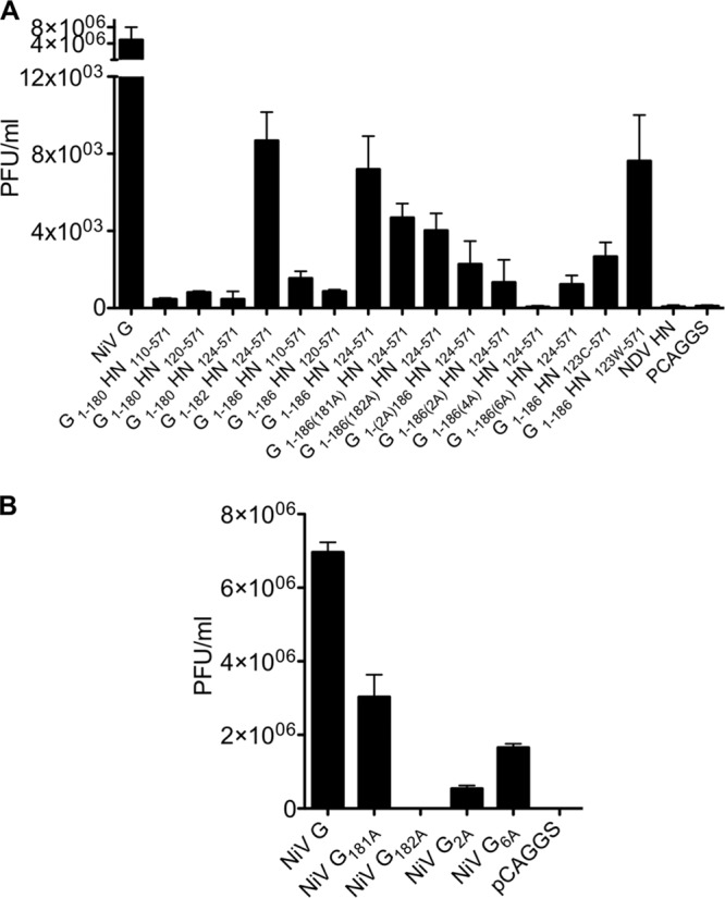
Chimeric envelope glycoproteins and the various NiV G mutant proteins on pseudotyped virions mediate infection. Monolayers of cells were infected with VSV-ΔG-NiV F pseudotyped virions with the indicated chimeric (A) or NiV G mutant (B) envelope glycoproteins. At 24 h postinfection, the PFU were determined as described in Materials and Methods. The values on the y axis reflect quantification of PFU/ml. The values are means (± the SD) of results from triplicate samples.
To evaluate the function of mutant NiV G molecules in a system that more closely mimics infection, the mutant G proteins were pseudotyped onto a recombinant VSV that expresses NiV F but lacks VSV G to generate the pseudotyped virus VSV-ΔG-NiVF/Gs bearing the mutant G proteins and NiV F. Entry of the pseudotyped virus into target cells (Fig. 14B) was quantified by plaque assay. Only the mutant G proteins that effectively promoted fusion were incorporated into infectious particles, except in the case of NiV G182A, which could be due to the lower expression level of NiV G182A.
NiV G can complement fusion activation of fusion impaired chimeric proteins.
We considered the possibility that NiV G could rescue the fusion of the impaired chimeric proteins bearing the NiV G stalk 1-186(6A). To address this question, we cotransfected the chimeric construct G1-186(6A)-HN124-571 with wt NiV G, along with NiV F, and assessed fusion, as in Fig. 6, in the presence of zanamivir to allow constant receptor engagement. Since the RBCs used in the assay lack the receptor for NiV G, binding is mediated only by the NDV globular domain of the chimeric protein. However, F activation can occur only through the wt NiV G stalk since the chimeric protein's stalk is impaired in F activation. Coexpression of the defective protein with the NDV head [G1-186(6A)-HN124-571], wt NiV G, and NiV F permitted fusion to occur (Fig. 15), suggesting that the NDV globular domain transmits the fusion activation signal to the stalk of another monomer in a oligomeric complex of NiV G and chimeric protein.
Fig 15.
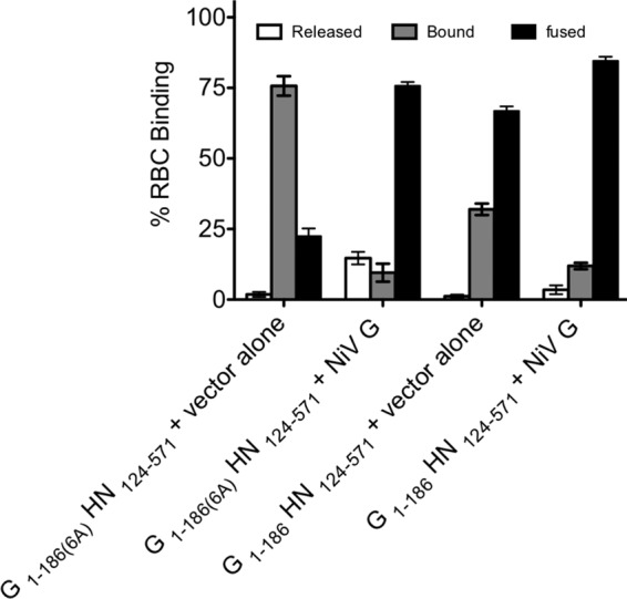
cis-Complementation of the F-triggering activity of the fusion-deficient chimeric proteins by wt NiV G. 293T cells coexpressing NiV G and F with the NiV-NDV chimeric binding protein were allowed to bind to receptor-bearing RBCs at 4°C in the absence (A) or presence (B) of zanamivir. Zanamivir was added to activate the NDV HN site II. Unbound RBCs were then washed, and standard medium without (A) or with (B) zanamivir, for the activation of binding site II, was added at 37°C for 120 min. The values on the y axis reflect quantification of RBCs that were (i) released (□), (ii) bound (▩), or (iii) fused (■). Note that there is no fusion (■) in panel A and that there are no released cells (□) in panel B. The values are means (± the standard errors) of results from two experiments.
DISCUSSION
For paramyxoviruses, we have proposed a model for the role of the receptor binding protein during the fusion process that partially unites the two disparate models previously applied to viruses that bind a proteinaceous receptor and viruses that bind sialic acid receptors. The role of the receptor binding protein has been considered to be mainly repressive for viruses such as measles virus and NiV, where clamping the F protein prevents it from assuming a postfusion structure before the virus engages its proteinaceous receptor (10, 12, 36, 47, 57–61). In the case of sialic acid binding receptor binding paramyxoviruses, such as HPIV and NDV, engagement of the receptor binding protein by the host cell receptor was thought to initiate the interaction between the receptor binding protein and the F protein, triggering F-mediated fusion. This model attributed an active role for the receptor binding protein in F triggering and did not require that the receptor binding protein and the F protein be physically associated prior to receptor engagement. We have shown that for at least several paramyxoviruses from each type, both mechanisms are at play: before the viruses engage receptor, the receptor binding proteins stabilize the F protein to prevent premature activation, and the switch to an active role in triggering the F protein to fuse occurs upon receptor engagement (17). The existence of a common mechanism for signal transmission between the receptor binding protein's head and its stalk in both sialic-acid-dependent and non-sialic-acid-dependent receptor binding proteins was demonstrated (27, 28) using a chimeric receptor binding protein that has the receptor binding head of a receptor binding protein from a sialic acid-binding virus (NDV) and the receptor binding stalk from a protein-binding virus (NiV). The NiV G stalk activated NiV F after receiving the signal from the receptor-engaged NDV HN head. In the present study, we used this and similar chimeric receptor binding molecules to define the specific region in the receptor binding protein(s), and the specific properties conferred by that region, that are required for fusion promotion.
The stalk region of the paramyxovirus receptor binding protein is important for fusion activation (7, 62). Here, we established that the intervening region between the globular head domain and the stalk is key for transmission of the activating signal that is initiated by engagement of receptor. G1-180-HN124-571 and the chimeric binding proteins with mutations in the 181 and 182 residues were defective in activation of NiV F despite the presence of zanamivir and activation of site II, indicating the importance of proper communication between the globular domain and the stalk. Constant binding of the HN globular domain to its receptor is necessary but not sufficient for activation of F, and this was particularly evident for the chimeric glycoproteins (Fig. 6B). A minimal connecting region between stalk and globular head was identified to be necessary for fusion activation by the chimeric receptor binding proteins studied here. Residues 181 and 182 in the stalk of NiV G, which are part of this head-stalk interface region, were found to be essential for transmission of the activating signal. Do these two residues function as a connector region between the head and the stalk, thereby contributing to the transmission of the receptor-engaged state signal, or are they required for interaction of the stalk with F? Future experiments where this particular region is exchanged with the connector region of other paramyxoviruses may help answer this question. The importance of the residues at positions 181 and 182 of the chimeric receptor binding proteins was confirmed in the context of the NiV G protein, where, intriguingly, the change at position 182 enhanced fusion promotion. It appears that residues 181 and 182 are critical for the signal transmission from the stalk to the fusion protein of NiV in the chimeric protein and in authentic NiV G. Although a previous publication proposed that chimeric G1-180-HN124-571 failed to activate fusion due to the lack of a clamping domain from the NiV G globular head (36), we noted this chimera promoted fusion and contend that, while elements of the “clamp model” are correct, the element of the receptor binding protein that stabilizes the F protein before receptor engagement is purely in the stalk.
The chimeric receptor binding protein experiments provide support for the notion that Cys in the 123 position, which impairs signal transmission through the stalk domain, leads to disulfide bond formation and reduction in flexibility at the globular heads. In NDV, Cys123 from two neighboring receptor binding proteins form a native disulfide bond. It is reasonable to propose that a disulfide bond between the two subunits can also exist in G1-186-HN123C-571; however, we have not been able to demonstrate this disulfide bond in the chimeric protein experimentally due to the large number of disulfide bonds in the protein and the technical challenge in demonstrating individual ones.
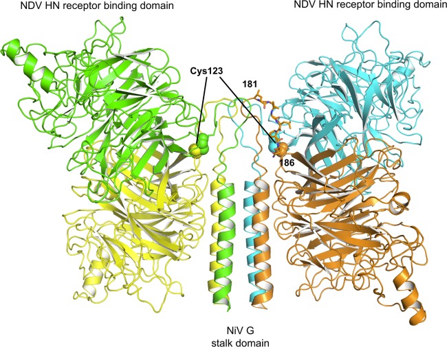
To address this issue, a model of the chimeric protein was generated. The stalk domain structures of NDV (63) and PIV5 (64) have recently been solved, and our chimeric proteins were modeled based on the NDV structure (Fig. 16). The stalk contains a portion of the Nipah G stalk (amino acids 148 to 186) and was modeled from the NDV structure (PDB ID 3T1E) (63) based on sequence homology. It also contains the NDV receptor binding domain (amino acids 123 to 571). The loop connecting the domains was manually built, and the model was energy minimized. Each of the four subunits is colored differently, and the LVGLPN region at positions 181 to 186 from one subunit (orange) is shown as a peptide in all-atom stick representation (Fig. 16). In this model, the two cysteines are in close proximity and would permit formation of a disulfide bridge. We propose that formation of a disulfide bridge at this position may prevent conformational changes that are required for the fusion activation function. Future work will explore whether there are mechanical constraints imposed by a disulfide bridge on fusion activation.
Fig 16.
Model of the NDV receptor binding domain (residues 123 to 571) and a portion of the NiV G stalk (residues 148 to 186). The stalk was modeled from the NDV structure (PDB ID 3T1E) based on sequence homology. The chimera model, like NDV HN, is a tetrameric complex formed by a dimer of dimers. The four subunits of the chimeric binding protein are colored differently and the LVGLPN region from residues 181 to 186 from one subunit (orange one) is shown as a peptide in an all-atom stick representation. Two Cys123 residues (ball representation) within NDV dimer are in close proximity for intermolecule disulfide bridge.
The oligomeric state of paramyxovirus receptor binding proteins has been shown to be essential for fusion promotion for NiV (44) and has been suggested to be critical for fusion promotion by HPIV3 (38), measles virus (65, 66), and other paramyxoviruses (24). It was unknown whether, within a tetramer, the receptor-engaged signal can be transmitted from the head of one monomer via the stalk of a different monomer, or whether each monomer acts individually in terms of signal transmission; the chimeric receptor binding proteins provided the answer. When chimeric G1-186(6A)-HN124-571, which bears all six alanines in the connecting region and is defective in fusion activation, was coexpressed with wt NiV G, fusion promotion activity of the oligomeric protein was rescued (Fig. 15). The NDV globular domain transmitted the receptor-engaged fusion signal to F via the functional stalk of wt NiV G, indicating that one monomer head can transmit the signal to a different stalk and suggesting that simultaneous engagement of all heads of an oligomer may not be necessary. In fact, for measles virus (MV), while neither a receptor binding protein (H) defective in receptor binding nor an H defective in fusion activation complemented MV F in fusion, when both defective proteins were cotransfected with MV F, fusion ensued (65), supporting the notion that not every head must be receptor engaged in order to transmit the signal to F.
The intervening region between the head and stalk domains of the receptor binding proteins we described, at positions 181 to 186 (for NiV), could provide a new target for antiviral drugs or neutralizing antibodies. Interrupting the cross talk between the globular domain and the stalk of the receptor binding protein can interfere with fusion mediated by the F protein and halt infection. For the receptor binding proteins described here, we show striking protein-protein interaction modularity; the inter- and intraprotein regulatory regions of several paramyxoviruses can be interchanged and still retain function in the presence of a correct connecting region. Chimeric proteins that are functional in the context of viral infection may also provide a new tool for use in developing safe attenuated vaccines against these lethal viruses.
ACKNOWLEDGMENTS
We are grateful to Dan and Nancy Paduano for support of innovative research projects, to Ashton Kutcher and Jonathan Ledecky for their support, and to the Friedman Family Foundation for the renovation of our laboratories at Weill Cornell Medical College. We thank Jacob Moscona-Skolnik for critical readings of the manuscript. We acknowledge the flow cytometry support from Sergei Rudchenko in the Flow Cytometry Facility of the Hospital for Special Surgery/Weill Cornell Medical College. We acknowledge the Northeast Center of Excellence for Bio-Defense and Emerging Infections Disease Research's Proteomics Core for peptide synthesis and purification.
This study was supported by the National Institutes of Health (NIAID and NIBIB), Northeast Center of Excellence for Biodefense and Emerging Infectious Disease Research, grants U54AI057158 to M.P. and A.M. (principal investigator for Center of Excellence grant, W. I. Lipkin), grant R01AI31971 to A.M., grants R21EBO11707 and R21AI100292-01 to M.P., and a Friedman Research Scholar in Pediatric Infectious Diseases Award to M.P. R.X. and I.A.W. were supported in part by NIH R56 AI099275 (I.A.W.) and the Skaggs Institute for Chemical Biology.
Footnotes
Published ahead of print 31 July 2013
This is publication 21556 from The Scripps Research Institute.
REFERENCES
- 1.Eckert DM, Kim PS. 2001. Mechanisms of viral membrane fusion and its inhibition. Annu. Rev. Biochem. 70:777–810. [DOI] [PubMed] [Google Scholar]
- 2.White JM, Delos SE, Brecher M, Schornberg K. 2008. Structures and mechanisms of viral membrane fusion proteins: multiple variations on a common theme. Crit. Rev. Biochem. Mol. Biol. 43:189–219. [DOI] [PMC free article] [PubMed] [Google Scholar]
- 3.Harrison SC. 2008. Viral membrane fusion. Nat. Struct. Mol. Biol. 15:690–698. [DOI] [PMC free article] [PubMed] [Google Scholar]
- 4.Weissenhorn W, Hinz A, Gaudin Y. 2007. Virus membrane fusion. FEBS Lett. 581:2150–2155. [DOI] [PMC free article] [PubMed] [Google Scholar]
- 5.Sapir A, Avinoam O, Podbilewicz B, Chernomordik LV. 2008. Viral and developmental cell fusion mechanisms: conservation and divergence. Dev. Cell 14:11–21. [DOI] [PMC free article] [PubMed] [Google Scholar]
- 6.Plattet P, Plemper RK. 2013. Envelope protein dynamics in paramyxovirus entry. mBio 4:e00413–13. 10.1128/mBio.00413-13. [DOI] [PMC free article] [PubMed] [Google Scholar]
- 7.Porotto M, Murrell M, Greengard O, Moscona A. 2003. Triggering of human parainfluenza virus 3 fusion protein (F) by the hemagglutinin-neuraminidase (HN): an HN mutation diminishing the rate of F activation and fusion. J. Virol. 77:3647–3654. [DOI] [PMC free article] [PubMed] [Google Scholar]
- 8.Moscona A. 2005. Entry of parainfluenza virus into cells as a target for interrupting childhood respiratory disease. J. Clin. Invest. 115:1688–1698. [DOI] [PMC free article] [PubMed] [Google Scholar]
- 9.Lamb RA, Paterson RG, Jardetzky TS. 2006. Paramyxovirus membrane fusion: lessons from the F and HN atomic structures. Virology 344:30–37. [DOI] [PMC free article] [PubMed] [Google Scholar]
- 10.Iorio RM, Melanson VR, Mahon PJ. 2009. Glycoprotein interactions in paramyxovirus fusion. Future Virol. 4:335–351. [DOI] [PMC free article] [PubMed] [Google Scholar]
- 11.Dutch RE. 2010. Entry and fusion of emerging paramyxoviruses. PLoS Pathog. 6:e1000881. 10.1371/journal.ppat.1000881. [DOI] [PMC free article] [PubMed] [Google Scholar]
- 12.Lee B, Ataman ZA. 2011. Modes of paramyxovirus fusion: a henipavirus perspective. Trends Microbiol. 19:389–399. [DOI] [PMC free article] [PubMed] [Google Scholar]
- 13.Aguilar HC, Iorio RM. 2012. Henipavirus membrane fusion and viral entry. Curr. Top. Microbiol. Immunol. 359:79–94. [DOI] [PubMed] [Google Scholar]
- 14.Broder CC. 2012. Henipavirus outbreaks to antivirals: the current status of potential therapeutics. Curr. Opin. Virol. 2:176–187. [DOI] [PMC free article] [PubMed] [Google Scholar]
- 15.Chang A, Dutch RE. 2012. Paramyxovirus fusion and entry: multiple paths to a common end. Viruses 4:613–636. [DOI] [PMC free article] [PubMed] [Google Scholar]
- 16.Liljeroos L, Krzyzaniak MA, Helenius A, Butcher SJ. 2013. Architecture of respiratory syncytial virus revealed by electron cryotomography. Proc. Natl. Acad. Sci. U. S. A. 110:11133–11138. [DOI] [PMC free article] [PubMed] [Google Scholar]
- 17.Porotto M, Salah ZW, Gui L, Devito I, Jurgens EM, Lu H, Yokoyama CC, Palermo LM, Lee KK, Moscona A. 2012. Regulation of paramyxovirus fusion activation: the hemagglutinin-neuraminidase protein stabilizes the fusion protein in a pretriggered state. J. Virol. 86:12838–12848. [DOI] [PMC free article] [PubMed] [Google Scholar]
- 18.Russell CJ, Jardetzky TS, Lamb RA. 2001. Membrane fusion machines of paramyxoviruses: capture of intermediates of fusion. EMBO J. 20:4024–4034. [DOI] [PMC free article] [PubMed] [Google Scholar]
- 19.Porotto M, Murrell M, Greengard O, Doctor L, Moscona A. 2005. Influence of the human parainfluenza virus 3 attachment protein's neuraminidase activity on its capacity to activate the fusion protein. J. Virol. 79:2383–2392. [DOI] [PMC free article] [PubMed] [Google Scholar]
- 20.Palermo LM, Porotto M, Greengard O, Moscona A. 2007. Fusion promotion by a paramyxovirus hemagglutinin-neuraminidase protein: pH modulation of receptor avidity of binding sites I and II. J. Virol. 81:9152–9161. [DOI] [PMC free article] [PubMed] [Google Scholar]
- 21.Crennell S, Takimoto T, Portner A, Taylor G. 2000. Crystal structure of the multifunctional paramyxovirus hemagglutinin-neuraminidase. Nat. Struct. Biol. 7:1068–1074. [DOI] [PubMed] [Google Scholar]
- 22.Zaitsev V, von Itzstein M, Groves D, Kiefel M, Takimoto T, Portner A, Taylor G. 2004. Second sialic acid binding site in Newcastle disease virus hemagglutinin-neuraminidase: implications for fusion. J. Virol. 78:3733–3741. [DOI] [PMC free article] [PubMed] [Google Scholar]
- 23.Lawrence MC, Borg NA, Streltsov VA, Pilling PA, Epa VC, Varghese JN, McKimm-Breschkin JL, Colman PM. 2004. Structure of the haemagglutinin-neuraminidase from human parainfluenza virus type III. J. Mol. Biol. 335:1343–1357. [DOI] [PubMed] [Google Scholar]
- 24.Yuan P, Thompson TB, Wurzburg BA, Paterson RG, Lamb RA, Jardetzky TS. 2005. Structural studies of the parainfluenza virus 5 hemagglutinin-neuraminidase tetramer in complex with its receptor, sialyllactose. Structure 13:803–815. [DOI] [PubMed] [Google Scholar]
- 25.Porotto M, Fornabaio M, Greengard O, Murrell MT, Kellogg GE, Moscona A. 2006. Paramyxovirus receptor-binding molecules: engagement of one site on the hemagglutinin-neuraminidase protein modulates activity at the second site. J. Virol. 80:1204–1213. [DOI] [PMC free article] [PubMed] [Google Scholar]
- 26.Mahon PJ, Mirza AM, Iorio RM. 2011. Role of the two sialic acid binding sites on the Newcastle disease virus HN protein in triggering the interaction with the F protein required for the promotion of fusion. J. Virol. 85:12079–12082. [DOI] [PMC free article] [PubMed] [Google Scholar]
- 27.Porotto M, Salah Z, Devito I, Talekar A, Palmer SG, Xu R, Wilson IA, Moscona A. 2012. The second receptor binding site of the globular head of the Newcastle disease virus (NDV) hemagglutinin-neuraminidase activates the stalk of multiple paramyxovirus receptor binding proteins to trigger fusion. J. Virol. 86:5730–5741. [DOI] [PMC free article] [PubMed] [Google Scholar]
- 28.Porotto M, Devito I, Palmer SG, Jurgens EM, Yee JL, Yokoyama CC, Pessi A, Moscona A. 2011. Spring-loaded model revisited: paramyxovirus fusion requires engagement of a receptor binding protein beyond initial triggering of the fusion protein. J. Virol. 85:12867–12880. [DOI] [PMC free article] [PubMed] [Google Scholar]
- 29.Chattopadhyay A, Rose JK. 2011. Complementing defective viruses that express separate paramyxovirus glycoproteins provide a new vaccine vector approach. J. Virol. 85:2004–2011. [DOI] [PMC free article] [PubMed] [Google Scholar]
- 30.Porotto M, Carta P, Deng Y, Kellogg G, Whitt M, Lu M, Mungall B, Moscona A. 2007. Molecular determinants of antiviral potency of paramyxovirus entry inhibitors. J. Virol. 81:10567–10574. [DOI] [PMC free article] [PubMed] [Google Scholar]
- 31.Obrig TG, Culp WJ, McKeehan WL, Hardesty B. 1971. The mechanism by which cycloheximide and related glutarimide antibiotics inhibit peptide synthesis on reticulocyte ribosomes. J. Biol. Chem. 246:174–181. [PubMed] [Google Scholar]
- 32.Porotto M, Greengard O, Poltoratskaia N, Horga M-A, Moscona A. 2001. Human parainfluenza virus type 3 HN-receptor interaction: the effect of 4-GU-DANA on a neuraminidase-deficient variant. J. Virol. 76:7481–7488. [DOI] [PMC free article] [PubMed] [Google Scholar]
- 33.Talekar A, Pessi A, Glickman F, Sengupta U, Briese T, Whitt MA, Mathieu C, Horvat B, Moscona A, Porotto M. 2012. Rapid screening for entry inhibitors of highly pathogenic viruses under low-level biocontainment. PLoS One 7:e30538. 10.1371/journal.pone.0030538. [DOI] [PMC free article] [PubMed] [Google Scholar]
- 34.Moosmann P, Rusconi S. 1996. Alpha complementation of LacZ in mammalian cells. Nucleic Acids Res. 24:1171–1172. [DOI] [PMC free article] [PubMed] [Google Scholar]
- 35.Porotto M, Fornabaio M, Kellogg GE, Moscona A. 2007. A second receptor binding site on human parainfluenza virus type 3 hemagglutinin-neuraminidase contributes to activation of the fusion mechanism. J. Virol. 81:3216–3228. [DOI] [PMC free article] [PubMed] [Google Scholar]
- 36.Mirza AM, Aguilar HC, Zhu Q, Mahon PJ, Rota PA, Lee B, Iorio RM. 2011. Triggering of the Newcastle disease virus fusion protein by a chimeric attachment protein that binds to Nipah virus receptors. J. Biol. Chem. 286:17851–17860. [DOI] [PMC free article] [PubMed] [Google Scholar]
- 37.Deng R, Wang Z, Mahon PJ, Marinello M, Mirza A, Iorio RM. 1999. Mutations in the Newcastle disease virus hemagglutinin-neuraminidase protein that interfere with its ability to interact with the homologous F protein in the promotion of fusion. Virology 253:43–54. [DOI] [PubMed] [Google Scholar]
- 38.Porotto M, Palmer SG, Palermo LM, Moscona A. 2012. Mechanism of fusion triggering by human parainfluenza virus type III: communication between viral glycoproteins during entry. J. Biol. Chem. 287:778–793. [DOI] [PMC free article] [PubMed] [Google Scholar]
- 39.Porotto M, Yokoyama C, Palermo LM, Mungall B, Aljofan M, Cortese R, Pessi A, Moscona A. 2010. Viral entry inhibitors targeted to the membrane site of action. J. Virol. 84:6760–6768. [DOI] [PMC free article] [PubMed] [Google Scholar]
- 40.Porotto M, Rockx B, Yokoyama C, Talekar A, DeVito I, Palermo L, Liu J, Cortese R, Lu M, Feldmann H, Pessi A, Moscona A. 2010. Inhibition of Nipah virus infection in vivo: targeting an early stage of paramyxovirus fusion activation during viral entry. PLoS Pathog. 6:e1001168. 10.1371/journal.ppat.1001168. [DOI] [PMC free article] [PubMed] [Google Scholar]
- 41.Kim YH, Donald JE, Grigoryan G, Leser GP, Fadeev AY, Lamb RA, DeGrado WF. 2011. Capture and imaging of a prehairpin fusion intermediate of the paramyxovirus PIV5. Proc. Natl. Acad. Sci. U. S. A. 108:20992–20997. [DOI] [PMC free article] [PubMed] [Google Scholar]
- 42.Smith EC, Dutch RE. 2010. Side chain packing below the fusion peptide strongly modulates triggering of the Hendra virus F protein. J. Virol. 84:10928–10932. [DOI] [PMC free article] [PubMed] [Google Scholar]
- 43.Aguilar HC, Aspericueta V, Robinson LR, Aanensen KE, Lee B. 2010. A quantitative and kinetic fusion protein-triggering assay can discern distinct steps in the Nipah virus membrane fusion cascade. J. Virol. 84:8033–8041. [DOI] [PMC free article] [PubMed] [Google Scholar]
- 44.Maar D, Harmon B, Chu D, Schulz B, Aguilar HC, Lee B, Negrete OA. 2012. Cysteines in the stalk of the Nipah virus G glycoprotein are located in a distinct subdomain critical for fusion activation. J. Virol. 86:6632–6642. [DOI] [PMC free article] [PubMed] [Google Scholar]
- 45.Porotto M, Yi F, Moscona A, LaVan DA. 2011. Synthetic protocells interact with viral nanomachinery and inactivate pathogenic human virus. PLoS One 6:e16874. 10.1371/journal.pone.0016874. [DOI] [PMC free article] [PubMed] [Google Scholar]
- 46.Bishop KA, Stantchev TS, Hickey AC, Khetawat D, Bossart KN, Krasnoperov V, Gill P, Feng YR, Wang L, Eaton BT, Wang LF, Broder CC. 2007. Identification of Hendra virus G glycoprotein residues that are critical for receptor binding. J. Virol. 81:5893–5901. [DOI] [PMC free article] [PubMed] [Google Scholar]
- 47.Bishop KA, Hickey AC, Khetawat D, Patch JR, Bossart KN, Zhu Z, Wang LF, Dimitrov DS, Broder CC. 2008. Residues in the stalk domain of the Hendra virus g glycoprotein modulate conformational changes associated with receptor binding. J. Virol. 82:11398–11409. [DOI] [PMC free article] [PubMed] [Google Scholar]
- 48.Negrete OA, Levroney EL, Aguilar HC, Bertolotti-Ciarlet A, Nazarian R, Tajyar S, Lee B. 2005. EphrinB2 is the entry receptor for Nipah virus, an emergent deadly paramyxovirus. Nature 436:401–405. [DOI] [PubMed] [Google Scholar]
- 49.Negrete OA, Wolf MC, Aguilar HC, Enterlein S, Wang W, Muhlberger E, Su SV, Bertolotti-Ciarlet A, Flick R, Lee B. 2006. Two key residues in EphrinB3 are critical for its use as an alternative receptor for Nipah virus. PLoS Pathog. 2:e7. 10.1371/journal.ppat.0020007. [DOI] [PMC free article] [PubMed] [Google Scholar]
- 50.Negrete OA, Chu D, Aguilar HC, Lee B. 2007. Single amino acid changes in the Nipah and Hendra virus attachment glycoproteins distinguish EphrinB2 from EphrinB3 usage. J. Virol. 81:10804–10814. [DOI] [PMC free article] [PubMed] [Google Scholar]
- 51.Bonaparte MI, Dimitrov AS, Bossart KN, Crameri G, Mungall BA, Bishop KA, Choudhry V, Dimitrov DS, Wang LF, Eaton BT, Broder CC. 2005. Ephrin-B2 ligand is a functional receptor for Hendra virus and Nipah virus. Proc. Natl. Acad. Sci. U. S. A. 102:10652–10657. [DOI] [PMC free article] [PubMed] [Google Scholar]
- 52.Mahon PJ, Mirza AM, Musich TA, Iorio RM. 2008. Engineered intermonomeric disulfide bonds in the globular domain of Newcastle disease virus hemagglutinin-neuraminidase protein: implications for the mechanism of fusion promotion. J. Virol. 82:10386–10396. [DOI] [PMC free article] [PubMed] [Google Scholar]
- 53.Sheehan JP, Iorio RM, Syddall RJ, Glickman RL, Bratt MA. 1987. Reducing agent-sensitive dimerization of the hemagglutinin-neuraminidase glycoprotein of Newcastle disease virus correlates with the presence of cysteine at residue 123. Virology 161:603–606. [DOI] [PubMed] [Google Scholar]
- 54.Lamb R, Parks G. Paramyxoviridae: viruses and their replication, p 1449–1496. In Knipe D, Howley P. (ed), Fields virology, 5th ed. vol 1 Lippincott/Williams & Wilkins, Philadelphia, PA. [Google Scholar]
- 55.Pager CT, Craft WW, Jr, Patch J, Dutch RE. 2006. A mature and fusogenic form of the Nipah virus fusion protein requires proteolytic processing by cathepsin L. Virology 346:251–257. [DOI] [PMC free article] [PubMed] [Google Scholar]
- 56.Pager CT, Dutch RE. 2005. Cathepsin L is involved in proteolytic processing of the Hendra virus fusion protein. J. Virol. 79:12714–12720. [DOI] [PMC free article] [PubMed] [Google Scholar]
- 57.Navaratnarajah CK, Oezguen N, Rupp L, Kay L, Leonard VH, Braun W, Cattaneo R. 2011. The heads of the measles virus attachment protein move to transmit the fusion-triggering signal. Nat. Struct. Mol. Biol. 18:128–134. [DOI] [PMC free article] [PubMed] [Google Scholar]
- 58.Corey EA, Iorio RM. 2007. Mutations in the stalk of the measles virus hemagglutinin protein decrease fusion but do not interfere with virus-specific interaction with the homologous fusion protein. J. Virol. 81:9900–9910. [DOI] [PMC free article] [PubMed] [Google Scholar]
- 59.Aguilar HC, Matreyek KA, Filone CM, Hashimi ST, Levroney EL, Negrete OA, Bertolotti-Ciarlet A, Choi DY, McHardy I, Fulcher JA, Su SV, Wolf MC, Kohatsu L, Baum LG, Lee B. 2006. N-glycans on Nipah virus fusion protein protect against neutralization but reduce membrane fusion and viral entry. J. Virol. 80:4878–4889. [DOI] [PMC free article] [PubMed] [Google Scholar]
- 60.Plemper RK, Hammond AL, Gerlier D, Fielding AK, Cattaneo R. 2002. Strength of envelope protein interaction modulates cytopathicity of measles virus. J. Virol. 76:5051–5061. [DOI] [PMC free article] [PubMed] [Google Scholar]
- 61.Iorio RM, Mahon PJ. 2008. Paramyxoviruses: different receptors: different mechanisms of fusion. Trends Microbiol. 16:135–137. [DOI] [PMC free article] [PubMed] [Google Scholar]
- 62.Melanson VR, Iorio RM. 2004. Amino acid substitutions in the F-specific domain in the stalk of the Newcastle disease virus HN protein modulate fusion and interfere with its interaction with the F protein. J. Virol. 78:13053–13061. [DOI] [PMC free article] [PubMed] [Google Scholar]
- 63.Yuan P, Swanson KA, Leser GP, Paterson RG, Lamb RA, Jardetzky TS. 2011. Structure of the Newcastle disease virus hemagglutinin-neuraminidase (HN) ectodomain reveals a four-helix bundle stalk. Proc. Natl. Acad. Sci. U. S. A. 108:14920–14925. [DOI] [PMC free article] [PubMed] [Google Scholar]
- 64.Bose S, Welch BD, Kors CA, Yuan P, Jardetzky TS, Lamb RA. 2011. Structure and mutagenesis of the parainfluenza virus 5 hemagglutinin-neuraminidase stalk domain reveals a four-helix bundle and the role of the stalk in fusion promotion. J. Virol. 85:12855–12866. [DOI] [PMC free article] [PubMed] [Google Scholar]
- 65.Brindley MA, Plemper RK. 2010. Blue native PAGE and biomolecular complementation reveal a tetrameric or higher-order oligomer organization of the physiological measles virus attachment protein H. J. Virol. 84:12174–12184. [DOI] [PMC free article] [PubMed] [Google Scholar]
- 66.Plemper RK, Hammond AL, Cattaneo R. 2000. Characterization of a region of the measles virus hemagglutinin sufficient for its dimerization. J. Virol. 74:6485–6493. [DOI] [PMC free article] [PubMed] [Google Scholar]


