Abstract
We previously reported that influenza A/swine/Korea/1204/2009(H1N2) virus was virulent and transmissible in ferrets in which the respiratory-droplet-transmissible virus (CT-Sw/1204) had acquired simultaneous hemagglutinin (HAD225G) and neuraminidase (NAS315N) mutations. Incorporating these mutations into the nonpathogenic A/swine/Korea/1130/2009(H1N2, Sw/1130) virus consequently altered pathogenicity and growth in animal models but could not establish efficient transmission or noticeable disease. We therefore exploited various reassortants of these two viruses to better understand and identify other viral factors responsible for pathogenicity, transmissibility, or both. We found that possession of the CT-Sw/1204 tripartite viral polymerase enhanced replicative ability and pathogenicity in mice more significantly than did expression of individual polymerase subunit proteins. In ferrets, homologous expression of viral RNA polymerase complex genes in the context of the mutant Sw/1130 carrying the HA225G and NA315N modifications induced optimal replication in the upper nasal and lower respiratory tracts and also promoted efficient aerosol transmission to respiratory droplet contact ferrets. These data show that the synergistic function of the tripartite polymerase gene complex of CT-Sw/1204 is critically important for virulence and transmission independent of the surface glycoproteins. Sequence comparison results reveal putative differences that are likely to be responsible for variation in disease. Our findings may help elucidate previously undefined viral factors that could expand the host range and disease severity induced by triple-reassortant swine viruses, including the A(H1N1)pdm09 virus, and therefore further justify the ongoing development of novel antiviral drugs targeting the viral polymerase complex subunits.
INTRODUCTION
Influenza A viruses are major viral respiratory pathogens that cause significant morbidity and mortality worldwide in the form of annually recurring seasonal epidemics. These viruses continue to be a threat to public health because of their unpredictable nature and the constant threat of new pandemic viruses. Influenza A viruses belong to the Orthomyxoviridae family of viruses, whose viral genome exists as eight single-stranded RNA segments with negative orientation (1). The segmented nature of the influenza virus genome is a key driver of the enigmatic viral evolution and ecology patterns observed through the years, because it allows reassortment in a suitable host (e.g., pigs) coinfected with two or more viruses from different sources or lineages, which could lead to antigenic shift and the generation of pandemic viruses (2). Indeed, in early 2009, a novel reassortant influenza A H1N1 [A(H1N1)pdm09] virus, presumed to have come from pigs, caused the first human influenza pandemic of the 21st century (3, 4). The causative virus closely resembles the “triple-reassortant” swine H1 strains of the North American lineage that contain classical swine-like nucleoprotein (NP), nonstructural (NS) protein, human-like hemagglutinin (HA), basic polymerase 1 (PB1), and avian-like PB2 and acidic polymerase (PA) segments. However, A(H1N1)pdm09 had uniquely acquired the neuraminidase (NA) and matrix (M) segments of the Eurasian avian-like swine viruses (5, 6).
The emergence of the A(H1N1)pdm09 virus demonstrated that strains prevalent in swine populations can be the direct source of future influenza pandemics in humans. Some triple-reassortant swine viruses isolated in the United States before the 2009 pandemic induce severe disease in mice but are only moderately pathogenic in ferrets (7–9). However, their transmissibility by respiratory droplets (RD) in ferret models varies considerably depending on the HA and NA lineages (7). Recently, we have shown that the A/swine/Korea/1204/2009(H1N2) Korean swine virus, which is genetically related to North American strains and was similarly isolated before the 2009 pandemic, is virulent in ferrets and is efficiently transmitted to naive RD contacts (10). Viruses recovered from the RD contact ferrets (CT-Sw/1204) had acquired Asp-225-Gly (H3 numbering) and Ser-315-Asn (N2 numbering) mutations in their HA and NA proteins, respectively, contributing to the altered replicative ability, pathogenicity, and transmissibility. However, when HA225G and NA315N were incorporated into another H1N2 virus, A/swine/Korea/1130/2009 (Sw/1130), the same degree of pathogenesis and ease of RD transmission were not observed, indicating the genetic relevance of the internal gene constellation of CT-Sw/1204. In the present study, we attempted to further elucidate the viral factors that contribute to pathogenicity and transmissibility. To address these issues, we created a panel of reassortant CT-Sw/1204 and Sw/1130 viruses to compare disease phenotypes in mice and ferrets and found that the pathogenicity and transmissibility of the CT-Sw/1204 virus may have been innate to the field virus isolate. We provide evidence that the viral RNA polymerases are critical factors for altered pathogenicity and virus dissemination.
MATERIALS AND METHODS
Cells and viruses.
Madin-Darby canine kidney (MDCK) cells were grown in Eagle's minimum essential medium with Earle's salts (EMEM) (Lonza, Switzerland) containing 5% fetal bovine serum (FBS) (Gibco, NY), and 293T human embryonic kidney cells were grown in Dulbecco's modified Eagle's medium (DMEM) (Gibco) containing 10% FBS. Primary normal human bronchial epithelial (NHBE) cells were purchased from ScienCell Research Laboratories (Carlsbad, CA) and grown in bronchial epithelial cell growth medium (BEpiCM; Gibco) as defined and recommended by ScienCell. All cells were incubated at 37°C in 5% CO2.
Generation of recombinant and reassortant viruses.
The eight gene segments of the H1N2 swine viruses CT-Sw/1204 and Sw/1130 (10) were amplified and cloned into the pHW2000 plasmid vector to generate recombinant viruses through a plasmid-based reverse-genetics system as previously described (Tables 1 and 2) (11). All recombinant parental and reassortant viruses were rescued in cocultured 293T and MDCK cell mixtures transfected (TransIT-LT1; Mirus Bio) with the eight viral plasmids as indicated. The transfection medium was removed after 6 h and replaced with Opti-MEM I (Gibco). After 30 h, Opti-MEM I (500 μl) containing l-1-tosylamido-2-phenylethyl chloromethyl ketone (TPCK)-treated trypsin (0.2 μg/ml; Sigma-Aldrich) was added to the transfected cells. Supernatants were harvested 48 h posttransfection and used to inoculate MDCK cells for the production of stock viruses. The eight genes of each rescued virus were partially sequenced (Cosmo GeneTech, Seoul, Republic of Korea) to confirm the genotype and origin of the viral segments. The handling of viruses and virus rescue were performed in an enhanced biosafety level 3 (BSL3+) containment laboratory as approved by the Korean Centers for Disease Control and Prevention.
Table 1.
Replication and virulence of recombinant parental and reassortant Sw/1130 viruses in mice
| Recombinant virusa | Stock titer (TCID50/ml) | Lung viral titer (SD)b |
MLD50c | |
|---|---|---|---|---|
| 3 days p.i. | 5 days p.i. | |||
| Sw/1130 | 2.0 × 10⁶ | 2.2 (0.2) | 1.7 (0.1) | >6 |
| CT-Sw/1204HA | 1.0 × 10⁷ | 3.9 (0.3)d | 3.5 (0.3)d | 4.9 |
| CT-Sw/1204NA | 2.0 × 10⁶ | 2.4 (0.2) | 2.1 (0.2) | >6 |
| CT-Sw/1204PB2 | 1.0 × 10⁷ | 3.6 (0.2)d | 3.4 (0.3)e | 5.1 |
| CT-Sw/1204PB1 | 1.0 × 10⁶ | 1.9 (0.1) | - | >6 |
| CT-Sw/1204PA | 4.0 × 10⁶ | 3.7 (0.3)e | 4.2 (0.4)e | 5.3 |
| CT-Sw/1204NP | 2.0 × 10⁶ | 2.7 (0.2)e | 2.8 (0.2)e | 5.5 |
| CT-Sw/1204M | 2.0 × 10⁶ | 2.2 (0.2) | 2.1 (0.2) | >6 |
| CT-Sw/1204NS | 6.3 × 10⁶ | 2.4 (0.2) | 3.0 (0.3) | 5.5 |
| CT-Sw/1204HA-NA | 4.0 × 10⁷ | 3.7 (0.3)d | 3.7 (0.3)d | 4.5 |
| CT-Sw/12043P | 1.0 × 10⁷ | 4.9 (0.4)d | 3.7 (0.3)e | 4.7 |
| CT-Sw/1204vRNP | 2.0 × 10⁷ | 4.9 (0.5)d | 3.9 (0.3)e | 4.7 |
| CT-Sw/1204 | 3.2 × 10⁸ | 5.9 (0.5)d | 5.2 (0.4)e | 3.5 |
Reassortant virus names were assigned according to the origin of the exchanged genes (e.g., CT-Sw/1204HA denotes a reassortant bearing the HA of CT-Sw/1204 with the rest of its genes from Sw/1130). Parental strains are in boldface.
Obtained from mice inoculated intranasally with 104 TCID50 of the indicated virus; titers are expressed as log10 TCID50/g. The dash denotes negative detection (<1.5 log10 TCID50/g).
Determined by inoculating mice with 102 to 106 TCID50 of virus and expressed as the log10 TCID50 required to yield 1 LD50.
P < 0.005 comparing virus titers relative to Sw/1130-inoculated mice (two-tailed unpaired t test).
P < 0.05 comparing virus titers relative to Sw/1130-inoculated mice (two-tailed unpaired t test).
Table 2.
Replication and virulence of recombinant parental and reassortant CT-Sw/1204 viruses in mice
| Recombinant virusa | Stock titer (TCID50/ml) | Lung viral titer (SD)b |
MLD50c | |
|---|---|---|---|---|
| 3 days p.i. | 5 days p.i. | |||
| CT-Sw/1204 | 3.2 × 10⁸ | 6.5 (0.5) | 6.2 (0.6) | 3.5 |
| Sw/1130HA | 5.0 × 10⁶ | 4.8 (0.4)d | 4.5 (0.4)d | 4.8 |
| Sw/1130NA | 8.0 × 10⁷ | 6.3 (0.6) | 6.0 (0.6) | 3.7 |
| Sw/1130PB2 | 3.2 × 10⁷ | 5.5 (0.5)d | 5.1 (0.5)d | 4.3 |
| Sw/1130PB1 | 8.0 × 10⁷ | 6.5 (0.5) | 6.1 (0.6) | 3.5 |
| Sw/1130PA | 1.3 × 10⁷ | 6.0 (0.6)d | 5.6 (0.5)d | 4 |
| Sw/1130NP | 8.0 × 10⁷ | 6.1 (0.6) | 5.9 (0.6) | 3.7 |
| Sw/1130M | 1.3 × 10⁸ | 6.5 (0.6) | 5.9 (0.5) | 3.5 |
| Sw/1130NS | 6.3 × 10⁸ | 6.2 (0.6) | 6.3 (0.6) | 3.8 |
| Sw/1130HA-NA | 1.3 ×10⁷ | 5.1 (0.5)d | 4.8 (0.4)d | 5.2 |
| Sw/11303P | 6.3 × 10⁶ | 4.5 (0.4)d | 4.1 (0.3)d | 4.7 |
| Sw/1130vRNP | 6.3 × 10⁶ | 4.6 (0.4)d | 4.4 (0.4)d | 5.1 |
| Sw/1130 | 2.0 × 10⁶ | 2.9 (0.30d | 2.5 (0.2)d | > 6 |
Reassortant virus names were assigned according to the origin of the exchanged genes (e.g., Sw/1130HA denotes a reassortant bearing the HA of Sw/1130 with the rest of its genes from CT-Sw/1204). Parental strains are in boldface.
Obtained from mice inoculated intranasally with 105 TCID50 of the respective viruses; titers are expressed as log10 TCID50/g.
Determined by inoculating mice with 102 to 106 TCID50 of virus and expressed as the log10 TCID50 required to yield 1 LD50.
P < 0.05 comparing virus titers relative to CT-Sw/1204-inoculated mice (two-tailed unpaired t test).
Viral growth kinetics in vitro.
Growth characteristics in vitro were compared by inoculating MDCK cells at a multiplicity of infection (MOI) of 0.001 and NHBE cells at an MOI of 0.1. Infectious cell culture supernatants were collected at 6, 12, 24, 48, and 72 h postinoculation (p.i.) for virus titration. All 24 recombinant parental and reassortant viruses were examined in six-well plates (in 2.5 ml trypsinized medium), and supernatants were collected at the designated time points (at 200 μl). The 50% tissue culture infective doses (TCID50) of viruses in stock cultures, nasal washes, tissue homogenates, and culture supernatants were endpoint titrated in MDCK cells and calculated by using the Reed and Muench method (12), with results expressed as log10 TCID50 per ml of supernatant or log10 TCID50 per gram of tissue, as appropriate. The limit of virus detection was 2.5 log10 TCID50 (in vitro) or 1.5 log10 TCID50 (in vivo) per unit sample tested, as indicated. Student's t test was used for statistical comparisons and calculated using GraphPad Prism software (version 6); the threshold of significance was set at a P value of <0.05.
Experimental inoculation of mice and ferrets.
Groups of 16 6-week-old female BALB/c mice (weighing ≥18 g/mouse; Samtaco, Seoul, Republic of Korea) were anesthetized and inoculated intranasally with 104 or 105 TCID50 of the designated virus (50-μl total volume). Three mice from each inoculation group were humanely killed 3 and 5 days p.i. for lung viral titrations; the remaining 10 mice of each group were monitored daily for up to 14 days for weight change, morbidity, and mortality. Mice that lost 30% or more of their starting body weight were humanely killed. The 50% lethal doses (LD50) of viruses that killed more than 50% of the mice were determined in additional groups of mice that were inoculated intranasally with 10-fold serial dilutions (102 to 106 TCID50/50 μl) of virus.
Groups of three 14- to 16-week-old female outbred ferrets (Mustela putorius furo; Wuxi Sangosho Pet Park Co., China) that were seronegative for influenza virus exposure/infection by hemagglutination inhibition (HI) and NP enzyme-linked immunosorbent assays (ELISAs) were inoculated intranasally (300 μl/nostril) and intratracheally (400 μl) with the indicated recombinant virus (105.5 TCID50/ml) as previously described (10). At 1 day p.i., each inoculated ferret was paired with a seronegative ferret (in a 1:1 setup) in barrier-separated transmission isolators (35 mm apart) that permitted only aerosol contact. At 5 days p.i., 1 of the 3 infected ferrets in each group was humanely killed for virus detection in various tissues (trachea, lung, spleen, intestine, and brain) and for lung histopathologic examination. Signs of morbidity (e.g., weight change, fever, or sneezing) were monitored daily for 14 days. Each test group consisted of 2 infected and 2 RD contact ferrets. Viral growth in the upper respiratory tract was determined by collecting nasal washes from inoculated ferrets on days 1, 3, 5, 7, and 9 p.i. or daily up to 12 days p.i. from RD contact ferrets and then performing endpoint titration in MDCK cells.
Blood samples were collected from mice and ferrets before inoculation and at 18 to 21 days p.i. HI assays using 0.7% turkey red blood cells were used to analyze seroreactivity according to standard methods (13). Animal experiments were performed in a BSL3+ containment facility approved by the Korea Centers for Disease Control and Prevention, and general animal care guidelines required by the Institutional Animal Care and Use Committee of Chungbuk National University, Cheongju, Republic of Korea, were followed.
Mini-genome replication assays.
Luciferase mini-genome reporter assays were performed using influenza A virus M-driven luciferase reporter plasmid (pHW72-Luci_M) (14). Briefly, 293T cells were prepared in 24-well plates 24 h before use and transfected with 0.1 μg each of pHW72-Luci_M, pHW2000-PB2, pHW2000-PB1, pHW2000-PA, pHW2000-NP, and pCMV-β-galactosidase plasmids. After 6 h, the transfection medium was replaced with fresh DMEM containing 10% FBS. After 24 h, the cells were washed with PBS and lysed for 30 min with 100 μl of lysis buffer (Promega). The cell lysates were then harvested, and luciferase activity was assayed in triplicate by using the Promega luciferase assay system. The results were normalized against the β-galactosidase activity level of the cells.
Lung pathological examination.
Portions of tissue samples harvested from ferrets on day 5 postinoculation were fixed in 10% neutral buffered formalin. The tissues were then processed for paraffin embedding, sectioned (4 μm thick), and examined in the pathology laboratory of Chungbuk National University Hospital by using standard hematoxylin and eosin staining and light microscopy (Olympus IX71; ×40 and ×200 magnifications).
RESULTS
Virus rescue and viability in vitro.
The inability of the Sw/1130 virus to induce severe clinical disease in mice and ferrets provided a framework with which to identify other viral factors of the CT-Sw/1204 virus that are required to cause noticeable morbidity. To determine which of the viral gene segments of CT-Sw/1204 other than HA225G and NA315N contributed to pathogenicity, we used Sw/1130 and CT-Sw/1204 to generate a series of reassortant viruses and compared their characteristics to those of the recombinant parental strains (Tables 1 and 2). Reassortant viruses were named according to the origin of their exchanged genes. For example, CT-Sw/1204HA denotes a reassortant bearing the HA of CT-Sw/1204 with the rest of its genes from Sw/1130. To examine independently the effects of the surface glycoproteins (HA and NA in combination), the tripartite viral RNA polymerase (3P) components (PA, PB1, and PB2), and the RNA polymerase components together with NP (i.e., viral ribonucleoprotein complex [vRNP]: 3P plus NP), we also generated CT-Sw/1204HA-NA, CT-Sw/12043P, and CT-Sw/1204vRNP reassortant viruses.
The stock titer of the parental Sw/1130 recombinant virus was low (2.0 × 10⁶ TCID50/ml) but was increased by the incorporation of the CT-Sw/1204 HA, PB2, HA-NA, 3P, and vRNP genes (i.e., stock titers reached at least 107 TCID50/ml) (Table 1). Among these recombinant viruses, the CT-Sw/1204PB1 virus was particularly difficult to rescue. Although it was eventually rescued, it produced the lowest virus stock titers (8.0 × 10⁵ TCID50/ml). However, most recombinant viruses in the background of CT-Sw/1204 produced higher stock titers (reaching up to 108 TCID50/ml) in MDCK cells than did those in the Sw/1130 background. Replacing the HA, PB2, PA, HA-NA, 3P, and vRNP segments of CT-Sw/1204 with those from Sw/1130 reduced virus titers by at least 1.0 ×101.5 TCID50/ml (Table 2).
Comparison of the replicative abilities of recombinant viruses in vitro.
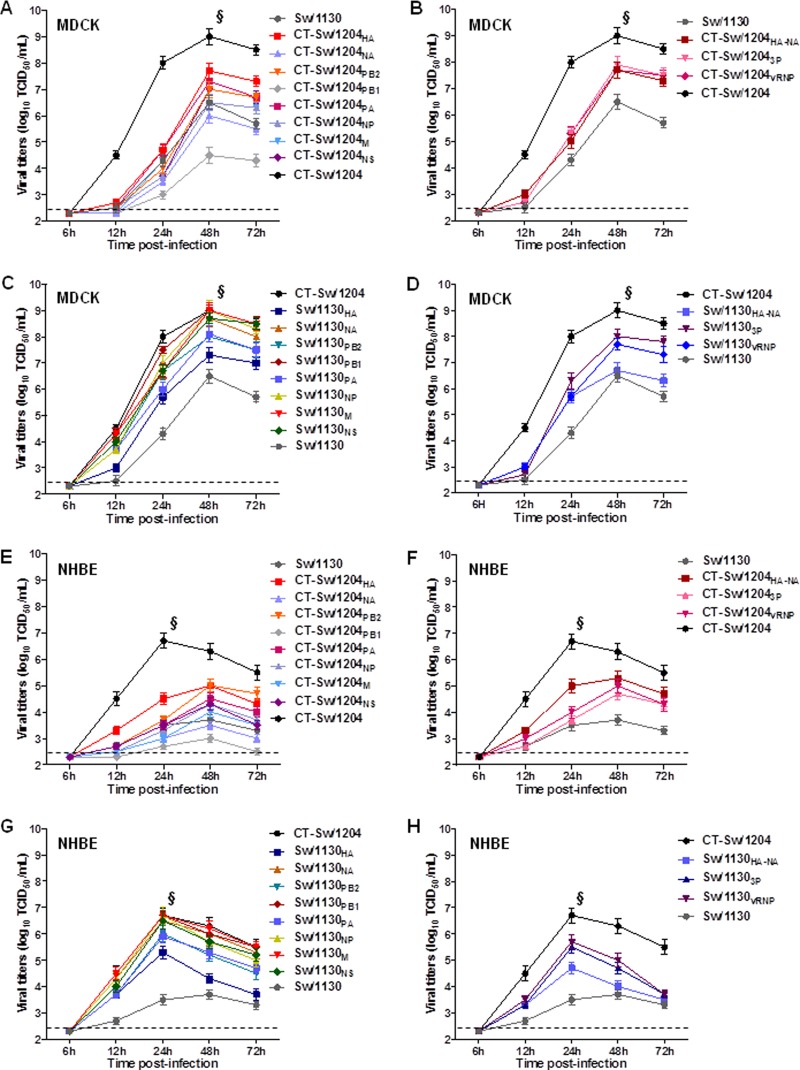
We first examined whether swapping the corresponding viral segments would affect viral growth kinetics in cell culture by comparing the amounts of infectious virus released in culture supernatants. In MDCK cells, the parental CT-Sw/1204 virus exhibited rapid growth, yielding mean peak titers that were at least 100-fold higher than those of the Sw/1130 virus (P < 0.001) (Fig. 1A). Viruses possessing the HA of CT-Sw/1204 (i.e., CT-Sw/1204HA and CT-Sw/1204HA-NA) replicated more efficiently than did Sw/1130 (peak titers of 7.7 to 8.0 log10 TCID50/ml versus 6.5 log10TCID50/ml) (Fig. 1A and B), whereas the Sw/1130HA and Sw/1130HA-NA viruses yielded lower mean peak titers than the parental CT-Sw/1204 virus (6.7 to 7.3 log10 TCID50/ml versus 9.0 log10 TCID50/ml) (Fig. 1C and D). The CT-Sw/1204PB1 recombinant had the lowest replication kinetics (4.5 log10 TCID50/ml mean peak titer), consistent with its low infectivity in these cells (Fig. 1A and Table 1).
Fig 1.
Replicative abilities of recombinant viruses in cultured cells. MDCK (A to D) and primary NHBE (E to H) cells were infected with recombinant parental or reassortant virus. Virus titers in aliquots of harvested supernatants were determined at the indicated time points. The replicative abilities of parental viruses were compared with those of reassortants carrying segments from CT-Sw/1204 (A, B, E, and F) or Sw/1130 (C, D, G, and H). The values are means (± standard deviations [SD]) from three independently performed experiments at 37°C. The dashed lines indicate the detection limit (2.5 log10 TCID50/ml). §, P < 0.001 relative to peak titers of recombinant parental Sw/1130 virus.
Possession of the CT-Sw/1204 PB2, PA, 3P, or vRNP increased the replication efficiency of the Sw/1130 virus, reaching at least 7.0 log10 TCID50/ml mean peak titers at 48 h p.i. (Fig. 1A and B), suggesting that these segments are essential for the optimal replication of CT-Sw/1204 in MDCK cells, apart from HA or HA-NA. In the background of the CT-Sw/1204 virus, Sw/1130PB1, Sw/1130NP, Sw/1130M, and Sw/1130NS displayed replicative abilities commensurate with those of CT-Sw/1204, indicating that replacing these segments does not significantly affect viral growth kinetics in vitro (Fig. 1C and D). However, Sw/1130PB2, Sw/1130PA, Sw/11303P, and Sw/1130vRNP reassortants had slightly lower mean peak titers (7.7 to 8.0 log10 TCID50/ml).
In primary NHBE cells, similar replication trends were observed. CT-Sw/1204 rapidly grew to high titers at 24 h p.i., whereas Sw/1130 produced lower viral titers that peaked at 48 h (P < 0.001) (Fig. 1E). Among reassortant viruses, swapping the PB2 and PA segments marginally altered the replication of the corresponding parental strains; however, greater differences were observed with the homologous substitution of the viral RNA polymerases (i.e., CT-Sw/12043P and CT-Sw/1204vRNP reassortants) (Fig. 1E to H). Regardless, more significantly different titers were produced when HA or HA-NA was replaced. Overall, growth of CT-Sw/1204 and Sw/1130 differed significantly in vitro, and substituting the viral surface genes, except for NA alone, subsequently affected in vitro viral growth.
The heterotrimeric viral RNA polymerase of CT-Sw/1204, independent of HA and NA, contributes to efficient replication and pathogenicity in mice.
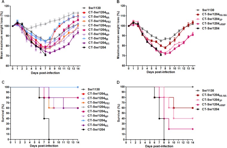
To determine whether the apparent variable growth kinetics in cultured cells also translate into distinct virus characteristics in animal models, we evaluated the replicative abilities and pathogenicities of the parental and reassortant viruses in groups of BALB/c mice. First, the highest possible standard dose for the recombinants possessing the CT-Sw/1204 viral segments in the background of Sw/1130 were used to inoculate groups of mice for determinations of morbidity and virus replication in the lungs (Table 1 and Fig. 2). The parental Sw/1130 recombinant virus produced significantly lower viral titers in harvested mouse lungs than did the recombinant CT-Sw/1204 virus (2.2 log10 TCID50/g and 5.9 log10 TCID50/g, respectively; P = 0.0021) and did not induce appreciable signs of morbidity (Table 1).
Fig 2.
Pathogenicities of recombinant Sw/1130 reassortant viruses carrying CT-Sw/1204 segments in mice. Ten mice were inoculated intranasally with the indicated viruses. All groups were observed for morbidity (A and B) and mortality (C and D) for 14 days. The error bars indicate SD.
Incorporation of PB1 from CT-Sw/1204 into the Sw/1130 background did not produce high viral titers in mouse lungs at 3 or 5 days p.i.; the reassortant virus (CT-Sw/1204PB1) could not be recovered from any mice at 5 days p.i., indicating attenuation, which coincided with its low level of in vitro replication (Fig. 1A). However, expression of HA or HA-NA from CT-Sw/1204 increased replication of Sw/1130 by almost 100-fold (P = 0.002 and 0.004, respectively) (Table 1). Inoculated mice experienced at least 23% body weight reductions, and 40% of the mice died during the 14-day observation period after inoculation (Fig. 2A to D). Apart from those reassortants, possession of the CT-Sw/1204 PB2, PA, NP, NS, 3P, or vRNP complex significantly elevated virus replication (Table 1). All six of the reassortant viruses caused severe disease and lethality; however, the CT-Sw/12043P and CT-Sw/1204vRNP reassortants appeared to be the most pathogenic, with 60% to 80% of mice experimentally inoculated with the viruses dying within 10 days (Fig. 2). The CT-Sw/1204NS reassortant also had a higher mean lung virus titer at 5 days p.i. than did the parental strain, but this difference was not statistically significant (P = 0.0853), although 40% of the inoculated mice died.
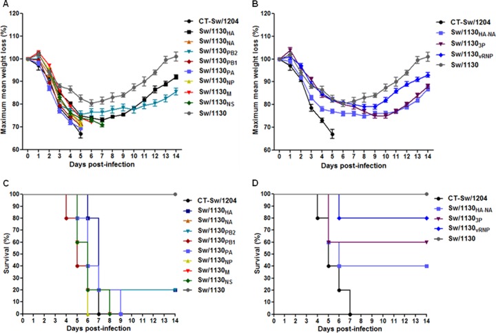
We next assessed the growth kinetics of reassortant CT-Sw/1204 viruses bearing segments of the low-pathogenicity Sw/1130 virus in groups of mice to determine their virulence (Table 2 and Fig. 3). Yielding results similar to those discussed above, the parental CT-Sw/1204 virus replicated efficiently in mouse lungs, producing viral titers at 3 days p.i. that were at least 1,000-fold higher than those of the Sw/1130 virus (6.5 log10 TCID50/g versus 2.9 log10 TCID50/g; P = 0.0075) (Table 2). Replication of single-reassortant viruses containing the NA, PB1, NP, M, or NS of Sw/1130 did not significantly differ from that of the parental CT-Sw/1204 virus; however, the reassortants induced rapid reduction of mean body weight and started killing mice as early as 4 days p.i. (Fig. 3A and B). The death of mice infected with Sw/1130NP or Sw/1130NS was delayed by 1 or 2 days, but all mice eventually succumbed by 9 days p.i. In contrast to the lethal outcome induced by infection with these viruses, inoculation with Sw/1130HA induced significantly lower lung viral titers at both of the time points tested (P = 0.008), which corresponded to <25% weight loss and 20% survival rates (Fig. 3A and C). Although Sw/1130PB2 and Sw/1130PA reassortant viruses also induced significantly lower titers in mouse lungs (Table 2), only the PB2 replacement could rescue 20% of the mice infected with the higher dose of virus. Sw/11303P and Sw/1130vRNP reassortants had viral titers that were significantly lower than those of CT-Sw/1204 (Table 2). Hampered viral growth correlated with the reduced signs of clinical disease and weight loss that resulted in survival of 60% and 80% of inoculated mice, respectively (Fig. 3). These findings showed that the reciprocal constellation of the Sw/11303P or Sw/1130vRNP segment is sufficient to alter the replication of CT-Sw/1204 despite the presence of HA and NA.
Fig 3.
Pathogenicities of recombinant CT-Sw/1204 reassortant viruses carrying Sw/1130 segments in mice. Ten mice were inoculated intranasally with the indicated viruses. All groups were observed for morbidity (A and B) and mortality (C and D) for 14 days. The error bars indicate SD.
Efficient viral growth mediates virulence of recombinant viruses.
We also determined the LD50 in mice inoculated with 10-fold serial dilutions of the recombinant viruses (Tables 1 and 2). The fifty percent mouse lethal doses (MLD50s) of the recombinant parental CT-Sw/1204 and Sw/1130 viruses were 3.5 log10 TCID50 and 6 log10 TCID50, respectively. As expected, the Sw/1130HA and Sw/1130HA-NA reassortant viruses had the greatest decrease in virulence (the MLD50 increased from 3.5 log10 TCID50 to 5.2 log10 TCID50), but these changes still could not completely attenuate CT-Sw/1204, indicating that the remaining viral gene segments contributed to virulence (Table 2). Recombinant CT-Sw/1204 viruses expressing the NA, PB1, NP, M, or NS of Sw/1130 retained the virulent phenotype (MLD50s ranged from 3.5 log10 TCID50 to 3.8 log10 TCID50), whereas reassortant viruses carrying its PB2 or PA had moderately attenuated virulence (4.0 log10 TCID50 to 4.3 log10 TCID50). Incorporation of the vRNA polymerase subunit of Sw/1130, irrespective of the source of NP, further dampened virulence (by 4.7 MLD50 to 5.0 MLD50) relative to that observed with the individual Sw/1130PB2 or Sw/1130PA replacement. Conversely, possession of the CT-Sw/1204 3P or vRNP complex increased virulence in mice, although the virulence was still considerably lower than that of the intact CT-Sw/1204 parental virus (4.7 MLD50s versus 3.5 MLD50s) (Table 1). These results are consistent with findings in the viral growth experiments in which CT-Sw/12043P and CT-Sw/1204vRNP reassortants, apart from CT-Sw/1204HA and CT-Sw/1204HA-NA, replicated to higher titers in mouse lungs than did the parental Sw/1130 strain. Thus, the efficient viral growth induced by the viral polymerases contributed to the virulence of the CT-Sw/1204 virus.
As previously stated, the triple-reassortant internal gene cascades of CT-Sw/1204 and Sw/1130 share approximately 91% genetic identity, with no indications of further genetic reassortment with other viruses of different lineages. However, comparing the deduced amino acid sequences revealed molecular differences between corresponding viral segments of each virus, mostly in PB2, PA, and NS1, but these changes did not appear to be associated with markers that define pathogenicity or alter the host range (data not shown). Sequence variations possessed by CT-Sw/1204 were also different from those of an ancestral North American triple-reassortant swine H3N2 (A/swine/Texas/4199/1998) and a prototypical A(H1N1)pdm09 (A/California/04/2009) virus. However, some of these sites appear to have base residues similar to those of the 1918 (H1N1), 1957 (H2N2), and 1968 (H3N2) pandemic viruses, suggesting that acquisition of molecular adaptive mechanisms occurs during persistent circulation in swine hosts.
Mini-genome replication assays for various combinations of reconstituted vRNP gene complexes.
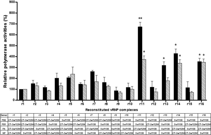
The influenza virus vRNP complex, encoded by the PA, PB1, PB2, and NP genes, is responsible for the transcription and replication of the viral RNA genome. To obtain functional correlates that could help elucidate the mechanism underlying the observed differences in replication efficiency and pathogenic phenotype, we exploited a mini-genome replication reporter assay, which detects any potential contribution of the vRNP complex genes individually or synergistically. The polymerase activities of 16 possible combinations of vRNPs of CT-Sw/1204 and Sw/1130 origin were examined at 33°C or 37°C (Fig. 4), temperatures simulating those in the human upper and lower respiratory tracts (15). Under both thermal conditions, the intact CT-Sw/1204 vRNP complex (Fig. 4, r1) had higher polymerase transcription and replication efficiencies than did the reconstituted Sw/1130 (r9) (33% to 50% difference). Replacing the NP of CT-Sw/1204 with that of Sw/1130 (r2) increased polymerase activity by at least 30%, indicating that the viral origin of segment 5 in our study could not markedly affect the synergistic transcription and replication efficiencies of the 3 viral polymerase segments. Possession of PB1 (r4) or PB1-NP (r7) from Sw/1130 improved the polymerase activities of the CT-Sw/1204 virus by approximately 125% at 33°C and 30% to 70% at 37°C. Conversely, the vRNP complex carrying PA (r11) from CT-Sw/1204 consistently induced the highest activities among the various RNP combinations under any thermal conditions examined. Acquisition of CT-Sw/1204 PB2 (r13) resulted in an approximately 130% increase in polymerase activity over that of Sw/1130 at 33°C but only an approximately 70% increase at 37°C, whereas the CT-Sw/1204 NP (r10) or PB1 (r12) could not significantly elevate in vitro replication and transcription at any given temperature (Fig. 4). Coexpression of CT-Sw/1204 PA with NP (r14) or PB1 (r16), but not with PB2 (as seen with r6 and r8), also significantly elevated the polymerase activity of Sw/1130, stressing the importance of the CT-Sw/1204 PA for the enhancement of transcription and replication activities.
Fig 4.
In vitro polymerase activities of different viral RNP gene combinations. Four expression plasmids (PB2, PB1, PA, and NP) were used to yield the 16 RNP gene combinations of CT-Sw/1204 and Sw/1130, together with pHW72-Luci_M, which carried the reporter luciferase gene. The transfected 293 cells were assayed for luciferase activity after 24 h of incubation at 33°C (upper respiratory tract temperature; solid bars) or 37°C (lower respiratory tract temperature; hatched bars). The values shown are means and standard errors of the mean of the results of 3 independent experiments performed in triplicate, which are standardized to the activity of the expression plasmids for the CT-Sw/1204 vRNP complex proteins (100%). Segments derived from CT-Sw/1204 are shown in italics. “r” denotes reconstituted. *, P < 0.05, and **, P < 0.01 compared to polymerase activities relative to the homologous CT-Sw/1204 vRNP (r1) complex (two-tailed unpaired t test).
The viral polymerase of CT-Sw/1204 promotes RD transmission in ferrets.
Introduction of the HA225G and NA315N found in CT-Sw/1204 into the Sw/1130 virus increases pathogenicity in mice and replication in the upper respiratory tract of ferrets but does not cause efficient horizontal RD transmission (10), suggesting that these mutations are not sufficient to support virus dissemination through the air. To explore the potential role of the viral RNA polymerase subunit of CT-Sw/1204 in RD contact transmission, we generated 2 reassortant viruses in the genetic background of the Sw/1130 (M-Sw/1130) virus mutants that also have the HA225G and NA315N mutations: CT-Sw/12043PM-Sw/1130 and CT-Sw/1204vRNPM-Sw/1130. For comparison, the double-reassortant CT-Sw/1204HA-NA virus in the context of the parental Sw/1130 was also tested.
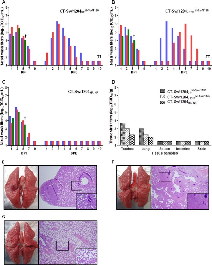
Groups of 3 ferrets were inoculated intranasally and intratracheally (as before) with 105.5 TCID50/ml of the respective reassortant viruses. All strains replicated reasonably well in the upper respiratory tract for up to 7 days p.i., with maximum nasal wash titers of at least 4.6 log10 TCID50/ml (Fig. 5). All ferrets inoculated with the CT-Sw/12043PM-Sw/1130 and CT-Sw/1204vRNPM-Sw/1130 reassortants had moderate to severe signs of influenza marked by infrequent sneezing, a 10% to 15% mean body weight reduction, and temperature elevations of up to 2.3°C. In contrast, inoculation of the CT-Sw/1204HA-NA reassortant induced milder clinical disease, as demonstrated by 3.6% mean body weight losses and temperature elevations of only 1.6°C. Among the 3 recombinant viruses examined, only CT-Sw/12043PM-Sw/1130 and CT-Sw/1204vRNPM-Sw/1130 were able to establish RD transmission to 2 seronegative RD contacts individually paired with inoculated ferrets (Fig. 5A to C). Exposure to CT-Sw/12043PM-Sw/1130 resulted in positive detection in nasal washes as early as 1 day postexposure (p.e.) (Fig. 5A), whereas CT-Sw/1204vRNPM-Sw/1130 had delayed transmission kinetics, requiring 2 to 4 days p.e. for detection in both RD contacts (Fig. 5B). In tissues obtained at 5 days p.i. from 1 of the experimentally infected ferrets, CT-Sw/12043PM-Sw/1130 and CT-Sw/1204vRNPM-Sw/1130 had higher growth capacity in the trachea and lungs than did CT-Sw/1204HA-NA (Fig. 5D). No infectious virions were recovered in any of the other tissue specimens tested (spleen, intestine, and brain). Furthermore, histopathologic examination of lung tissue sections indicated that CT-Sw/12043PM-Sw/1130 and CT-Sw/1204vRNPM-Sw/1130 induced more visible macroscopic lesions, which were accompanied by prominent accumulation of immune cells (Fig. 5E to G). All experimentally infected ferrets and RD contacts except those paired with the CT-Sw/1204HA-NA inoculation group demonstrated seroconversion at 21 days p.i. (or 20 days p.e.).
Fig 5.
Replication and transmission of recombinant viruses in ferrets. (A to C) Groups of 3 ferrets were inoculated with the CT-Sw/12043PM-Sw/1130 (A), CT-Sw/1204vRNPM-Sw/1130 (B), or CT-Sw/1204HA-NA (C) reassortant virus. One of three inoculated ferrets was sacrificed 5 days p.i. (†) for tissue viral titration (D) and histopathology (E). RD contacts were housed adjacent to the inoculated ferrets 1 day p.i. for viral exposure (1:1 setup). Each of the transmission test groups consisted of two directly infected animals and two RD contacts. Nasal wash titers are shown for individual ferrets (as indicated by the colors). The limit of virus detection was 1.5 log10 TCID50/ml per g. ‡, seroconversion of RD contacts after exposure to inoculated ferrets as measured by hemagglutinin inhibition assays. (E to G) Hematoxylin and eosin staining of lung tissue from ferrets infected with CT-Sw/12043PM-Sw/1130 (E), CT-Sw/1204vRNPM-Sw/1130 (F), and CT-Sw/1204HA-NA (G) are shown at low (×40) and high (×200; insets) magnifications.
DISCUSSION
Here, we used reassortant viruses to determine which other viral factors contributed to the growth, pathogenicity, and transmissibility of the CT-Sw/1204 H1N2 virus. Substitution of the isogenic CT-Sw/1204 HA alone or in combination with NA into the closely related Sw/1130 H1N2 virus enhanced replication and pathogenicity in mice, confirming the role of the HA225G and NA315N mutations found in CT-Sw/1204 after a single passage in ferrets (10). However, the reciprocal expression of Sw/1130 HA or HA-NA in the background of CT-Sw/1204 could not render the recombinant reassortant virus completely attenuated, implying that the remaining viral segments are required for pathogenicity. Indeed, we show that possession of the individual CT-Sw/1204 internal gene segments conferred variable effects on replication and pathogenicity, implying that they play critical roles in these areas apart from the contribution of HA-NA.
We found that the PB2, PA, NP, and NS genes of CT-Sw/1204 could alter the growth and pathogenic phenotype of Sw/1130 to some extent in mice. Conversely, reassortant CT-Sw/1204 viruses containing PB2 or PA of Sw/1130, but not PB1, could attenuate clinical disease. However, beyond these results, we demonstrated that the combined expression of the viral RNA polymerase PA, PB1, and PB2 genes of CT-Sw/1204 as a subunit, regardless of whether the NP was derived from CT-Sw/1204 or Sw/1130, had a prominent role in inducing efficient viral replication and lethality to mice. Therefore, our findings suggest that replication efficiency mediated by the epistatic gene interaction of the heterotrimeric RNA polymerase genes of CT-Sw/1204, rather than their individual expression, confers high pathogenicity independent of the balanced activity of the surface glycoproteins (HA225G-NA315N).
The influenza virus polymerase complex genes have been implicated in optimal growth and virulence of the 1918 pandemic virus (16–18) and of highly pathogenic avian influenza (HPAI) H5N1 viruses (19–22). As such, viral pathogenicity is generally attributed to a high level of replicative ability in a given host. Not surprisingly, expression of the homologous polymerase complex genes from CT-Sw/1204 in the context of the HA225G and NA315N mutations in Sw/1130 (i.e., CT-Sw/12043PM-Sw/1130 and CT-Sw/1204vRNPM-Sw/1130) increased replication and invasive infection in the lungs of inoculated ferrets relative to that of the CT-Sw/1204HA-NA reassortant. However, neither CT-Sw/12043PM-Sw/1130 nor CT-Sw/1204vRNPM-Sw/1130 was as virulent as the parental CT-Sw/1204 virus in this host (10), indicating the requirement for other viral factors in ferrets. Nonstructural protein 1 (NS1), encoded by the NS gene, confers efficient replication on human influenza A viruses and is a virulence determinant of HPAI H5N1 viruses (23–25). Noting that CT-Sw/1204 NS has the potential to increase viral growth and elevate pathogenicity, we speculate that segment 8 could also contribute to virulence in concert with the tripartite viral RNA polymerases. NS1 could interact with cognate PA and NP proteins to stabilize the CPSF30-NS1 protein complex (26) or with the vRNP complexes through NP (27), presumably to suppress antiviral responses or to regulate transcriptional activity, respectively. Thus, these interactions could be beneficial because the polymerase genes generally work in concert with the NS and HA genes to produce virulent viruses (28). Therefore, the overall polymerase functionality and its context within the other viral genes could be important in determining the ability of the CT-Sw/1204 virus to induce lethality in ferrets, and acquisition of the HA225G and NA315N mutations exacerbates disease. Overall, these results show that the high pathogenicity of the CT-Sw/1204 virus involves interplay of multiple viral factors, further exemplifying the polygenic nature of virulence among influenza A viruses.
Respiratory droplet transmission has been mostly attributed to the receptor specificity of HA in H1 viruses (29, 30) or to balanced HA-NA activity (31, 32). However, the polymerase complex genes facilitate virus dissemination through the air (33, 34). We found that CT-Sw/12043PM-Sw/1130 or CT-Sw/1204vRNPM-Sw/1130 promoted infection of RD contact ferrets, implying the indispensability of the tripartite viral RNA polymerases of CT-Sw/1204 for attaining RD transmission. Similarly, PB2 of the 1918 pandemic virus was required to support RD transmission of a recombinant avian virus carrying the 1918 HA (34). Possession of the CT-Sw/1204 HA-NA in the genetic background of Sw/1130 did not lead to efficient transmission, consistent with our previous results indicating that the HA and NA modifications alone are inadequate to mediate virus dissemination through the air. Hence, our findings confirm the notion that aerosol transmissibility may not always depend on receptor specificity or balanced activities of the surface glycoproteins in a given host (34).
The reconstituted vRNP complex of CT-Sw/1204 consistently had higher polymerase activity than did Sw/1130 at temperatures correlating with those in both regions of the human respiratory tract (15). Such selective advantage may have facilitated viral replication competence, allowing reassortant viruses carrying the CT-Sw/1204 polymerase segments to be readily available in the upper nasal cavity of infected ferrets for efficient transmission. It is apparent, therefore, that HA225G and NA315N in CT-Sw/1204 contributed to virulence and transmission by allowing virus dissemination in the upper and lower respiratory tract and that the heterotrimeric polymerase gene segments remained active in such tissue environments, facilitating robust replication.
The viral polymerase can mediate adaptation of influenza viruses in a new host (35). The internal gene segments of CT-Sw/1204 and Sw/1130 have notable amino acid differences, which are likely responsible for the altered growth, pathogenicity, and transmissibility of these strains. Of particular interest are positions 64 in PB2, 100 and 337 in PA, and 215 in NS1, all of which have been identified as persistent sites for adaptation and host shifting of influenza viruses, including in the 1918, 1957, and 1968 pandemic strains and H5N1 viruses (36–38). Although PB1 is a key component of the viral RNA polymerase complex functionally and structurally, replication impairment due to possession of CT-Sw/1204 PB1 alone suggests that the genetic modifications in segment 2 are deleterious in the context of the parental recombinant Sw/1130 virus. However, PB1 appears to function better with segments 1 and 3 from CT-Sw/1204, indicating its indispensability for the tripartite polymerase complex. Further studies will shed light on the specific role of these mutations in CT-Sw/1204 or other triple-reassortant swine viruses, including the genetically related A(H1N1)pdm09 virus.
Altogether, the data generated in this study confirm the importance of the HA225G and NA315N mutations to the CT-Sw/1204 virus. However, we also provide proof that the synergistic function of the homologous polymerase gene complex is critically important for the virulence and transmission of CT-Sw/1204. Because this report demonstrates that some viruses in the field may have made subtle, modest adaptations that could alter disease impact, continued monitoring is highly recommended to manage risks associated with the emergence of strains that could become significant threats to public health. Along with the growing number of reports on the importance of the viral polymerase for replication, pathogenesis, and dissemination, our results justify the development of novel antiviral drugs aimed at curbing the functions of the viral polymerase complex subunits.
ACKNOWLEDGMENTS
This work was partly supported by grant A103001 from the Korea Healthcare Technology R&D Project, Ministry of Health and Welfare, Republic of Korea, by a National Agenda Project grant from the Korea Research Council of Fundamental Science and Technology, and by the KRIBB Initiative Program (KGM3111013). Robert G. Webster and Richard J. Webby are supported by contract no. HHSN266200700005C from the National Institute of Allergy and Infectious Diseases, National Institutes of Health, Department of Health and Human Services, and by the American Lebanese Syrian Associated Charities.
We thank Eun-Hye Choi, Seong-Mi Hong, and Arun Decano for their excellent technical assistance. We greatly appreciate the careful editing of our manuscript by Cherise Guess.
Footnotes
Published ahead of print 17 July 2013
REFERENCES
- 1.Webster RG, Bean WJ, Gorman OT, Chambers TM, Kawaoka Y. 1992. Evolution and ecology of influenza A viruses. Microbiol. Rev. 56:152–179 [DOI] [PMC free article] [PubMed] [Google Scholar]
- 2.Laver WG, Webster RG. 1973. Studies on the origin of pandemic influenza. 3. Evidence implicating duck and equine influenza viruses as possible progenitors of the Hong Kong strain of human influenza. Virology 51:383–391 [DOI] [PubMed] [Google Scholar]
- 3.Cohen J, Enserink M. 2009. Swine flu. After delays, WHO agrees: the 2009 pandemic has begun. Science 324:1496–1497 [DOI] [PubMed] [Google Scholar]
- 4.Dawood FS, Jain S, Finelli L, Shaw MW, Lindstrom S, Garten RJ, Gubareva LV, Xu X, Bridges CB, Uyeki TM. 2009. Emergence of a novel swine-origin influenza A (H1N1) virus in humans. N. Engl. J. Med. 360:2605–2615 [DOI] [PubMed] [Google Scholar]
- 5.Garten RJ, Davis CT, Russell CA, Shu B, Lindstrom S, Balish A, Sessions WM, Xu X, Skepner E, Deyde V, Okomo-Adhiambo M, Gubareva L, Barnes J, Smith CB, Emery SL, Hillman MJ, Rivailler P, Smagala J, de Graaf M, Burke DF, Fouchier RA, Pappas C, Alpuche-Aranda CM, Lopez-Gatell H, Olivera H, Lopez I, Myers CA, Faix D, Blair PJ, Yu C, Keene KM, Dotson PD, Jr, Boxrud D, Sambol AR, Abid SH, St George K, Bannerman T, Moore AL, Stringer DJ, Blevins P, Demmler-Harrison GJ, Ginsberg M, Kriner P, Waterman S, Smole S, Guevara HF, Belongia EA, Clark PA, Beatrice ST, Donis R, Katz J, Finelli L, Bridges CB, Shaw M, Jernigan DB, Uyeki TM, Smith DJ, Klimov AI, Cox NJ. 2009. Antigenic and genetic characteristics of swine-origin 2009 A(H1N1) influenza viruses circulating in humans. Science 325:197–201 [DOI] [PMC free article] [PubMed] [Google Scholar]
- 6.Trifonov V, Khiabanian H, Greenbaum B, Rabadan R. 2009. Origin of the recent swine influenza A(H1N1) virus infecting humans. Euro Surveill. 14:19193. [PubMed] [Google Scholar]
- 7.Barman S, Krylov PS, Fabrizio TP, Franks J, Turner JC, Seiler P, Wang D, Rehg JE, Erickson GA, Gramer M, Webster RG, Webby RJ. 2012. Pathogenicity and transmissibility of North American triple reassortant swine influenza A viruses in ferrets. PLoS Pathog. 8:e1002791. 10.1371/journal.ppat.1002791 [DOI] [PMC free article] [PubMed] [Google Scholar]
- 8.Belser JA, Gustin KM, Maines TR, Blau DM, Zaki SR, Katz JM, Tumpey TM. 2011. Pathogenesis and transmission of triple-reassortant swine H1N1 influenza viruses isolated before the 2009 H1N1 pandemic. J. Virol. 85:1563–1572 [DOI] [PMC free article] [PubMed] [Google Scholar]
- 9.Belser JA, Wadford DA, Pappas C, Gustin KM, Maines TR, Pearce MB, Zeng H, Swayne DE, Pantin-Jackwood M, Katz JM, Tumpey TM. 2010. Pathogenesis of pandemic influenza A (H1N1) and triple-reassortant swine influenza A (H1) viruses in mice. J. Virol. 84:4194–4203 [DOI] [PMC free article] [PubMed] [Google Scholar]
- 10.Pascua PN, Song MS, Lee JH, Baek YH, Kwon HI, Park SJ, Choi EH, Lim GJ, Lee OJ, Kim SW, Kim CJ, Sung MH, Kim MH, Yoon SW, Govorkova EA, Webby RJ, Webster RG, Choi YK. 2012. Virulence and transmissibility of H1N2 influenza virus in ferrets imply the continuing threat of triple-reassortant swine viruses. Proc. Natl. Acad. Sci. U. S. A. 109:15900–15905 [DOI] [PMC free article] [PubMed] [Google Scholar]
- 11.Hoffmann E, Neumann G, Kawaoka Y, Hobom G, Webster RG. 2000. A DNA transfection system for generation of influenza A virus from eight plasmids. Proc. Natl. Acad. Sci. U. S. A. 97:6108–6113 [DOI] [PMC free article] [PubMed] [Google Scholar]
- 12.Reed LJ, Muench H. 1938. A simple method of estimating fifty percent endpoints. Am. J. Hyg. 27:493–497 [Google Scholar]
- 13.Palmer DF, Dowdle WR, Coleman MT, Schild GC. 1975. Advanced laboratory techniques for influenza diagnosis. Immun. Ser. 6:25–45 [Google Scholar]
- 14.Song MS, Pascua PN, Lee JH, Baek YH, Park KJ, Kwon HI, Park SJ, Kim CJ, Kim H, Webby RJ, Webster RG, Choi YK. 2011. Virulence and genetic compatibility of polymerase reassortant viruses derived from the pandemic (H1N1) 2009 influenza virus and circulating influenza A viruses. J. Virol. 85:6275–6286 [DOI] [PMC free article] [PubMed] [Google Scholar]
- 15.Massin P, van der Werf S, Naffakh N. 2001. Residue 627 of PB2 is a determinant of cold sensitivity in RNA replication of avian influenza viruses. J. Virol. 75:5398–5404 [DOI] [PMC free article] [PubMed] [Google Scholar]
- 16.Pappas C, Aguilar PV, Basler CF, Solorzano A, Zeng H, Perrone LA, Palese P, Garcia-Sastre A, Katz JM, Tumpey TM. 2008. Single gene reassortants identify a critical role for PB1, HA, and NA in the high virulence of the 1918 pandemic influenza virus. Proc. Natl. Acad. Sci. U. S. A. 105:3064–3069 [DOI] [PMC free article] [PubMed] [Google Scholar]
- 17.Watanabe T, Tisoncik-Go J, Tchitchek N, Watanabe S, Benecke AG, Katze MG, Kawaoka Y. 2013. 1918 influenza virus hemagglutinin (HA) and the viral RNA polymerase complex enhance viral pathogenicity, but only HA induces aberrant host responses in mice. J. Virol. 87:5239–5254 [DOI] [PMC free article] [PubMed] [Google Scholar]
- 18.Watanabe T, Watanabe S, Shinya K, Kim JH, Hatta M, Kawaoka Y. 2009. Viral RNA polymerase complex promotes optimal growth of 1918 virus in the lower respiratory tract of ferrets. Proc. Natl. Acad. Sci. U. S. A. 106:588–592 [DOI] [PMC free article] [PubMed] [Google Scholar]
- 19.Hatta M, Gao P, Halfmann P, Kawaoka Y. 2001. Molecular basis for high virulence of Hong Kong H5N1 influenza A viruses. Science 293:1840–1842 [DOI] [PubMed] [Google Scholar]
- 20.Li Z, Chen H, Jiao P, Deng G, Tian G, Li Y, Hoffmann E, Webster RG, Matsuoka Y, Yu K. 2005. Molecular basis of replication of duck H5N1 influenza viruses in a mammalian mouse model. J. Virol. 79:12058–12064 [DOI] [PMC free article] [PubMed] [Google Scholar]
- 21.Maines TR, Chen LM, Belser JA, Van HN, Smith E, Donis RO, Tumpey TM, Katz JM. 2011. Multiple genes contribute to the virulent phenotype observed in ferrets of an H5N1 influenza virus isolated from Thailand in 2004. Virology 413:226–230 [DOI] [PubMed] [Google Scholar]
- 22.Salomon R, Franks J, Govorkova EA, Ilyushina NA, Yen HL, Hulse-Post DJ, Humberd J, Trichet M, Rehg JE, Webby RJ, Webster RG, Hoffmann E. 2006. The polymerase complex genes contribute to the high virulence of the human H5N1 influenza virus isolate A/Vietnam/1203/04. J. Exp. Med. 203:689–697 [DOI] [PMC free article] [PubMed] [Google Scholar]
- 23.Das K, Ma LC, Xiao R, Radvansky B, Aramini J, Zhao L, Marklund J, Kuo RL, Twu KY, Arnold E, Krug RM, Montelione GT. 2008. Structural basis for suppression of a host antiviral response by influenza A virus. Proc. Natl. Acad. Sci. U. S. A. 105:13093–13098 [DOI] [PMC free article] [PubMed] [Google Scholar]
- 24.Talon J, Horvath CM, Polley R, Basler CF, Muster T, Palese P, Garcia-Sastre A. 2000. Activation of interferon regulatory factor 3 is inhibited by the influenza A virus NS1 protein. J. Virol. 74:7989–7996 [DOI] [PMC free article] [PubMed] [Google Scholar]
- 25.Twu KY, Kuo RL, Marklund J, Krug RM. 2007. The H5N1 influenza virus NS genes selected after 1998 enhance virus replication in mammalian cells. J. Virol. 81:8112–8121 [DOI] [PMC free article] [PubMed] [Google Scholar]
- 26.Kuo RL, Krug RM. 2009. Influenza A virus polymerase is an integral component of the CPSF30-NS1A protein complex in infected cells. J. Virol. 83:1611–1616 [DOI] [PMC free article] [PubMed] [Google Scholar]
- 27.Robb NC, Chase G, Bier K, Vreede FT, Shaw PC, Naffakh N, Schwemmle M, Fodor E. 2011. The influenza A virus NS1 protein interacts with the nucleoprotein of viral ribonucleoprotein complexes. J. Virol. 85:5228–5231 [DOI] [PMC free article] [PubMed] [Google Scholar]
- 28.Hulse-Post DJ, Franks J, Boyd K, Salomon R, Hoffmann E, Yen HL, Webby RJ, Walker D, Nguyen TD, Webster RG. 2007. Molecular changes in the polymerase genes (PA and PB1) associated with high pathogenicity of H5N1 influenza virus in mallard ducks. J. Virol. 81:8515–8524 [DOI] [PMC free article] [PubMed] [Google Scholar]
- 29.Jayaraman A, Pappas C, Raman R, Belser JA, Viswanathan K, Shriver Z, Tumpey TM, Sasisekharan R. 2011. A single base-pair change in 2009 H1N1 hemagglutinin increases human receptor affinity and leads to efficient airborne viral transmission in ferrets. PLoS One 6:e17616. 10.1371/journal.pone.0017616 [DOI] [PMC free article] [PubMed] [Google Scholar]
- 30.Tumpey TM, Maines TR, Van HN, Glaser L, Solorzano A, Pappas C, Cox NJ, Swayne DE, Palese P, Katz JM, Garcia-Sastre A. 2007. A two-amino acid change in the hemagglutinin of the 1918 influenza virus abolishes transmission. Science 315:655–659 [DOI] [PubMed] [Google Scholar]
- 31.Xu R, Zhu X, McBride R, Nycholat CM, Yu W, Paulson JC, Wilson IA. 2012. Functional balance of the hemagglutinin and neuraminidase activities accompanies the emergence of the 2009 H1N1 influenza pandemic. J. Virol. 86:9221–9232 [DOI] [PMC free article] [PubMed] [Google Scholar]
- 32.Yen HL, Liang CH, Wu CY, Forrest HL, Ferguson A, Choy KT, Jones J, Wong DD, Cheung PP, Hsu CH, Li OT, Yuen KM, Chan RW, Poon LL, Chan MC, Nicholls JM, Krauss S, Wong CH, Guan Y, Webster RG, Webby RJ, Peiris M. 2011. Hemagglutinin-neuraminidase balance confers respiratory-droplet transmissibility of the pandemic H1N1 influenza virus in ferrets. Proc. Natl. Acad. Sci. U. S. A. 108:14264–14269 [DOI] [PMC free article] [PubMed] [Google Scholar]
- 33.Steel J, Lowen AC, Mubareka S, Palese P. 2009. Transmission of influenza virus in a mammalian host is increased by PB2 amino acids 627K or 627E/701N. PLoS Pathog. 5:e1000252. 10.1371/journal.ppat.1000252 [DOI] [PMC free article] [PubMed] [Google Scholar]
- 34.Van Hoeven N, Pappas C, Belser JA, Maines TR, Zeng H, Garcia-Sastre A, Sasisekharan R, Katz JM, Tumpey TM. 2009. Human HA and polymerase subunit PB2 proteins confer transmission of an avian influenza virus through the air. Proc. Natl. Acad. Sci. U. S. A. 106:3366–3371 [DOI] [PMC free article] [PubMed] [Google Scholar]
- 35.Gabriel G, Dauber B, Wolff T, Planz O, Klenk HD, Stech J. 2005. The viral polymerase mediates adaptation of an avian influenza virus to a mammalian host. Proc. Natl. Acad. Sci. U. S. A. 102:18590–18595 [DOI] [PMC free article] [PubMed] [Google Scholar]
- 36.Finkelstein DB, Mukatira S, Mehta PK, Obenauer JC, Su X, Webster RG, Naeve CW. 2007. Persistent host markers in pandemic and H5N1 influenza viruses. J. Virol. 81:10292–10299 [DOI] [PMC free article] [PubMed] [Google Scholar]
- 37.Tamuri AU, Dos RM, Hay AJ, Goldstein RA. 2009. Identifying changes in selective constraints: host shifts in influenza. PLoS Comput. Biol. 5:e1000564. 10.1371/journal.pcbi.1000564 [DOI] [PMC free article] [PubMed] [Google Scholar]
- 38.Taubenberger JK, Reid AH, Lourens RM, Wang R, Jin G, Fanning TG. 2005. Characterization of the 1918 influenza virus polymerase genes. Nature 437:889–893 [DOI] [PubMed] [Google Scholar]


