Abstract
In martial arts and contact sports, strikes are often trained in two different ways: with and without impacts. This study aims to compare the electromyographical activity (EMG) of the triceps brachii (TB), biceps brachii (BB) and brachioradialis (BR) muscles during strikes with and without impacts. Eight Yau-Man Kung Fu practitioners participated in the experiment. Each participant performed 5 sequences of 5 consecutive KF Yau-Man palm strikes with no impact intercalated with 5 sequences of 5 repetitions targeting a KF training shield. Surface EMG signals were obtained from the TB, BB, and RB for 3.0 seconds using an eight-channel module with a total amplifier gain of 2000 and sampled at 3500 Hz. The EMG analyses were done in the time (rms) and frequency (wavelet) domains. For the frequency domain, Morlet wavelet power spectra were obtained and an original method was used to quantify statistically significant regions on the power spectra. The results both in the time and frequency domains indicate a higher TB and BR muscle activity for the strikes with impacts. No significant difference was found for the BB in the two different scenarios. In addition, the results show that the wavelet power spectra pattern for the three analysed muscles obtained from the strikes with and without impacts were similar.
Key points.
EMG analysis of a sequence of Kung Fu strikes demonstrates higher Triceps Brachii and Brachioradialis muscle activity for strikes with impact than strikes without impact.
An original reliable method for quantifying EMG wavelet transform results is presented.
EMG wavelet power spectra describe muscle roles during a Kung Fu sequence of strikes.
Key words: Electromyography, wavelet transform, impact, martial arts, biomechanics
Introduction
Chinese Martial Arts has a history of thousands of years. During the past 1500 years, many different styles of Kung Fu (KF) have been developed in China and several of them are still being practiced. The Yau-Man style of KF was developed during the Ch’ing Dynasty (1644-1911) with the purpose to help Chinese revolutionaries in the war against the Manchu invaders (Neto et. al, 2007). The Yau-Man KF was developed from the collective knowledge of several other ancient styles. The strikes of Yau-Man KF differ from most other combat sports’ strikes because they usually begin closer to the target or opponent. Other distinguished characteristic of the Yau-Man strikes is that the movements are terminated before full extension of the arms (Neto et al., 2007).
In most martial arts and contact sports strikes are often trained in two different ways: with and without impacts. In the last couple of years, there have been studies that focused on strikes with impacts (Neto et al., 2007; Walilko et. al., 2005) and without impacts (Neto and Magini, 2007; Ribeiro et al., 2006). However, the authors are not aware of any article that compared either physiological or biomechanical variables in both scenarios. This study aims to compare the EMG of the triceps brachii (TB), biceps brachii (BB) and brachioradialis (BR) muscles during strikes with and without impacts.
Methods
Eight Kung Fu Yau-Man practitioners (training experience of 2.8 ± 3 years; 1 year minimum) were selected to participate in the experiment. The participants’ average height, mass and age were 1.76 (SD = 0.05) m, 75 (SD = 9.2) Kg, and 21.8 (SD = 5.9) years. Each participant performed 5 sequences of 5 consecutive KF Yau-Man palm strikes without impact intercalated with 5 sequences of 5 repetitions targeting a KF training shield. A detailed description of this movement can be found on Neto et al., 2007. Surface EMG signals were obtained from the biceps brachii (BB), brachioradialis (BR) (two of the main antagonist muscles) and triceps brachii (TB) (main agonist muscle) at the dominant side of the body. The participants were allowed to position themselves in relation to the shield and to adjust its height as they wished.
This methodology was approved by the University of Vale do Paraíba Ethics in Research Committee (Protocol #: L008/2005/CEP) and all participants or legal responsible provided their informed written consent.
Materials
The surface EMG signals were obtained according to standard procedures (De Luca, 1997) for 3.0 seconds using an eight-channel module (model EMG800C, EMGSystem, Brazil) with a total amplifier gain of 2000 and sampled at 3500 Hz. A 12 bits AD converter digitalized the analogue signals with a sampling frequency of anti-aliasing of 2.0 kHz for each channel and an input range of 5mV. Active bipolar superficial electrodes consisting of two rectangular parallel bars of Ag/AgCl (1 cm in length, 0.2 cm in width and separated by 1 cm) were used and coupled to a rectangular acrylic resin capsule 2.2 cm in length, 1.9 cm in width and 0.6 cm in high with an internal amplifier (with gain of 20) in order to reduce the effects of electromagnetic interference and other noises. After shaving and cleaning the skin with alcohol, the electrodes were placed on the muscles guided by bone prominences and the route of the muscle fibbers. EMG sensors and sensor placement procedures were in agreement with recommendations resulted from SENIAM studies (Hermens and Freriks, 2000).
Data analyses
All EMG data was processed off-line with Matlab 7.0.1 (MathWorks Inc) following standard procedures (De Luca, 1997). The EMG signals were filtered (Butterworth order 4, band pass 50-500Hz) to eliminate noise from the movement of artefacts and to consider the band of the power spectrum of higher energy. The lower frequency threshold was chosen because of the greater arm speed in martial arts strikes compared to most other types of arm movements (Neto and Magini, 2007).
The EMG signals were analysed in the time and frequency domains. For the time domain analysis the EMG signals were full wave rectified and then linear smoothed using a low pass Butterworth order 4 filter with a cut frequency of 14 Hz (Bolgla and Uhl, 2007). The mean amplitude for this linear envelope was determined and it was used to normalize the EMG signals from which root mean square (rms) values were calculated. For the frequency domain, the EMG signals were normalized by the standard deviation. The frequency domain analyses were done by Morlet wavelet. Wavelet power spectra were generated using the algorithm developed by C. Torrence and G. Compo (1998 - available at URL: http://paos.colorado.edu/research/wavelets). The wavelet power spectra can be seen as a flat three-dimensional graph; the y-axis gives the period (s), the x-axis gives the time (s) and different colours indicate the different power values (V2). Another information given in the spectra is the statistically significant regions of the spectra, which are delimited by thick contours. An algorithm was developed in Matlab 7.0.1 (MathWorks Inc.) to determine the sum of the significant power (SSP) on the wavelet power spectra. SSP values were calculated adding up power values that fell inside the significance contours.
Statistics
The comparisons of rms and SSP values from the strikes with and without impact were done using a balanced Multivariate Analyses of Variance for repeated measures. The Turkey method was used for a pair wise comparison of means. Intrasubject coefficient of variation (IACV) for each muscle with and without impact were calculated by dividing the standard deviation of the SPS and rms values for the five sequences of strikes for each participant by its respective mean. Intersubject coefficient of variation (IECV) were calculated for each muscle with and without impact by dividing the standard deviation of the SPS and rms values for all sequences of strikes by their respective means. All statistical analyses were conducted using MINITAB® 14.12.0 (Minitab Inc.) at the 0.05 level of significance. The power spectra thick contour encloses regions of greater than 95% confidence in the qui-square test for a red-noise process with a lag-1 coefficient of 0.72 (Torrence and Compo, 1998).
Results
Both rms (see Table 1) and SSP (see Table 2) results indicate a significant higher TB and BR muscle activity for the strikes with impacts considering the repeated measures for all eight participants (p < 0.01 for both rms and SSP). No significant difference was found in the EMG of the BB for the two different scenarios (p = 0.26 for rms and p = 0.12 for SSP). The average of all 48 values of SSP IACV (0.074 ± 0.043) was smaller than the corresponding average rms IACV (0.095 SD 0.088) (p = 0.03 for paired t-test). The average of all 6 values of SSP IECV (0.153 ± 0.041) was also smaller than the corresponding average rms IECV (0.275 ± 0.139), however because of the sample sizes the statistic reliability is lower (p = 0.05 for paired t-test).
Table 1.
Average values of rms for each participant’s sequence of strikes with and without impact plus intrasubject coefficient of variation (IACV) and total mean plus intersubject coefficient of variation (IECV).
| rms(IACV) | Triceps Brachii | Biceps Brachii | Brachioradialis | |||
|---|---|---|---|---|---|---|
| Subjects | Without Impact | With Impact |
Without Impact | With Impact |
Without Impact | With Impact |
| 1 | 2.56 (.11) | 2.96 (.13) | 2.48 (.05) | 2.09 (.09) | 2.56 (.03) | 4.14 (.08) |
| 2 | 2.24 (.06) | 2.59 (.05 | 2.04 (.09) | 2.01 (.08) | 2.40 (.13) | 2.64 (.05) |
| 3 | 2.50 (.06) | 2.68 (.03) | 2.97 (.05) | 2.81 (.06) | 2.88 (.05) | 2.81 (.07) |
| 4 | 3.18 (.09) | 3.42 (.02) | 2.89 (.04) | 2.89 (.03) | 2.55 (.14) | 2.77 (.02) |
| 5 | 1.98 (.10) | 3.14 (.25) | 3.57 (.42) | 6.13 (.43) | 2.06 (.04) | 3.04 (.18) |
| 6 | 3.45 (.02) | 3.59 (.05) | 2.62 (.06) | 2.73 (.02) | 3.61 (.06) | 3.63 (.05) |
| 7 | 3.06 (.13) | 2.99 (.08) | 3.55 (.21) | 3.28 (.16) | 3.37 (.20) | 3.40 (.07) |
| 8 | 2.12 (.10) | 4.22 (.07) | 1.66 (.03) | 1.64 (.01) | 1.84 (.07) | 3.21 (.15) |
| Mean (IECV) | 2.62 (.21) | 3.18 (.18) | 2.72 (.31) | 2.95 (.54) | 2.66 (.24) | 3.18 (.17) |
Table 2.
Average values of SSP (105V) for each participant’s sequence of strikes with and without impact plus intrasubject coefficient of variation (IACV) and total mean plus intersubject coefficient of variation (IECV).
| SSP(IACV) | Triceps Brachii | Biceps Brachii | Brachioradialis | |||
|---|---|---|---|---|---|---|
| Subjects | Without Impact | With Impact |
Without Impact | With Impact |
Without Impact | With Impact |
| 1 | 8.53 (.05) | 9.11 (.19) | 7.46 (.07) | 7.36 (.15) | 9.11 (.05) | 9.49 (.07) |
| 2 | 9.60 (.10) | 9.95 (.02) | 8.60 (.04) | 9.54 (.03) | 7.74 (.09) | 8.61 (.09) |
| 3 | 7.21 (.07) | 7.32 (.04) | 7.43 (.05) | 7.07 (.07) | 7.48 (.06) | 7.74 (.08) |
| 4 | 8.19 (.03) | 8.11 (.05) | 7.02 (.05) | 6.97 (.03) | 7.89 (.03) | 7.69 (.07) |
| 5 | 9.79 (.06) | 11.3 (.18) | 9.87 (.07) | 9.86 (.16) | 6.90 (.09) | 7.45 (.13) |
| 6 | 5.91 (.03) | 5.80 (.06) | 8.33 (.03) | 8.44 (.08) | 7.24 (.06) | 7.62 (.07) |
| 7 | 7.75 (.08) | 7.99 (.05) | 7.12 (.13) | 7.57 (.09) | 7.83 (.09) | 9.18 (.16) |
| 8 | 9.16 (.14)) | 10.1 (.05) | 8.48 (.03) | 9.32 (.06) | 8.40 (.01) | 9.27 (.07) |
| Mean (IECV) | 8.29 (.17) | 8.69 (.22) | 8.04 (.13) | 8.28 (.16) | 7.84 (.10) | 8.43 (.14) |
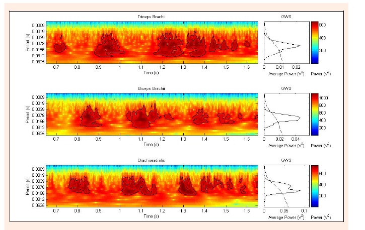
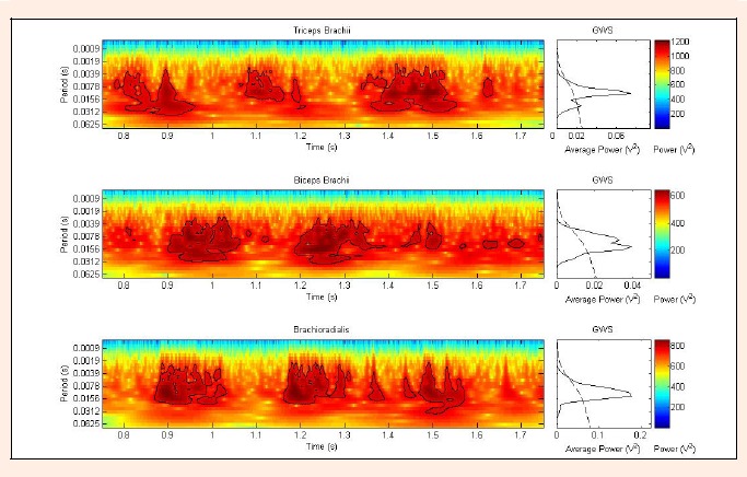
Although it was collected three seconds of EMG signals from the muscles, in average, only a period of one second presented significant contours on the power spectra. The results show that the wavelet power spectra pattern for the three analysed muscles obtained from the strikes with and without impacts were similar (see Figure 1 and Figure 2). For all strikes, it was observed that the TB was the first muscle to present a significant contour on their spectra. The TB spectra present three main regions of significant contour(s). The third region in general presents more than one contour and it extends more in time. The BB and BR presented very similar spectra. They also presented three distinct main regions containing significant contour(s); the regions were shifted by approximately 100-200 ms in relation to the regions in the TB spectra.
Figure 1.

Wavelet transform power spectrum for the TB, BB and BR for one of the sequence of strikes without impact. A colour scale gives the power (V2) of the periods; intensity increases from dark blue to dark red. The thick contour on the spectra encloses significant regions ( =0.05). Global Wavelet Spectrum (GWS) is the graph of power versus period for the whole duration of the strike analysed. Peaks above the dashed line in the GWS graphs are significant ( = 0.05).
Figure 2.

Similar to the wavelet transform power spectrum of Fig. 1 but for a sequence of strikes with impact.
Discussion
The coefficients of variation found for the rms values are in agreement with values reported by other researchers (Bolgla and Uhl, 2007). According to many researchers, the normalization method used in this experiment to calculate the values of rms provides lower IACV and IECV (Burden et al., 2003; Knutson et al., 1994). A comparison of the rms and the original SSP methods used in this study revealed that the SSP method resulted in lower IACV and IECV (see Tables 1 and 2). Lower IACV indicates greater reliability of the measurements, while the advantages of using methods that result in lower IECV are well documented in the literature (Yang and Winter, 1984). These results confirm the validity of the SSP method used. Since there were only six data points for the comparison of the IECV of the rms and SSP, further studies should be done to confirm the hypothesis that the SSP method may be a more stable and precise method for measuring significant muscle activity than the rms method.
The reason for the higher TB (an agonist muscle for the strike) activity in the strikes with impact might be more psychological than biomechanical, possibly due to greater participant’s motivation when performing the strikes with impact. The reason for the higher BR activity, on the other hand, is probably due to the importance of this muscle in stabilizing the wrist and elbow joints when receiving the reaction force from the impact.
The result that an approximate time of only one second presented significant contours on the power spectra suggests that the participants took approximately one second to perform the sequences of five palm strikes. This result is in agreement with previous high-speed camera study that found the duration of one palm strike to be in the order of 200 ms (Neto et al., 2007).
The wavelet power spectra from the three analysed muscles (Figure 1 and 2) illustrate the muscle roles during the five strikes performed in each sequence. The first significant contour on the TB powers spectra demonstrates a muscle activity responsible for pushing the right hand during the first palm strike. This region is followed in time by a significant contour both in the BB and BR spectra, which correspond to muscle activity responsible to brake the right hand’s movement and pull it back while the left hand is being pushed during the first left hand palm strike. After that, the same pattern in wavelet power spectra repeats for the second right hand palm strike followed by the second left hand palm strike. For the third right hand palm, more extended significant regions in the spectra can be seen, which implies longer muscles’ activity. It can be speculated that, for both strikes with and without impact, the participants keep tightening their muscles once the arm stops. These isometric muscles’ contractions, once the movement stops, are often performed in martial arts.
Conclusion
This study compares the EMG activity of the TB, BB and BR muscles during strikes with and without impacts. The EMG analyses were done in the time and frequency domains. For the frequency domain, an original method was used to quantify statistically significant regions on the wavelet power spectra, and it was proven be very reliable. The results indicate that there are greater TB and BR muscle activities for the strikes with impacts. The reasons for this greater muscle activity are expected to be both psychological, such as greater motivation, and biomechanical, such as the need for stabilizing the joints during the impacts. In addition, the results show that the wavelet power spectra pattern for the three analysed muscles obtained from the strikes with and without impacts were similar. The results suggest that training forms or “katas ”should not replace training with pads. Although in both types of training the movements performed present very similar muscle activation characteristics, the magnitude of the contraction of some muscle are greater for the movements done with pads. In consequence, the relative time spend in each type of training should be determined depending on the application. For example, for participants that need to increase strength either for combats or breaking demonstrations, training with pads should be more relevant than without pads. In addition, the methodology developed in this article has other applications, such as muscle control and rehabilitation evaluations for athletes and non-athletes.
Acknowledgments
The authors would like to thank CNPq and CAPES for financial support, Prof. Maurício Bolzan, Regiane Albertini de Carvalho, Thais Helena de Freitas and Ana Carolina Marzullo for helping obtaining the data, and all volunteers.
Biographies

Osmar Pinto NETO
Employment
PhD student, Institute of Research Development, Univap, Brazil.
Degree
MSc, BSc.Hons
Research interests
Biomechanics of Sports.
E-mail: osmar@univap.br

Marcio MAGINI
Employment
Director, Department of Computer Science, Univap, Brazil.
Degree
PhD, MSc, BSc
Research interests
Mathematical modelling of biological systems.
E-mail: magini@univap.br

Marcos T.T. PACHECO
Employment
Director, Institute of Research and Development, Univap, Brazil.
Degree
PhD, MSc, BSc
Research interests
Biomedical engineering.
E-mail: mtadeu@univap.br
References
- Bolgla L.A., Uhl T.L. (2007) Reliability of electromyographic normalization methods for evaluating the hip musculature. Journal of Electromyography and Kinesiology 17, 102-111 [DOI] [PubMed] [Google Scholar]
- Burden A.M., Trew M., Baltzopoulos V. (2003) Normalisation of gait EMGs: a re-examination. Journal of Electromyography and Kinesiology 13, 519-32 [DOI] [PubMed] [Google Scholar]
- De Luca C.J. (1997) The use of electromyography in biomechanics. Journal of Applied Biomechanics 13, 135-163 [Google Scholar]
- Hermens H. J., Freriks B. (2000) Development of recommendations for SEMG sensors and sensor placement procedures. Journal of Electromyography and Kinesiology 10(5), 361-374 [DOI] [PubMed] [Google Scholar]
- Knutson L.M., Soderburg G.L., Ballantyne B.T., Clarke W.R. (1994) A study of various normalization procedures for within day electromyographic data. Journal of Electromyography and Kinesiology 4(1), 47-59 [DOI] [PubMed] [Google Scholar]
- Neto O.P., Magini M. (2007) Electromiographic and kinematic characteristics of Kung Fu Yau-Man palm strike. Journal of Electromyography and Kinesiology, in press, doi:10.1016/j.jelekin.2007.03.009 [DOI] [PubMed] [Google Scholar]
- Neto O. P., Magini M., Saba M.M.F. (2007) The role of effective mass and hand speed in the performance of Kung Fu athletes compared to non-practitioners. Journal of Applied Biomechanics 23, 139-148 [DOI] [PubMed] [Google Scholar]
- Ribeiro J.L., de Castro B.O.S.D., Rosa C.S., Baptista R.R., Oliveira A.R. (2006) Heart rate and blood lactate responses to Changquan and Daoshu forms of modern Wushu. Journal of Sports Science and Medicine 5(CSSI), 1-4 [PMC free article] [PubMed] [Google Scholar]
- Torrence C., Compo G.P. (1998) A Practical Guide to Wavelet Analysis. Bulletin of the American Meteorological Society 79(1), 61-78 [Google Scholar]
- Walilko T. J., Viano D. C., Bir C. A. (2005) Biomechanics of the head for Olympic boxer punches to the face. British Journal of Sports Medicine 39, 710-719 [DOI] [PMC free article] [PubMed] [Google Scholar]
- Yang J.F., Winter D.A. (1984) Electromyographic amplitude normalization methods: improving their sensitivity as diagnostic tools in gait analysis. Archives of Physical Medicine and Rehabilitation 65, 517-521 [PubMed] [Google Scholar]


