Abstract
Background:
This study was designed to investigate the effect of vitiligo on the retinal nerve fiber layer (RNFL) thickness.
Materials and Methods:
This prospective study was conducted in the Department of Ophthalmology at Kırıkkale University during 2010 and 2011. Sixty eight eyes of 34 vitiligo patients were included in the study. Eighty four eyes were served as control. RNFL thickness was measured by scanning laser polarimetry (Nerve Fiber Analyzer, GDx VCC: 5.3.3; Laser Diagnostic Technologies, San Diego, CA, USA).
Results:
The mean duration of vitiligo was found to be 9.8 ± 2.3 years. The mean average RNFL thickness outside the disc margin was significantly lower in the right eyes of vitiligo group in comparison to the controls (P = 0.02). The mean average thickness of RNFL beneath the measuring ellipse in the superior sector of both eyes were significantly lower than the controls (P = 0.004, P = 0.008, respectively). The topographical distributions of RNFL thickness in superior, inferior, nasal and temporal quadrants were similar for two groups.
Conclusion:
RNFL thickness seems to be unaffected in vitiligo patients.
Keywords: Nerve fiber layer, retina, thickness, vitiligo
INTRODUCTION
Vitiligo is an acquired, chronic pigmentation disorder characterized by white patches corresponding to a substantial loss of functioning epidermal and hair follicle melanocytes.[1] Few studies in the literature have reported ocular findings in vitiligo patients and possible associations between these two entities. Some degree of retinal pigment findings have been found as the major feature in the eyes of vitiligo patients.[2,3,4,5,6] Vitiligo patients with more extensive skin involvement and a longer disease duration were found to exhibit altered visual evoked potentials and an abnormal electro-oculographic findings.[7]
In a recent study, Rogosic et al. have proposed a possible association between vitiligo duration and glaucoma development.[8] Assessment of the retinal nerve fiber layer (RNFL) thickness by scanning laser polarimetry appears to be sensitive and specific for the evaluation of glaucomatous damage, especially early during the course of the disease.[9,10,11]
In the current literature, there is no report investigating the effect of vitiligo on RNFL. Therefore, In this study we measured the RNFL thickness in order to find out a structural effect of vitiligo on to the RNFL.
MATERIALS AND METHODS
This prospective study was conducted in the Department of Ophthalmology at Kırıkkale University, Kırıkkale, Turkey during 2010 and 2011. The Ethics Comitee of Kırıkkale University approved the study protocol (Research Project No. 562/10) and informed consents were obtained from all participants. The patient group included 68 eyes of 34 vitiligo patients. A total number of 42 subjects matched for age, and sex were recruited as controls.
For each patient, a thorough medical history including the medications was obtained and a general physical examination was performed by the same dermatologist to exclude any reason for secondary causes of vitiligo. Both vitiligo patients and control group underwent a complete ophthalmologic examination, including best corrected visual acuity and refraction, intraocular pressure measurment, ocular movements and biomicroscopy of the anterior and posterior segments of the eye.
Excluded were patients with previous neurological disease, previous ocular surgery, glaucoma, ocular hypertension, markedly diminished visual acuity and retinal disorders or systemic disorders affecting eye such as, diabetes mellitus and hypertension. Randomly assigned 42 eyes volunteers of similar age, and sex distribution were taken as the control group. The simple randomization was achieved by including a study coordinator who was not involved with the investigative part of the research. Same exclusion criteria were also valid for the control group.
RNFL thickness was measured by scanning laser polarimetry (Nerve Fiber Analyzer, GDx VCC: 5.3.3; Laser Diagnostic Technologies, San Diego, CA, USA) for both groups. The measurement of RNFL thickness was carried out by the same examiner. The examinations were carried out in the same room for all subjects. The pupils were undilated.
The software device measures and then gives a series of parameters related to RNFL thickness. For each subject, more than one good-quality images were acquired to obtain the best image that would be used for the measurement of RNFL thickness. For all subjects mean RNFL thickness values were computed for all of the retina as well as superior (120 °), inferior (120 °), temporal (70 °) and nasal (50 °) segments and compared with the control group.
Statistical analysis
The statistical analysis was performed using the Statistical Package for Social Sciences program. Analysis of variance was used to evaluate the statistical significance when comparing the two groups. P values less than 0.05 were considered as statistically significant.
RESULTS
The study included 34 (15 male, 19 female) vitiligo patients and 42 (20 male, 22 female) controls. There was no difference in gender between the two groups. The mean age in the patient group was 33.47 ± 12.34 (range 17-57) years compared to 30 ± 8.51 (range 22-53) years in the control group. There was no statistically significant difference in mean age between the vitiligo group and control group (P = 0.31). The mean duration of vitiligo was found to be 9.8 ± 2.3 years.
All vitiligo patients were receiving phototherapy with psoralen ultraviolet A. Vitiligo was generalized type in all patients. Five patients were found to have ocular findings, including peripapillary atrophy in three patients, focal hipopigmentation in one patient and diffuse hipopigmentation in one patient.
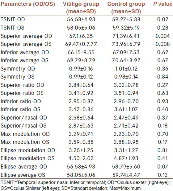
Table 1 shows the mean values for all parameters associated with RNFL thickness for both vitiligo and control groups.
Table 1.
Distribution of retinal nerve fiber layer thickness parameters in vitiligo patients and controls

The mean average RNFL thickness outside the disc margin was significantly lower than in the right eyes of vitiligo group in comparison to the controls (F (1-74) = 5.20, 95% confidence interval (CI) for mean 56.87-59.27, P = 0.02). The mean average thickness of RNFL beneath the measuring ellipse in the superior sector of both eyes were significantly lower than the controls (F (1-74) = 8.70, 95% CI for mean 67.97-70.99, P = 0.004, F (1-74) = 7.30, 95% CI for mean 70.25-73.66, P = 0.008, respectively). The topographical distributions of RNFL in the superior, inferior, nasal, and temporal quadrants were similar between the vitiligo group and control group.
DISCUSSION
Although the exact etiology of vitiligo is unknown, it has become quite clear in recent reports that genetic, immunological, neural and self-destructive mechanisms are involved in its pathogenesis. Whatever the primary injury is, antibodies directed against antigens of the melanocyte system were found to be destructive to the melanocytes in a vast majority of patients with vitiligo.[1] It is evident that multiple ocular abnormalities like uveal involvement, peripapillary atrophy, alterations of retinal pigment epithelium, focal and diffuse hypopigmentation may be found in patients with vitiligo.[2,3,4,5,6] Many of the ocular findings are non-specific and may be associated with immunological processes in the disease.
In a study by Rogosic et al., it was proposed that there may be a possible association between vitiligo duration and glaucoma development.[8] As previously mentioned, scanning laser polarimetry is a reliable method for the diagnosis and follow-up of glaucoma and related retinal damage. Measurements are compared to a normative database to provide quick, easy, and objective analysis.[9,10,11,12,13,14,15] Therefore, we measured the RNFL thickness with scanning laser polarimetry in vitiligo patients to detect a possible structural effect of the disease on to the neural retina.
In this study, the results revealed a little amount of significant evidence about the alterations of RNFL thickness in patients with vitiligo when compared with control group. The mean average RNFL thickness outside the disc margin was significantly lower in the right eyes of vitiligo group in comparison to the controls. The mean average thickness of RNFL beneath the measuring ellipse in the superior sector of both eyes was significantly lower than the controls. These differences need further investigations before considering them as early changes in RNFL thickness. The topographical distribution of RNFL in superior, inferior, nasal, and temporal quadrants were similar between vitiligo patients and control subjects.
To the best of our knowledge, this is the first study to focus on the possible alterations in RNFL thickness in vitiligo patients. We may conclude that the RNFL thickness seems to be unaffected in vitiligo patients. However, further data with larger series and longer disease durations might provide information to enlighten the association between vitiligo and glaucoma adequately. As our sample group is small, this study may provide only preliminary information that might pioneer clinical research in this field.
Footnotes
Source of Support: None
Conflict of Interest: None declared.
REFERENCES
- 1.Alikhan A, Felsten LM, Daly M, Petronic-Rosic V. Vitiligo: A comprehensive overview Part I. Introduction, epidemiology, quality of life, diagnosis, differential diagnosis, associations, histopathology, etiology, and work-up. J Am Acad Dermatol. 2011;65:473–91. doi: 10.1016/j.jaad.2010.11.061. [DOI] [PubMed] [Google Scholar]
- 2.Schwartz R. Vitiligo: A sign of systemic disease. Indian J Dermatol Venereol Leprol. 2006;72:68–71. doi: 10.4103/0378-6323.19730. [DOI] [PubMed] [Google Scholar]
- 3.Cowan CL, Jr, Halder RM, Grimes PE, Chakrabarti SG, Kenney JA., Jr Ocular disturbances in vitiligo. J Am Acad Dermatol. 1986;15:17–24. doi: 10.1016/s0190-9622(86)70136-9. [DOI] [PubMed] [Google Scholar]
- 4.Park S, Albert DM, Bolognia JL. Ocular manifestations of pigmentary disorders. Dermatol Clin. 1992;10:609–22. [PubMed] [Google Scholar]
- 5.Biswas G, Barbhuiya JN, Biswas MC, Islam MN, Dutta S. Clinical pattern of ocular manifestations in vitiligo. J Indian Med Assoc. 2003;101:478–80. [PubMed] [Google Scholar]
- 6.Bulbul Baskan E, Baykara M, Ercan I, Tunalı S, Yücel A. Vitiligo and ocular findings: A study on possible associations. J Eur Acad Dermatol Venereol. 2006;20:829–33. doi: 10.1111/j.1468-3083.2006.01655.x. [DOI] [PubMed] [Google Scholar]
- 7.Perossini M, Turio E, Perossini T, Cei G, Barachini P. Vitiligo: Ocular and electrophysiological findings. G Ital Dermatol Venereol. 2010;145:141–9. [PubMed] [Google Scholar]
- 8.Rogosić V, Bojić L, Puizina-Ivić N, Vanjaka-Rogosić L, Titlić M, Kovacević D, et al. Vitiligo and glaucoma: An association or a coincidence? A pilot study. Acta Dermatovenerol Croat. 2010;18:21–6. [PubMed] [Google Scholar]
- 9.Weinreb RN, Shakiba S, Zangwill L. Scanning laser polarimetry to measure the nerve fiber layer of normal and glaucomatous eyes. Am J Ophthalmol. 1995;119:627–36. doi: 10.1016/s0002-9394(14)70221-1. [DOI] [PubMed] [Google Scholar]
- 10.Medeiros FA, Vizzeri G, Zangwill LM, Alencar LM, Sample PA, Weinreb RN. Comparison of retinal nerve fiber layer and optic disc imaging for diagnosing glaucoma in patients suspected of having the disease. Ophthalmology. 2008;115:1340–6. doi: 10.1016/j.ophtha.2007.11.008. [DOI] [PMC free article] [PubMed] [Google Scholar]
- 11.Zheng W, Baohua C, Qun C, Zhi Q, Hong D. Retinal nerve fiber layer images captured by GDx-VCC in early diagnosis of glaucoma. Ophthalmologica. 2008;222:17–20. doi: 10.1159/000109273. [DOI] [PubMed] [Google Scholar]
- 12.Chi QM, Tomita G, Inazumi K, Hayakawa T, Ido T, Kitazawa Y. Evaluation of the effect of aging on the retinal nerve fiber layer thickness using scanning laser polarimetry. J Glaucoma. 1995;4:406–13. [PubMed] [Google Scholar]
- 13.Zangwill L, Berry CA, Garden VS, Weinreb RN. Reproducibility of retardation measurements with the nerve fiber analyzer II. J Glaucoma. 1997;6:384–9. [PubMed] [Google Scholar]
- 14.Hoh ST, Ishikawa H, Greenfield DS, Liebmann JM, Chew SJ, Ritch R. Peripapillary nerve fiber layer thickness measurement reproducibility using scanning laser polarimetry. J Glaucoma. 1998;7:12–5. [PubMed] [Google Scholar]
- 15.Rhee DJ, Greenfield DS, Chen PP, Schiffman J. Reproducibility of retinal nerve fiber layer thickness measurements using scanning laser polarimetry in pseudophakic eyes. Ophthalmic Surg Lasers. 2002;33:117–22. [PubMed] [Google Scholar]


