Abstract
Our recent study shows a pivotal role of Dmp1 in quenching hyperproliferative signals from HER2 to the Arf-p53 pathway as a safety mechanism to prevent breast carcinogenesis. To directly demonstrate the role of Dmp1 in preventing HER2/neu-driven oncogenic transformation, we established Flag-Dmp1α transgenic mice (MDTG) under the control of the mouse mammary tumor virus (MMTV) promoter. The mice were viable but exhibited poorly developed mammary glands with markedly reduced milk production; thus more than half of parous females were unable to support the lives of new born pups. The mammary glands of the MDTG mice had very low Ki-67 expression but high levels of Arf, Ink4a, p53, and p21Cip1, markers of senescence and accelerated aging. In all strains of generated MDTG;neu mice, tumor development was significantly delayed with decreased tumor weight. Tumors from MDTG;neu mice expressed Flag-Dmp1α and Ki-67 in a mutually exclusive fashion indicating that transgenic Dmp1α prevented tumor growth in vivo. Genomic DNA analyses showed that the Dmp1α transgene was partially lost in half of the MDTG;neu tumors, and Western blot analyses showed Dmp1α protein downregulation in 80% of the cases. Our data demonstrate critical roles of Dmp1 in preventing mammary tumorigenesis and raise the possibility of treating breast cancer by restoring Dmp1α expression.
Introduction
Breast cancer is one of the most important public health issues in the United States and most industrialized countries [1]–[4]. In the U.S., an estimated 210,000 women will be diagnosed with breast cancer in 2013 [2], making it the most common cancer in U.S. women, and second only to lung cancer in cancer-related death. Approximately 70% of human breast tumors express hormone receptors, the estrogen receptor (ER) and/or progesterone receptor (PR). ER is the primary transcription factor driving oncogenesis in hormone receptor-positive breast cancers, and thus usually responsive to adjuvant hormonal therapy with anti-estrogens or aromatase inhibitors, giving a more favorable prognosis [1]. Conversely, ER-negative tumors are frequently associated with more aggressive disease with poorer clinical outcomes, including amplification of HER2 or c-Myc oncogenes or gain-of-function mutation of p53 [1], [4]. BRCA1/2 are high-penetrance breast cancer predisposition genes identified by genome-wide linkage analysis and positional cloning. Mutations of so-called low penetrance breast cancer genes functionally related to BRCA1/2, such as CHEK2, ATM, BRIP1, and PALB2, are rare, but confer an intermediate risk of the disease [5].
The c-ErbB2 gene (HER2) has been identified in the human genome 17q21 and encodes a polypeptide with a kinase domain highly homologous to that of the epidermal growth factor (EGF) receptor [6]. The human c-ErbB2 gene (HER2) is an equivalent of the rat neu gene, detected in a series of rat neuro/glioblastomas [6]. HER2/neu encodes a receptor-type tyrosine kinase that belongs to the EGFR family [7]–[9]. It is overexpressed in ∼50% of human breast cancers, primarily due to gene amplification (30%) [9], although protein overexpression without gene amplification is also found in ∼20% of breast cancer cases. HER2/neu overexpression is prominent in metastatic lesions, and thus associated with poor clinical outcomes [7]–[9]. It has been shown that phosphatidylinositol-3′-kinase (PI3K) and serine/threonine kinase Akt/protein kinase B play critical roles in oncogenic HER2/neu signaling [10]. Aberrant HER2/neu expression initially causes cell proliferation, but eventually leads to cell cycle arrest or senescence in normal cells to prevent their malignant transformation, in which Dmp1 plays a critical role [11].
A valine to glutamic acid substitution in the trans-membrane domain of a rat neu mutant results in the constitutive aggregation and activation of the receptor in the absence of ligand [12] (reviewed in [13]). In human breast cancers overexpressing HER2, the same trans-membrane point mutation of HER2/neu has not been reported, but its activated splicing variants have been reported in tumors [14], [15]. Transgenic mice expressing activated neu under the control of mouse mammary tumor virus promoter (MMTV-neu) develop multifocal mammary tumors at a median age of 7 months with high potential of lung metastasis [16]. Mice bearing the wild-type ErbB2 allele under the control of the MMTV promoter (MMTV-ErbB2) have also been established [17]. The usefulness of the MMTV-driven transgenic mice as models of breast cancer has been emphasized since the detection of MMTV env-like sequence in ∼40% of human breast cancers [13], [18]. In contrast to the rapid tumor progression observed in several transgenic strains carrying the activated neu transgene, wild-type neu expression in the mammary epithelium results in the development of focal mammary tumors with longer latency than those with constitutively active neu (8–12 months vs. 6–7 months) [16], [17]. Interestingly, many of the tumor-bearing transgenic mice developed secondary metastatic lesions in lung indicating that wild-type neu overexpression can induce metastatic disease after long latency [17].
Dmp1 (a cyclin D binding myb-like protein 1; also called Dmtf1) is a transcription factor originally isolated in a yeast two-hybrid screen of a murine T-lymphocyte library with cyclin D2 as bait [19]. Dmp1 shows its activity as a tumor suppressor by directly binding to the Arf promoter to activate its gene expression, and thereby induces p53-dependent cell cycle arrest [20], [21] (reviewed in [22], [23]). Our recent study indicates that Dmp1 directly binds to p53 and neutralizes the negative regulation of p53 by Mdm2, especially in epithelial and hematopoietic cells [24]. Dmp1-null murine embryonic fibroblasts (MEFs) give rise to immortalized cell lines that retain wild-type p19Arf and p53, and are transformed by oncogenic Ras alone, suggesting that the activity of the Arf-p53 pathway is significantly subverted in Dmp1-deficient cells [25], [26]. The murine Dmp1 promoter is efficiently activated by oncogenic Ras and HER2 and repressed by E2Fs-mediated mitogenic signals and NF-κB-mediated genotoxic signals [27]–[29], suggesting that the promoter receives both positive and negative regulation.
Both Dmp1−/− and Dmp1+/− mice were prone to tumor development when newborn pups were treated with dimethylbenzanthracene or ionizing radiation [25], [26]. Dmp1, p53, and p21Cip1 were induced in pre-malignant lesions of MMTV-neu mice to prevent incipient cancer cells from transformation. Selective Dmp1 deletion and/or Tbx2/Pokemon overexpression was found in >50% of wild-type HER2/neu carcinomas while the involvement of Arf, Mdm2, or p53 was rare [11]. Tumors induced by the Eµ-Myc, K-Ras and HER2 transgenes were greatly accelerated in both Dmp1+/− and Dmp1−/− backgrounds with no differences between groups lacking one or two Dmp1 alleles, suggesting haploid-insufficiency of Dmp1 in tumor suppression in these mouse models of human cancers [11], [26], [30]. Mammary tumors from MMTV-neu; Dmp1+/−, Dmp1−/− mice showed significant downregulation of Arf and p21Cip1, with p53 inactivity and more aggressive phenotypes than tumors with intact Dmp1 [11].
The hDMP1 gene is located on chromosome 7q21, a region often deleted in breast cancer and hematopoietic malignancies [31]–[34]. We recently found that loss of heterozygosity (LOH) of hDMP1 was present in ∼35% of non-small cell lung carcinomas [30], [35], [36]. The hDMP1 locus encodes at least three splicing variants, i.e. hDMP1α, β, and γ [32]. The full-length hDMP1α gene corresponds to the murine Dmp1 that positively regulates the Arf-p53 pathway. On the other hand, the hDMP1β and γ isoforms lack the DNA-binding domain, and hDMP1β is dominant-negative for hDMP1α on myeloid differentiation and CD13 induction [32].
To elucidate the role of human DMP1 (hDMP1) in breast cancer, we recently analyzed 110 tumor and normal pairs of human breast cancer samples for the alterations (gene loss or amplification) of the hDMP1-ARF-Hdm2-p53 pathway with follow up of clinical outcomes. LOH of the hDMP1 locus was found in 42% of human breast cancers, while that of INK4a/ARF and p53 were found in 20% and 34%, respectively. Amplification of the Hdm2 locus was found in 13% of the samples, which was independent of LOH for hDMP1, INK4a/ARF, or p53 [37]. LOH for hDMP1 was mutually exclusive with that of INK4a/ARF and p53, and associated with low Ki67 index and diploidy of the nuclear DNA. Consistently, LOH for hDMP1 was associated with luminal A category and longer relapse-free survival, while that of p53 was associated with non-luminal A subgroup. Thus, loss of hDMP1 defined a new disease category with a potential prognostic value for breast cancer patients [37].
In this study, we established Flag-Dmp1α transgenic mice (MDTG) driven by the MMTV promoter to study the effects of Dmp1 expression in mammary gland development, function, and gene/protein expression in vivo. We crossed MDTG mice with MMTV-neu mice to demonstrate the role of Dmp1 in preventing HER2/neu-induced mammary carcinogenesis.
Materials and Methods
Creation of MMTV-Flag-Dmp1α Mice
To create transgenic mice that constitutively express Dmp1α in mammary glands, we cloned the Flag-tagged murine Dmp1α cDNA into the Hind III site of the vector MMTV-SV40-BSSK (a gift from Dr. Philip Leder, Harvard Medical School) and created MMTV-Flag-Dmp1α transgenic (MDTG) mice in the FVB/NJ background using the Transgenic Core of Wake Forest University Health Sciences. The term Flag-Dmp1 indicates Flag-Dmp1α hereafter. Briefly, the transgene construct was microinjected into the pronuclei of fertilized one-cell zygotes from FVB/NJ mice. These zygotes were reimplanted into pseudo-pregnant foster mothers, and the offspring were screened for the presence of the transgene by PCR. Carriers were bred to establish three independent transgenic lines, strains 76, 79, and 138.
Creation of MMTV-Flag-Dmp1α;neu (MDTG;neu) bitransgenic Mice and Whole Mount Preparation of Mammary Glands
One male MMTV-Flag-Dmp1α -transgenic mouse in each was crossed with two female MMTV-neu mice to obtain MDTG;neu double transgenic mice. We obtained more than 25 MDTG;neu bi-transgenic females for each MDTG strain. Mice were monitored daily, sacrificed in a CO2 chamber when moribund, and tumor tissues were resected from the mice for pathological and molecular genetic analyses. Whole mount sections of wild-type and MDTG mammary glands were prepared from 12-week-old nulliparous females as described previously [38].
Analysis of Mammary Tumors Obtained from the Bi-transgenic Mice
Mice were monitored daily for mammary tumor development. We detected tumors when they reached 3mm in size, and sacrificed the mice 4 weeks after the first tumor was found. After CO2 asphyxiation, mammary tumors were dissected from mice to weigh, and then we conducted histopathological and biochemical analyses. All experimental procedures were conducted according to a protocol approved by the Institutional Animal Care and Use Committee of the Wake Forest University Health Sciences.
Gene Expression Analyses and Gene Copy Number Assay for Flag-Dmp1α in Mouse Tails and Tumors
Quantitation of Flag-Dmp1, p19Arf, p16Ink4a, and p21Cip1 mRNAs was conducted by real-time PCR Taqman assay by ABI7500 (Applied Biosystems) using β-actin as an internal control (28, 29). The assays for mouse Arf (p19Arf-cDNA), Ink4a (Ink4a-cDNA-G), and β-actin (mactbEx3_4) were custom-designed at ABI. For mouse p21Cip1, Mm01303209_m1 was used. Gene copy number assay for Dmp1 was also performed by real-time PCR aiming at the exons deleted in Dmp1 knockout mice (Dmp1 Ex10-11-Ex11), using β-actin (mbactinex4-ANY) as an internal control. Tail DNAs from Dmp1+/+, Dmp1+ /−, and Dmp1−/− were used as two copies, one copy and zero-copy control.
Western Blotting
For Western blot analyses of MDTG;neu tumors, proteins were extracted with ice-cold EBC buffer with proteinase inhibitors (Calbiochem proteinase inhibitor cocktail III, leupeptin, AEBSF, and aprotinin) [19]. After gel electrophoresis and transfer to nitrocellulose membranes, proteins were visualized by immunoblotting using affinity-purified polyclonal antibodies to Flag-Dmp1 (Abcam ab21536), p53 (sc-6243G), Mdm2 (ab16896, #2A10, Abcam), p19Arf (sc-32748), p16Ink4a (sc-74401), p21CIP1/WAF1 (sc-397G), or β-actin (sc-1615, sc-47778), followed by incubation of the filters with horseradish peroxidase–conjugated second antibodies, and reaction with the enhanced chemiluminescence detection kit (Perkin-Elmer).
Immunohistochemical Staining
Immunohistochemical staining of tissues and tumors were conducted as described previously [28]. The following antibodies were used for immunohistochemistry with formalin-fixed, paraffin-embedded sections: Ki67 (SP6, NeoMarkers), αDDDDK Tag (Abcam ab21536), cleaved caspase 3 (#9661, Cell, Signaling), and HER2 (sc-284, Santa Cruz Biotech).
Statistical Analyses
Statistical differences of survival in MMTV-neu and MDTG;neu females (nulliparous) were analyzed by Medcalc software, Mariakerke, Belgium. Statistical analyses for all experiments were conducted using unpaired Student’s t-tests.
Results
Establishment of MMTV-Flag-Dmp1α Mice
To elucidate the roles of Dmp1α in mammary tumor development and prevention of HER2;neu-induced mammary carcinogenesis, we created transgenic mice that constitutively express the Flag-tagged murine Dmp1α gene under the control of the MMTV promoter (MMTV-Flag-Dmp1α, MDTG) (Fig. S1; strain 138 results, not shown). Real-time PCR study of the genomic DNA extracted from mouse tails showed that strain 76 had 18–42 (30.2+/−11.8), strain 79 had 31–45 (37.4+/−7.1), and strain 138 had 6–12 (8.0+/−4.0) copies of the Flag-Dmp1α transgene. Thus strains 76 and 79 were designated as high copy number MDTG strains, while strain 138 was low copy number strain.
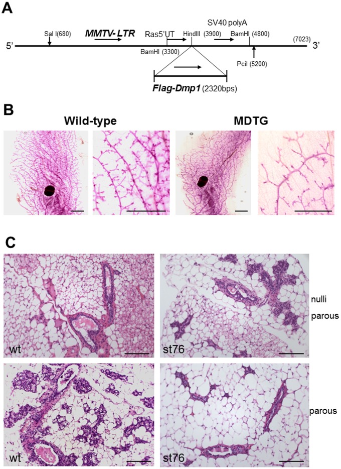
Compared to wild-type mice, the MDTG females showed significantly reduced (∼50% less) number of mammary glands and branching with poor alveolar sac development throughout the body, although the size of the germinal center remained the same (Fig. 1B). When stained with H&E, mammary glands (MMGs) from MDTG mice showed small and undilated tubules with little milk production in comparison to those of normal controls in null-parous mice (Fig. 1C, upper panels). The morphological difference of MMG between wild-type and MDTG mice was more prominent in parous females; they were well-branched and dilated with milk production in the former, but unopened or partially opened with little milk production in the latter (Fig. 1C, lower panels). Consistent with these findings, approximately 60% of MDTG parous females were unable to support their pups while wild-type FVB females were able, suggesting that MMG from MDTG females had defective function.
Figure 1. Creation of MMTV-Flag-Dmp1α mice.
A. The Flag-Dmp1 cDNA (BamHI fragment) was recovered from the pFLEX1-Dmp1 vector and was cloned into the HindIII site of the transgenic vector with MMTV promoter (MMTV-SV40-BSSK). B. Whole mammary gland mounts from 12-week-old nulliparous MDTG (strain 79) and wild-type mice. Scale bars show 2 mm. C. Photomicrographs of nulliparous and multi-parous female mammary glands (MMG) from wild-type and strain 76 MMTV-Flag-Dmp1 (MDTG) mice (12-week-old). Wild-type mammary glands were well-dilated with milk production, which became more prominent after getting pregnant. MDTG mammary glands, on the other hand remained small, unopened with little milk production. These mammary glands did not open well with very low milk production even after getting pregnant, making it difficult to feed their pups. The scale bars show 100 µm.
Tissue-specific Expression of Flag-Dmp1 mRNA and Protein in Mammary Tissues
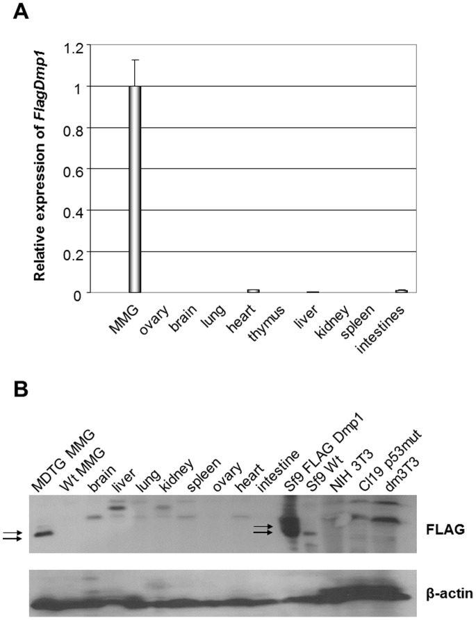
Gene expression studies as demonstrated by real-time PCR showed tissue-specific expression of transgenic Flag-Dmp1 mRNA in MMG from MDTG mouse strain 76 in comparison to other tissues (ovary, brain, lung, heart, thymus, liver, kidney, spleen, and intestines) from this transgenic mouse (Fig. 2A). We also analyzed the expression of the Flag-Dmp1 protein in tissues from MDTG mouse strain 79 and found tissue-specific expression of the Flag-Dmp1 protein in MMG from this transgenic mouse strain, confirming the selectivity of the promoter (Fig. 2B).
Figure 2. Expression of Flag-Dmp1 mRNA and proteins in tissues of MDTG mice.
A. Expression of Flag-Dmp1 mRNA in tissues from a mouse of MDTG (12-week-old, strain 76) as studied by real-time PCR. The level of Flag-Dmp1 in lactating mammary glands was defined as 1.0. B. Expression of the Flag-Dmp1 protein in tissues from a MDTG mouse (12-week-old, strain 76). Proteins were extracted in ice-cold EBC buffer (19) and 100 µg of proteins were analyzed by Western blotting with the antibody to the FLAG tag.
Expression of the Flag-Dmp1 Protein Decreased Cell Proliferation in MMG from MDTG Mice
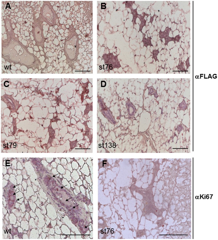
In immunohistochemical (IHC) studies using an antibody to Flag, we detected significant Flag-Dmp1 signals in mammary glands from all three strains of MDTG mice (Fig. 3B–D), but only background signals were observed in wild-type MMG (Fig. 3A). The Flag-Dmp1 intensity in MMG from strains 76 and 79 mice was higher than that from strain 138, reflecting Flag-Dmp1 transgene copy number differences among these strains. When blotting these tissues with an antibody for Ki67, a marker for cell proliferation, we detected 5–10 positive cells per MMG from wild-type nulliparous females but barely detected any in MDTG mice (Fig. 3E, F). The same patterns were obtained in females regardless of their age. These data suggest that MMG of MDTG remained unopened throughout their life due to strikingly decreased cell proliferation.
Figure 3. Immunohistochemical detection of Flag-Dmp1 and Ki67 in mammary glands of MDTG mice.
A, B, C, and D. Immunohistochemical detection of the Flag-Dmp1 protein in nulliparous transgenic MMG. A) wild-type, B) MDTG strain 76, C) MDTG strain 79, and D) MDTG strain 138. E, F. Immunohistochemical detection of Ki67 in nulliparous MMG from wild-type (E) and MDTG, strain 76 (F) female. Note that Ki67 signals were barely detectable in MMG from a MDTG mouse.
Induction of Ink4a/Arf proteins in MMG from MDTG mice
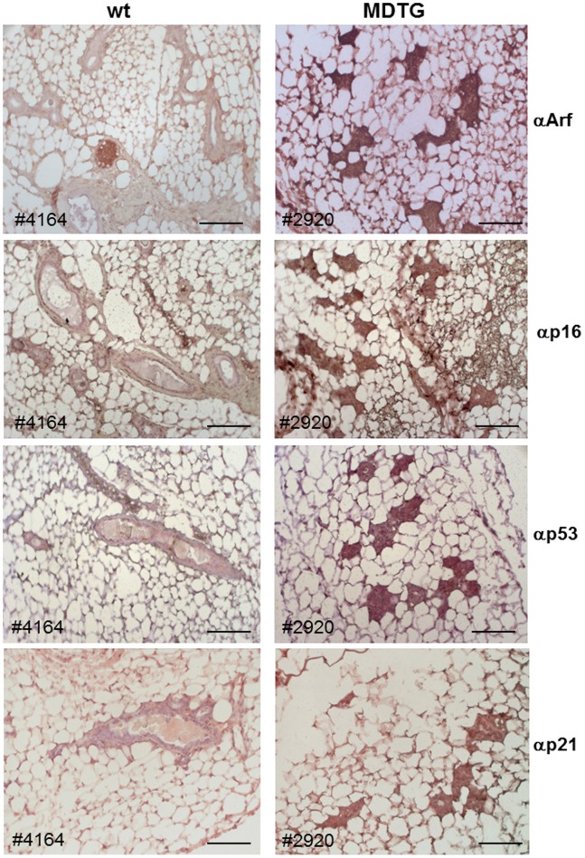
Our recent studies indicate that both p19Arf and p16Ink4a are direct transcriptional targets of Dmp1α [39]. To determine the mechanism of low Ki67 expression, we stained MMG from MDTG mice with antibodies to Arf, Ink4a, p53, and p21Cip1 (Fig. 4). The induction of Arf, Ink4a, and p21Cip1 mRNAs in the MMG from MDTG mice were confirmed by real-time PCR (230–1,025 folds for Arf; 2,500–28,400 folds for Ink4a, and 57–115 folds for p21). These data indicate that both p53 and Rb pathways were strongly activated in vivo when Dmp1 is highly expressed in MMG (Fig. 4). There was no difference in estrogen receptor α (ERα) and progesterone receptor (PR) expression between WT and MDTG mice (Fig. S2). We also found that the EGF ligand amphiregulin (Areg) [40] level was not significantly affected in MMG from MDTG mice while both EGF and TGFα were upregulated (Fig. S3), indicating that the regulation of Areg by Dmp1 [41] is tissue-dependent. The data indicate that high expression of Ink4a and Arf proteins, not lack of hormonal receptors or EGF ligands, contributed to the poor MMG development in MDTG females (Fig. 3).
Figure 4. Immunohistochemical detection of p19Arf, p16Ink4a, p53, and p21Cip1 in mammary glands of MDTG mice.
All of these proteins were induced in MMG from MDTG females suggesting the activation of the p53 and Rb pathways by Dmp1 in vivo. Scale bars show 100 µm.
HER2/Neu-driven Mammary Carcinogenesis is Significantly Delayed in MDTG;neu Double Transgenic Mice
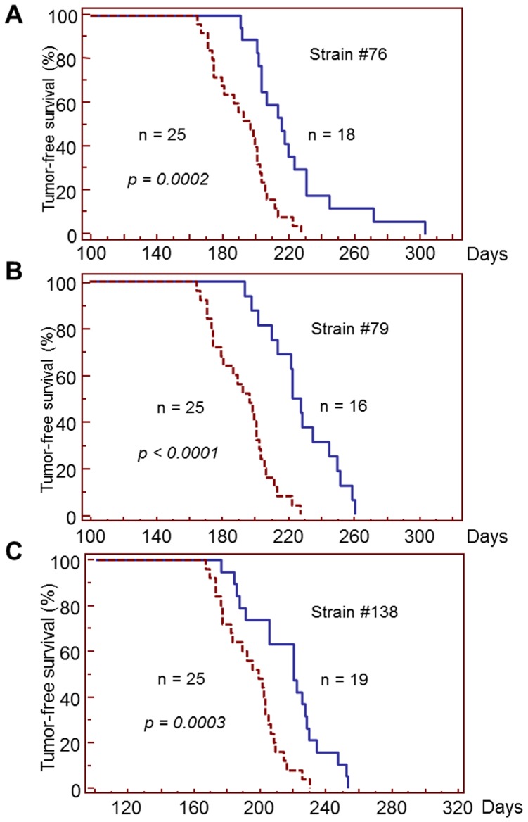
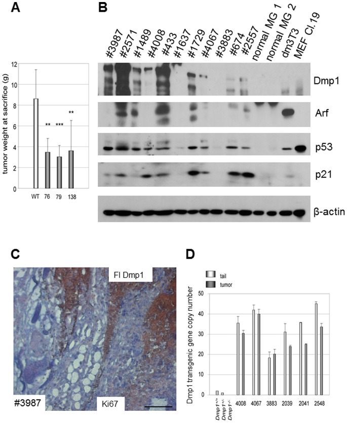
Our previous study showed that MMTV-neu-induced mammary tumorigenesis was significantly accelerated in Dmp1-deficient mice, indicating a critical role of Dmp1 as a mediator of aberrant HER2 signaling and the Arf-p53 pathway [11]. To determine the effect of high Dmp1α expression in mammary carcinogenesis by HER2/neu, we crossed MMTV-neu mice with MDTG mice. The generated 16–19 MDTG;neu bitransgenic female mice still developed mammary tumors; however, compared to MMTV-neu mice, MDTG;neu bitransgenic mice from all three strains exhibited significantly delayed tumor formation (Fig. 5). The median tumor/disease-free survival (DFS) for the MMTV-neu mice was 197 days (n = 25), but it was significantly extended in MDTG;neu mice to 216 days (from strain 76; n = 18, p = 0.0002), 223 days (from strain 79; n = 16, p<0.0001), and 211 days (from strain 138, n = 19; p = 0.0003). The average amount of tumors harvested at 4-week time point after the first tumor appearance was significantly decreased in bitransgenic mice from all three strains (MMTV-neu, 8.61+/−2.8 g (n = 10) vs. 3.48+/−1.32 g (n = 6, p = 0.0010) for strain 76; 3.08+/−1.08 g (n = 11, p<0.0001) for strain 79; 3.63+/−2.89 g, (n = 9, p = 0.0014) for strain 138) (Fig. 6A). Thus, not only was DFS extended, but also the absolute amount of tumors present when they died were decreased in MDTG;neu bitransgenic mice in comparison to non-MDTG;neu mice. These data suggest that transgenic expression of Dmp1α has significant effects on preventing HER2/neu-driven mammary carcinogenesis.
Figure 5. Disease-free survival (DFS) curves of MDTG;neu mice in each strain.
DFS curves for each strain of MDTG;neu females are shown. The p values were calculated in comparison to the survival of wild-type MMTV-neu mice (n = 25; medium survival, 197 days).
Figure 6. Mutually exclusive expression of Flag-Dmp1 and Ki67 in MDTG;neu tumors.
A. Total tumor weight harvested at sacrifice. The amount of tumors recovered at sacrifice (4 weeks after the first tumor was found) was significantly decreased in all three transgenic strains. B. Western blotting analyses of proteins expressed in MDTG;neu tumors. C. Mutually exclusive expression of Flag-Dmp1 and Ki67 in a MDTG;neu tumor (strain 76, #3987). Flag-Dmp1 was stained brown; Ki67 was stained blue. Scale bars show 100 µm. D. Gene copy number assay for Flag-Dmp1 in paired tails and MDTG;neu tumors showing partial deletion of the transgene in half of the cases.
Analysis of the p53 and Rb Pathways in Dmp1-resistant Tumors from MDTG; neu Mice
Although Dmp1 showed a significant role in inhibiting MMTV-neu-induced mammary carcinogenesis, all MDTG;neu mice died of tumors 1–2 months later than non-MDTG;neu transgenic mice. Thus, we investigated whether there was any molecular events that occurred in MDTG;neu mice during the course of tumor development. Western blot analyses for the MMG of MDTG;neu animals demonstrated that 2 of 11 tumors had high Flag-Dmp1 expression (#2571, #433), three had medium Flag-Dmp1 expression (#3987, #1489, #1729), and four had low expression, while Flag-Dmp1 was barely detectable in two tumors (#1637, #3883) (Fig. 6B, top). The Arf and p53 proteins well correlated with Flag-Dmp1 expression, suggesting that the Dmp1-Arf-p53 signaling was still functional in MDTG;neu tumor cells. The p21Cip1 levels correlated with Flag-Dmp1 levels except for the two tumors (#674, #2557), suggesting some other protein(s) induced p21Cip1 in these cases.
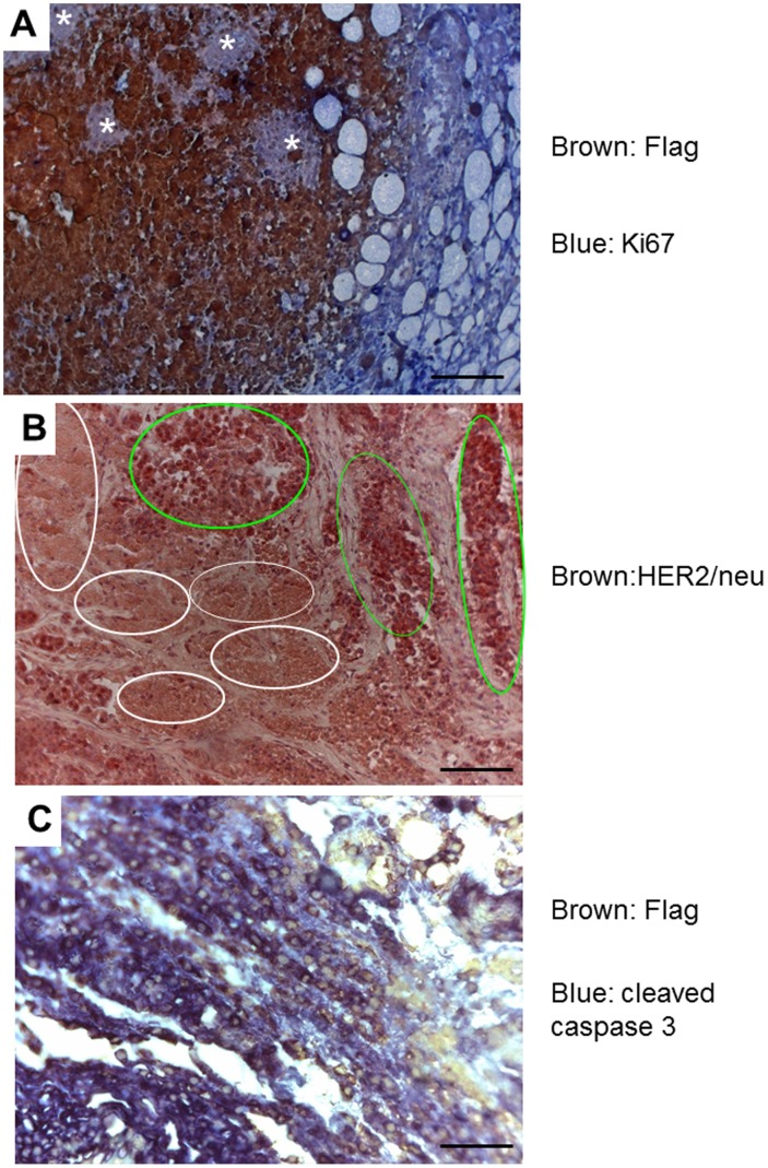
We then studied the cell proliferative status of MDTG;neu tumors. Double staining of MDTG;neu mammary tumors with Flag (brown) and Ki67 (blue) antibodies showed mutually exclusive pattern of their expression (Fig. 6C). This suggests that either cells expressing Flag-Dmp1 stopped proliferating or only tumor cells that succeeded in downregulating Flag-Dmp1 protein were growing. Consistently, we observed numerous areas of Ki67(+) Flag-Dmp1(−) cells (shown in stars, Fig. 7A), supporting our hypothesis that inactivation of the Flag-Dmp1 transgene is necessary for tumor cell proliferation. To further investigate the mechanisms of decreased Flag-Dmp1 expression in MDTG;neu tumors, we extracted genomic DNA from matched tail and tumor samples and calculated Flag-Dmp1 gene copy numbers by real-time PCR (Fig. 6D). The results showed that the transgenic Flag-Dmp1 was retained only in half of the MDTG;neu tumors (#4008, #4067, and #3883) and there was partial (20–25%) loss of the transgene in the other half of tumors. This is possibly because some HER2/neu-expressing cells that stochastically deleted the FlagDmp1 gene began to proliferate in vivo and emerged as tumors since the protein was interfering with neoplastic cell proliferation. We also observed that the Flag-Dmp1 mRNA was downregulated in some tumors in which the transgene was not deleted (data not shown). Thus, we conclude that both loss of the Flag-Dmp1 gene and downregulation of Flag-Dmp1 mRNA or protein contributed to generation of highly proliferative lesions, allowing HER2/neu tumors to progress.
Figure 7. Immunohistochemical studies of MDTG;neu tumors.
A. A MDTG;neu tumor (#3883) that shows areas of Flag-Dmp1 (brown) and islands of Dmp1-resistant tumor growth (white arrows). The Ki67-positive area is stained blue. B. Immunohistochemical staining of MDTG;neu tumor (#3987) with HER2 antibody. All of the tumor cells express the HER2/neu protein. Light green circles show areas of tumor growth while white circles show areas of tumor cell death. C. Double immunohistochemical staining of a MDTG;neu tumor (#3883) that shows significant overlap of Flag-Dmp1-positive cells (stained light brown in nuclei) and cleaved-caspase 3-positive cells (blue in the cytoplasm). A, B, C. Scale bars show 100 µm.
Finally, we stained tumors from MDTG;neu mice with an antibody to neu. We found that all tumors were positive for neu (Fig. 7B) with some areas showing stronger signals (light green circles) than the others (white circles), suggesting that significant areas of MDTG;neu tumor was in the process of cell death. To confirm this prediction, we double stained MDTG;neu tumors with antibodies to the Flag (brown) and cleaved caspase 3 (blue) (Fig. 7C). The Flag and caspase 3 staining signals were overlapping, indicating that Flag-Dmp1-positive area was undergoing apoptosis. Thus, high expression of Flag-Dmp1 induces the cell death of early stage neu tumors.
Discussion
In this study, we created transgenic mice that have tissue-specific expression of the Dmp1α gene in mammary glands, driven by the MMTV promoter. None of the transgenic females, neither nulliparous nor mono−/multi-parous, exhibited any malignant transformation within two years of observation period. The same observation was made also in males. The mammary glands in the females remained small and unopened with markedly reduced milk production in all cases, and expressed senescence markers such as Arf, p16, p21, and p53. Our data indicate that Dmp1 inhibits HER2/neu-mediated carcinogenesis in vivo. Since Arf, p16, and p53 themselves are tumor suppressor genes, they must have contributed to the significantly delayed mammary carcinogenesis in the MDTG;neu bitransgenic mice.
Previous studies suggest that the Arf gene is mostly activated by oncogenes, such as mutant Ras, HER2, E1A, v-abl, Myc, and E2F-1 [22], [42]. Thus, Arf is a major tumor suppressive molecule that diverts hyperproliferative signals to p53-mediated cell cycle arrest and/or apoptosis. As an activator of Arf, Dmp1 is unique in its role of achieving cell cycle arrest/senescence without causing cellular transformation in either cultured cells or in vivo. Our published studies have shown that the senescence response induced by mutant Ras or HER2 is mediated by Dmp1, while increased Myc or E2F-1 expression downregulates Dmp1 [27]. This suggests that Dmp1 is a mediator to convey oncogenic signals to the p53 pathway if they induce cell cycle arrest/senescence, but does not mediate the hyperproliferation signals for apoptosis, such as from Myc, E2F-1. We previously demonstrated that the Dmp1 promoter can be activated by TNFα, an endogenous pyrogen that induces fever, apoptotic cell death, and cachexia, and inhibits tumorigenesis [29]. Our recent study showed that the Dmp1 protein was induced in vivo in response to doxorubicin [24]; thus Dmp1 likely receives signals from sources other than oncogenes to activate both p53 and Rb pathways via Arf [21] and p16Ink4a [39]. It is currently unclear whether Dmp1 is involved in hypoxic response, osmotic pressure, malnutrition signaling, or hormonal signaling. The presence of 16 potential phosphorylation sites on Dmp1 raises the possibility of Dmp1 regulation via phosphorylation. Thus, further studies will be needed to elucidate the signaling pathways that lead to increased Dmp1 expression and activity.
We observed significant increase of DFS of MDTG;neu mice compared to MMTV-neu mice. This happened when MMTV-neu mice were crossed with all three MDTG transgenic founder lines. Interestingly the p values of the DFS were the smallest in strain 79 that had highest Flag-Dmp1α gene copy numbers: followed by strain 76 (high copy), and strain 138 (low copy), suggesting that the levels of Flag-Dmp1α transgene/protein expression matters for Dmp1 to show its biological effects. Dmp1 not only delayed HER2/neu-driven mammary carcinogenesis, but also decreased tumor volumes when the animals were sacrificed (Fig. 6A). Again the tumor-reducing effect was highest in strain 79, lowest in strain 138, suggesting that the effect of Dmp1 is dose-dependent. However it should be noted that mammary tumors arose later, regardless of Flag-Dmp1α copy numbers and the differences of transgene integration sites dependent on the strain. In our IHC studies, we observed that the Flag-Dmp1α and Ki67 protein expression were mutually exclusive, suggesting that Flag-Dmp1-positive areas were not proliferating and thereby preventing tumor progression in vivo. Moreover, we saw significant overlap between Flag-Dmp1-positive areas and cleaved caspase 3(+) areas in MDTG;neu tumors (Fig. 7C), indicating that Flag-Dmp1α induces apoptotic cell death in vivo. When we studied how the MDTG;neu animals eventually developed tumors, we observed reduced Flag-Dmp1 protein expression in MDTG;neu tumors in nearly 80% of cases. Partial loss of the Flag-Dmp1 transgene in tumors or downregulation of the Flag-Dmp1 mRNAs or proteins could contribute to this observation. The lack of correlation between the mRNA and protein levels of Flag-Dmp1 (data not shown) indicates that Flag-Dmp1 downregulation occurred at either transcriptional or protein level in the tumors of MDTG;neu mice. Indeed, we saw focal proliferation of HER2/neu tumors in islands lacking Flag-Dmp1 expression (Fig. 7A). Whether Dmp1 protein undergoes ubiquitin-mediated degradation remains to be determined.
In a related study, Yang et al. [43] created MMTV-p16INK4a transgenic mice to study the effects of p16INK4a on ErbB2-induced mammary tumorigenesis using MMTV-ErbB2 (wild-type; WT) rather than MMTV-neu (mutant) used in this study. The p16INK4a transgene dramatically delayed mammary tumorigenesis by ErbB2, indicating that ErbB2-mediated deregulation of cyclin D1/Cdk4/6 is a crucial step of tumor formation. Although p16INK4a might have a more prominent role than Flag-Dmp1 in preventing mammary carcinogenesis based on our results and the theirs, the difference can be simply attributed to the fact that mild wild-type ErbB2 mouse model [17], [43] was used by this group while ErbB2 mutant model [16] was used by us.
Our study shows highly expressed Dmp1 induces Ink4a/Arf, p53, and p21 in vivo. It has been reported that both Ink4a and Arf levels markedly increase in almost all rodent tissues with advanced age, while there is little change of other Cdk inhibitors such as p15, p18, and p19 [44]. This increase occurred in both epithelial and stromal cells of different lineages. This aging-related induction of Ink4a/Arf was attenuated in organs by caloric restriction with diminished expression of senescence markers, indicating that Ink4a/Arf expression themselves are biomarkers of aging [44]. Since Dmp1 does not have any oncogenic activity, we hypothesize that Dmp1 is also a biomarker of aging that transmits relevant signals to its downstream Ink4a/Arf genes. Whether Dmp1 is a bona fide regulator of aging needs to be addressed in the future by checking the expression of Ink4a/Arf and other Cdk inhibitors in tissues from Dmp1 wild-type and deficient mice.
Garcia-Cao et al. [45] established and characterized mice carrying supernumerary copies of the wild-type p53 gene as a large genomic transgene. These “super p53” mice, which carried p53-transgene alleles in addition to the two endogenous p53 alleles, exhibited an enhanced response to DNA damage, and importantly, were protected from cancer when compared to normal mice. It should be noted that constitutive activation of p53, such as chronic exposure to stress, could result in accelerated aging as demonstrated by Tyner et al [46]. In contrast, “super p53” mice showed a normal aging process despite having clearly increased p53 activity. A critical feature of the “super p53” mice is that the basal levels of p53 activity are not affected. This is important because further increase in gene dosage of p53 might eventually reach a threshold at which deleterious effects would be noticeable, such as defective tissue regeneration, growth atrophies, and premature aging. This suggests that increases in normally regulated p53, as in the “super p53” mice, could confer cancer protection without affecting aging, while constitutively high levels of active p53 or expression of a truncated carboxyl-terminal p53 promotes aging [44], [45]. Thus, cancer resistance could be enhanced by a simple genetic modification of p53 in the absence of undesirable effects. Likewise, Mathew et al. [47] generated a “super Ink4a/Arf” mouse strain carrying a transgenic copy of the entire Ink4a/Arf locus and demonstrated that modest increase in the activity of the Ink4a/Arf tumor suppressors also resulted in a beneficial cancer-resistant phenotype without affecting normal viability or aging. Likewise, it will be possible to prevent cancers by stimulating the Dmp1 activity or increasing its protein levels/activity without causing toxic effects of accelerated aging.
Four distinct stages of the breast epithelial cells are defined on the basis of the estrogen receptor profile. These are: (i) prepuberty, when both ERα and ERβ are present; (ii) pregnancy, when ERβ is present in the majority of epithelial cells, ERα-expression is low, and few cells express both receptors; (iii) lactation, when ERα and ERβ are both expressed in the majority of epithelial cells; and (iv) post lactation, when ERα is extremely low and there is little co-localization of the ERα/β receptors [48]. In mammary glands of the MDTG mice, ER or PR level did not decrease in comparison to that in wild-type. Collaborative roles of EGF receptor (EGFR) ligands in mammopoiesis and lactogenesis have been reported [40]. It was shown that triple knockout mice for the three EGFR ligands for EGF, Areg, and TGFα showed neonatal lethality due to the absence of milk production of the mother [40]. In the MMG of MDTG females, Areg expression did not significantly change while both EGF and TGFα were upregulated. We therefore conclude that markedly reduced proliferation of mammary epithelial cells in MDTG females were due to the Dmp1-mediated Ink4a/Arf overexpression rather than downregulation of hormonal receptors or EGF ligands.
In conclusion, we have established transgenic mice that specifically express the Dmp1α gene in the mammary glands. These animals show tumor suppressive effects on HER2/neu-driven tumorigenesis by inducing cell cycle arrest and/or apoptosis. These transgenic mice will be useful to predict tumor-suppressive effects of Dmp1 in other transgenic mice that are prone to mammary tumor development, and identify novel Dmp1 target genes/proteins in vivo. Molecules that specifically activate the Dmp1 promoter or the Dmp1α protein will be effective novel chemotherapeutic agents to induce regression of tumor growth in vivo.
Supporting Information
PCR-mediated amplification of the cloned cDNA using transgenic mouse tails. The results from strains 76 and 79 are shown.
(TIF)
Immunohistochemical staining of a wild-type (#4164) and MDTG (#2920, strain 79) MMG for hormone receptors. ER: estrogen receptor; PR: progesterone receptor.
(TIF)
Immunohistochemical staining of a wild-type (#4164) and MDTG (#2920, strain 79) MMG for EGFR ligands. Areg: Amphiregulin; EGF: Epidermal Growth Factor; TGFβ: Transforming Growth Factor beta.
(TIF)
Acknowledgments
We thank C. Sherr, M. Roussel, and P. Leder for plasmid DNAs. We are grateful to Takayuki Sugiyama and Fumitake Kai for assistance in mouse care. We also thank Nancy Kock for pathological diagnosis of H&E sections and immunohistochemically stained slides. Thanks are extended to Kenneth Grant for technical assistance with Zeiss Axioplan2 imaging and the Cell and Virus Vector Core Laboratory of the Comprehensive Cancer Center at WFUHS for provision of the access to materials of cell culture.
Funding Statement
K. Inoue has been supported by the American Cancer Society RSG-07-207-01-MGO, 5R01CA106314, and by Director’s Challenge Award #20595 from Wake Forest University Health Sciences. G. Sui has been supported by the American Cancer Society 116403-RSG-09-082-01-MGO and 5R01CA106314. P. Taneja was supported by the Susan G. Komen Foundation postdoctoral fellowship KG080179. D. Maglic has been supported by Department of Defense (DOD) pre-doctoral fellowship BC100907. S. Zhu was supported by institutional grant IRSC-GTS 37540. The funders had no role in study design, data collection and analysis, decision to publish, or preparation of the manuscript.
References
- 1.Ross JS, Hortobagyi GN (2005) Molecular Oncology of Breast Cancer. Publishers, Sudbury, Massachusetts.
- 2. Siegel R, Naishadham D, Jemal A (2013) Cancer statistics, 2013. CA Cancer J Clin 63: 11–30. [DOI] [PubMed] [Google Scholar]
- 3. Morrow PK, Hortobagyi GN (2009) Management of breast cancer in the genome era. Annu Rev Med 60: 153–165. [DOI] [PubMed] [Google Scholar]
- 4. Ross JS, Linette GP, Stec J, Clark E, Ayers M, et al. (2003) Breast cancer biomarkers and molecular medicine. Expert Rev Mol Diagn 3: 573–585. [DOI] [PubMed] [Google Scholar]
- 5. Turnbull C, Rahman N (2008) Genetic predisposition to breast cancer: past, present, and future. Annu Rev Genomics Hum Genet 9: 321–345. [DOI] [PubMed] [Google Scholar]
- 6. Yamamoto T, Ikawa S, Akiyama T, Semba K, Nomura N, et al. (1986) Similarity of protein encoded by the human c-erb-B-2 gene to epidermal growth factor receptor. Nature 319: 230–234. [DOI] [PubMed] [Google Scholar]
- 7. Holbro T, Civenni G, Hynes NE (2003) The ErbB receptors and their role in cancer progression. Exp Cell Res 284: 99–110. [DOI] [PubMed] [Google Scholar]
- 8. Hynes NE, Lane HA (2005) ERBB receptors and cancer: the complexity of targeted inhibitors. Nat Rev Cancer 5: 341–354. [DOI] [PubMed] [Google Scholar]
- 9. Zhou BP, Hung MC (2003) Dysregulation of cellular signaling by HER2/neu in breast cancer. Semin Oncol 30: 38–48. [DOI] [PubMed] [Google Scholar]
- 10. Citri A, Skaria KB, Yarden Y (2003) The deaf and the dumb: the biology of ErbB-2 and ErbB-3. Exp Cell Res 284: 54–65. [DOI] [PubMed] [Google Scholar]
- 11. Taneja P, Maglic D, Kai F, Sugiyama T, Kendig RD, et al. (2010) Critical roles of DMP1 in human epidermal growth factor receptor 2/neu-Arf-p53 signaling and breast cancer development. Cancer Res 70: 9084–9094. [DOI] [PMC free article] [PubMed] [Google Scholar]
- 12. Bargmann CI, Hung MC, Weinberg RA (1986) Multiple independent activations of the neu oncogene by a point mutation altering the transmembrane domain of p185. Cell 45: 649–657. [DOI] [PubMed] [Google Scholar]
- 13. Taneja P, Frazier DP, Kendig RD, Maglic D, Sugiyama T, et al. (2009) MMTV mouse models and the diagnostic values of MMTV-like sequences in human breast cancer. Expert Rev Mol Diagn 9: 423–440. [DOI] [PMC free article] [PubMed] [Google Scholar]
- 14. Kwong KY, Hung MC (1998) A novel splice variant of HER2 with increased transformation activity. Mol Carcinog 23: 62–68. [DOI] [PubMed] [Google Scholar]
- 15. Siegel PM, Ryan ED, Cardiff RD, Muller WJ (1999) Elevated expression of activated forms of Neu/ErbB-2 and ErbB-3 are involved in the induction of mammary tumors in transgenic mice: implications for human breast cancer. EMBO J 18: 2149–2164. [DOI] [PMC free article] [PubMed] [Google Scholar]
- 16. Muller WJ, Sinn E, Pattengale PK, Wallace R, Leder P (1988) Single-step induction of mammary adenocarcinoma in transgenic mice bearing the activated c-neu oncogene. Cell 54: 105–115. [DOI] [PubMed] [Google Scholar]
- 17. Guy CT, Webster MA, Schaller M, Parsons TJ, Cardiff RD, et al. (1992) Expression of the neu protooncogene in the mammary epithelium of transgenic mice induces metastatic disease. Proc Natl Acad Sci U S A 89: 10578–10582. [DOI] [PMC free article] [PubMed] [Google Scholar]
- 18. Amarante MK, Watanabe MA (2009) The possible involvement of virus in breast cancer. J Cancer Res Clin Oncol 135: 329–337. [DOI] [PubMed] [Google Scholar]
- 19. Hirai H, Sherr CJ (1996) Interaction of D-type cyclins with a novel myb-like transcription factor, DMP1. Mol Cell Biol 16: 6457–6467. [DOI] [PMC free article] [PubMed] [Google Scholar]
- 20. Inoue K, Sherr CJ (1998) Gene expression and cell cycle arrest mediated by transcription factor DMP1 is antagonized by D-type cyclins through a cyclin-dependent-kinase-independent mechanism. Mol Cell Biol 18: 1590–1600. [DOI] [PMC free article] [PubMed] [Google Scholar]
- 21. Inoue K, Roussel MF, Sherr CJ (1999) Induction of ARF tumor suppressor gene expression and cell cycle arrest by transcription factor DMP1. Proc Natl Acad Sci U S A 96: 3993–3998. [DOI] [PMC free article] [PubMed] [Google Scholar]
- 22. Inoue K, Mallakin A, Frazier DP (2007) Dmp1 and tumor suppression. Oncogene 26: 4329–4335. [DOI] [PMC free article] [PubMed] [Google Scholar]
- 23. Sugiyama T, Frazier DP, Taneja P, Kendig RD, Morgan RL, et al. (2008) Signal transduction involving the dmp1 transcription factor and its alteration in human cancer. Clin Med Oncol 2: 209–219. [DOI] [PMC free article] [PubMed] [Google Scholar]
- 24. Frazier DP, Kendig RD, Kai F, Maglic D, Sugiyama T, et al. (2012) Dmp1 physically interacts with p53 and positively regulates p53’s stability, nuclear localization, and function. Cancer Res 72: 1740–1750. [DOI] [PMC free article] [PubMed] [Google Scholar]
- 25. Inoue K, Wen R, Rehg JE, Adachi M, Cleveland JL, et al. (2000) Disruption of the ARF transcriptional activator DMP1 facilitates cell immortalization, Ras transformation, and tumorigenesis. Genes Dev 14: 1797–1809. [PMC free article] [PubMed] [Google Scholar]
- 26. Inoue K, Zindy F, Randle DH, Rehg JE, Sherr CJ (2001) Dmp1 is haplo-insufficient for tumor suppression and modifies the frequencies of Arf and p53 mutations in Myc-induced lymphomas. Genes Dev 15: 2934–2939. [DOI] [PMC free article] [PubMed] [Google Scholar]
- 27. Sreeramaneni R, Chaudhry A, McMahon M, Sherr CJ, Inoue K (2005) Ras-Raf-Arf signaling critically depends on the Dmp1 transcription factor. Mol Cell Biol 25: 220–232. [DOI] [PMC free article] [PubMed] [Google Scholar]
- 28. Mallakin A, Taneja P, Matise LA, Willingham MC, Inoue K (2006) Expression of Dmp1 in specific differentiated, nonproliferating cells and its regulation by E2Fs. Oncogene 25: 7703–7713. [DOI] [PMC free article] [PubMed] [Google Scholar]
- 29. Taneja P, Mallakin A, Matise LA, Frazier DP, Choudhary M, et al. (2007) Repression of Dmp1 and Arf transcription by anthracyclins: critical roles of the NF-kappaB subunit p65. Oncogene 26: 7457–7466. [DOI] [PMC free article] [PubMed] [Google Scholar]
- 30. Mallakin A, Sugiyama T, Taneja P, Matise LA, Frazier DP, et al. (2007) Mutually exclusive inactivation of DMP1 and ARF/p53 in lung cancer. Cancer Cell 12: 381–394. [DOI] [PMC free article] [PubMed] [Google Scholar]
- 31. Bodner SM, Naeve CW, Rakestraw KM, Jones BG, Valentine VA, et al. (1999) Cloning and chromosomal localization of the gene encoding human cyclin D-binding Myb-like protein (hDMP1). Gene 229: 223–228. [DOI] [PubMed] [Google Scholar]
- 32. Tschan MP, Fischer KM, Fung VS, Pirnia F, Borner MM, et al. (2003) Alternative splicing of the human cyclin D-binding Myb-like protein (hDMP1) yields a truncated protein isoform that alters macrophage differentiation patterns. J Biol Chem 278: 42750–42760. [DOI] [PubMed] [Google Scholar]
- 33. Bieche I, Champeme MH, Matifas F, Hacene K, Callahan R, et al. (1992) Loss of heterozygosity on chromosome 7q and aggressive primary breast cancer. Lancet 339: 139–143. [DOI] [PubMed] [Google Scholar]
- 34. Kristjansson AK, Eiriksdottir G, Ragnarsson G, Sigurdsson A, Gudmundsson J, et al. (1997) Loss of heterozygosity at chromosome 7q in human breast cancer: association with clinical variables. Anticancer Res 17: 93–98. [PubMed] [Google Scholar]
- 35. Inoue K, Sugiyama T, Taneja P, Morgan RL, Frazier DP (2008) Emerging roles of DMP1 in lung cancer. Cancer Res 68: 4487–4490. [DOI] [PMC free article] [PubMed] [Google Scholar]
- 36. Sugiyama T, Frazier DP, Taneja P, Morgan RL, Willingham MC, et al. (2008) Role of DMP1 and its future in lung cancer diagnostics. Expert Rev Mol Diagn 8: 435–447. [DOI] [PMC free article] [PubMed] [Google Scholar]
- 37. Maglic D, Zhu S, Fry EA, Taneja P, Kai F, et al. (2012) Prognostic value of the hDMP1-ARF-Hdm2-p53 pathway in breast cancer. Oncogene Aug 29 32(35): 4120–9. [DOI] [PMC free article] [PubMed] [Google Scholar]
- 38.Plante I, Stewart MK, Laird DW (2011) Evaluation of mammary gland development and function in mouse models. J Vis Exp Jul 21; (53). [DOI] [PMC free article] [PubMed]
- 39.Zhu S, Mott RT, Fry EA, Taneja P, Kulik G, et al.. (2013) Cooperation between cyclin D1 expression and Dmp1-loss in breast cancer. Am J Pathol Aug 11. [DOI] [PMC free article] [PubMed]
- 40. Luetteke NC, Qiu TH, Fenton SE, Troyer KL, Riedel RF, et al. (1999) Targeted inactivation of the EGF and amphiregulin genes reveals distinct roles for EGF receptor ligands in mouse mammary gland development. Development 126: 2739–2750. [DOI] [PubMed] [Google Scholar]
- 41. Mallakin A, Sugiyama T, Kai F, Taneja P, Kendig RD, et al. (2010) The Arf-inducing transcription factor Dmp1 encodes a transcriptional activator of amphiregulin, thrombospondin-1, JunB and Egr1. Int J Cancer 126: 1403–1416. [DOI] [PMC free article] [PubMed] [Google Scholar]
- 42. Sherr CJ, Bertwistle D, W DENB, Kuo ML, Sugimoto M, et al. (2005) p53-Dependent and -independent functions of the Arf tumor suppressor. Cold Spring Harb Symp Quant Biol 70: 129–137. [DOI] [PubMed] [Google Scholar]
- 43. Yang C, Ionescu-Tiba V, Burns K, Gadd M, Zukerberg L, et al. (2004) The role of the cyclin D1-dependent kinases in ErbB2-mediated breast cancer. Am J Pathol 164: 1031–1038. [DOI] [PMC free article] [PubMed] [Google Scholar]
- 44. Krishnamurthy J, Torrice C, Ramsey MR, Kovalev GI, Al-Regaiey K, et al. (2004) Ink4a/Arf expression is a biomarker of aging. J Clin Invest 114: 1299–1307. [DOI] [PMC free article] [PubMed] [Google Scholar]
- 45. Garcia-Cao I, Garcia-Cao M, Martin-Caballero J, Criado LM, Klatt P, et al. (2002) “Super p53” mice exhibit enhanced DNA damage response, are tumor resistant and age normally. EMBO J 21: 6225–6235. [DOI] [PMC free article] [PubMed] [Google Scholar]
- 46. Tyner SD, Venkatachalam S, Choi J, Jones S, Ghebranious N, et al. (2002) p53 mutant mice that display early ageing-associated phenotypes. Nature 415: 45–53. [DOI] [PubMed] [Google Scholar]
- 47. Matheu A, Pantoja C, Efeyan A, Criado LM, Martin-Caballero J, et al. (2004) Increased gene dosage of Ink4a/Arf results in cancer resistance and normal aging. Genes Dev 18: 2736–2746. [DOI] [PMC free article] [PubMed] [Google Scholar]
- 48. Saji S, Jensen EV, Nilsson S, Rylander T, Warner M, et al. (2000) Estrogen receptors alpha and beta in the rodent mammary gland. Proc Natl Acad Sci U S A 97: 337–342. [DOI] [PMC free article] [PubMed] [Google Scholar]
Associated Data
This section collects any data citations, data availability statements, or supplementary materials included in this article.
Supplementary Materials
PCR-mediated amplification of the cloned cDNA using transgenic mouse tails. The results from strains 76 and 79 are shown.
(TIF)
Immunohistochemical staining of a wild-type (#4164) and MDTG (#2920, strain 79) MMG for hormone receptors. ER: estrogen receptor; PR: progesterone receptor.
(TIF)
Immunohistochemical staining of a wild-type (#4164) and MDTG (#2920, strain 79) MMG for EGFR ligands. Areg: Amphiregulin; EGF: Epidermal Growth Factor; TGFβ: Transforming Growth Factor beta.
(TIF)


