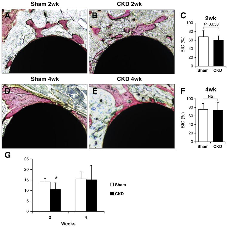
Fig. 4.
Histology and biomechanical test. (A&B) Representative images of the histological sections at 2-week healing. (C) Bone-implant contact ratio at 2-week healing. (D&E) Representative images of the histological sections at 4-week healing. (F) Bone-implant contact ratio at 4-week healing. (G) Resistance of push-in tests. *: p < 0.05.

