Abstract
Insects feeding on plant sap, blood, and other nutritionally incomplete diets are typically associated with mutualistic bacteria that supplement missing nutrients. Herbivorous mammal dung contains more than 86% cellulose and lacks amino acids essential for insect development and reproduction. Yet one of the most ecologically necessary and evolutionarily successful groups of beetles, the dung beetles (Scarabaeinae) feeds primarily, or exclusively, on dung. These associations suggest that dung beetles may benefit from mutualistic bacteria that provide nutrients missing from dung. The nesting behaviors of the female parent and the feeding behaviors of the larvae suggest that a microbiome could be vertically transmitted from the parental female to her offspring through the brood ball. Using sterile rearing and a combination of molecular and culture-based techniques, we examine transmission of the microbiome in the bull-headed dung beetle, Onthophagus taurus. Beetles were reared on autoclaved dung and the microbiome was characterized across development. A ~1425 bp region of the 16S rRNA identified Pseudomonadaceae, Enterobacteriaceae, and Comamonadaceae as the most common bacterial families across all life stages and populations, including cultured isolates from the 3rd instar digestive system. Finer level phylotyping analyses based on lepA and gyrB amplicons of cultured isolates placed the isolates closest to Enterobacter cloacae, Providencia stuartii, Pusillimonas sp., Pedobacter heparinus, and Lysinibacillus sphaericus. Scanning electron micrographs of brood balls constructed from sterile dung reveals secretions and microbes only in the chamber the female prepares for the egg. The use of autoclaved dung for rearing, the presence of microbes in the brood ball and offspring, and identical 16S rRNA sequences in both parent and offspring suggests that the O. taurus female parent transmits specific microbiome members to her offspring through the brood chamber. The transmission of the dung beetle microbiome highlights the maintenance and likely importance of this newly-characterized bacterial community.
Introduction
Dung beetles in the superfamily Scarabaeoidea have specialized on animal waste since the Jurassic period ~152 million years ago [1,2]. As critical decomposers in all temperate and tropical terrestrial ecosystems, dung beetles have evolved to feed on both dry [3,4] and wet dung of mammals in general, and large herbivorous mammals in particular [5], on every continent except Antarctica [6,7]. While many beetles have radiated onto dung as a food source, dung is a nutritionally incomplete diet. Dung lacks amino acids essential for insect metabolic needs including tryptophan, methionine, phenylalanine, histidine, and arginine [8]. The dung of herbivorous ruminants, such as cattle, deer, and buffalo, is more than 86% cellulose [8] — an indigestible polysaccharide for many eukaryotes. Despite these attributes, members of Diptera (flies) and Coleoptera (beetles) specialize on dung.
Although it may seem surprising that such diverse insects have radiated onto a nutritionally-poor resource, mutualistic symbioses between bacteria and their eukaryotic hosts allow animals to feed on a diversity of diets that would otherwise be inaccessible to the host. Such beneficial endosymbionts can provide essential amino acids and vitamins lacking in the host diet, or they can synthesize novel enzymes, such as cellulases and hydrolases, to degrade otherwise indigestible materials like cellulose, lignin, and chitin [9,10]. These mutualisms are seen in animals ranging from cellulose feeding vertebrates to wood-, sap-, and blood-feeding insects [9]. In insects, mutualistic endosymbionts frequently supplement the host with essential amino acids and vitamins missing from their food source [10,11]. This supplementation may be provided primarily by one symbiont species, as in the aphid-Buchnera system [12,13], or by a community of symbionts, such as in termites [14,15].
No matter the number of beneficial endosymbionts, the faithful transmission of these specific mutualists from parent to offspring is essential for offspring survival. There are two broad categories of transmission: vertical transmission, where symbionts are acquired from the parent, and horizontal transmission, where they are not [9,16]. In the dung beetle system, horizontal transmission could be from many sources including adult siblings, other insect taxa including other species of dung beetles, and host dung. Vertical transmission is the dominant transmission type in evolutionarily stable, obligate insect-bacterial nutritional mutualisms. Vertically transmitted endosymbionts are frequently transmitted transovarially, though alternate methods of transmission from parents to offspring are known [9,16]. These include endosymbiont transmission through proctodeal trophallaxis [17], milk glands [18,19], coprophagy [20], egg smearing [21,22] , capsules [23], and brood chambers [24].
Dung beetle diversity may in part be a result of microbial inhabitants that first allowed these scarab beetles to radiate onto and exploit niches that are otherwise inaccessible [25]. Within scarab beetles, specific microbes have been identified using 16S rRNA in a handful of beetle species that feed on living plant tissue [26,27] and humus [28] instead of dung [29,30]. Earlier work on two dung-feeding scarab beetles, Scarabaeus semipunctatus and Chironitis furcifer, identified culturable dung and cellulose degrading bacteria associated with the beetles, their brood balls, and their vertebrate dung source (i.e. sheep and cow, respectively) using dung agar plates [31]. We hypothesize that species of rolling and tunneling dung beetles that sequester individual eggs in brood balls of dung may provision their brood balls with a specific microbiome to aid in dung digestion. Our research focused on a more thorough description of the dung beetle microbiome by focusing on a species of tunneling dung beetle in the genus Onthophagus, which processes the dung of large herbivorous mammals.
Onthophagus is the most diverse tunneling dung beetle genus, with over 2,000 species described [32]. In the United States alone, there are at least 35 native Onthophagus spp. [32] and at least 5 more introduced, naturalized, non-native species [33–35]. All of these species are known to be dung specialists. Onthophagus taurus, the bull-headed dung beetle, is one of the most abundant dung beetles specialized on cattle dung. It is a native of the Mediterranean, was first found in Florida in 1974 [36], and has increased its distribution throughout the U.S. ever since [37]. Although native dung beetles such as O. hecate and O. pennsylvanicus are also found in agricultural habitats, O. taurus is the most abundant beetle in these settings. However, O. taurus is uncommon in non-agricultural ecosystems [37].
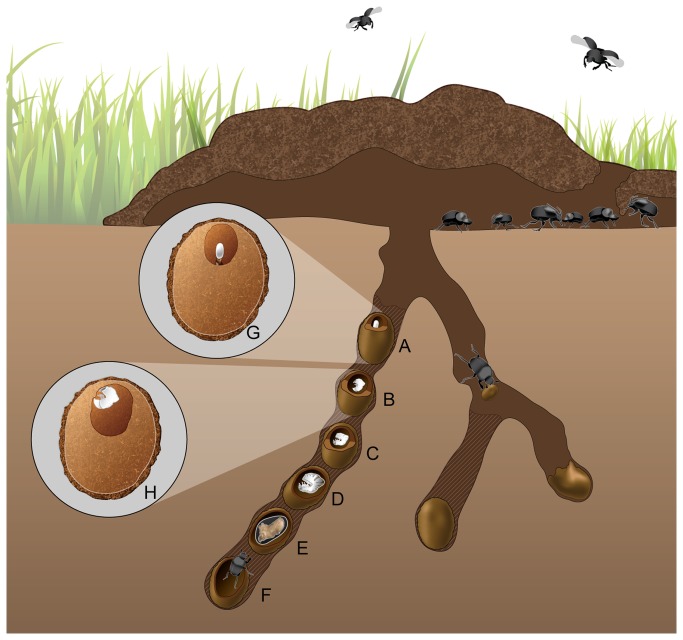
Onthophagus adults fly to a fresh dung pad where they use scoop-like mouthparts to filter the liquid portion of the dung for associated microbes [38] (Figure 1). Females then tunnel vertically in the soil underneath the dung pad where they form a series of brood balls for their offspring. Females move dung down to the ends of the tunnel where they pack dung into an oval brood ball with a brood chamber at one end of the brood ball (Figure 1). The female meticulously constructs a brood chamber lined with her own saliva where a single egg [39] is laid on top of a pedestal made of the adult female's own excrement [31,39] (Figure 1 A and G and 2 A). The entire juvenile portion of the life cycle - all 3 larval instars as well as pupation - occurs within this brood ball chamber [31,39] (Figure 1A-H). The larva hatches from the egg. Using its heavily sclerotized, toothed mandibles [38], the first instar larva immediately feeds on the dung pedestal, and then methodically alternates feeding on the solid, cellulose-rich portion of dung of the brood ball wall and its own excrement (Figure 1H) until pupation [31] (Figure 1E). Larvae pupate inside a pupation chamber constructed out of late larval fecal matter and left-over brood ball material in the remains of the brood ball (Figure 1E). After ~ 8 days at 25 °C the pupa ecloses into the filial adult, which remains inside the pupation chamber for at least several days until fully sclerotized. During dry or winter seasons, pupae will remain in their pupation chamber until the appropriate weather conditions are present. At this point during the breeding and nesting season, the adult digs to the soil surface to find food and mate [39] (Figure 1F).
Figure 1. O. taurus life cycle.
Adults fly into the dung pat to feed and mate. Beneath the dung pat, the juvenile life stages of Onthophagus taurus are isolated in the brood chamber of the brood balls constructed by the female beetles in tunnels. Females lay several brood balls in each tunnel that would all be at the same developmental stage. However, for illustrative purposes all life stages are represented in one tunnel. These stages include the: (A) egg, (B) 1st larval instar, (C) 2nd larval instar, (D) 3rd larval instar, (E) pupa, and (F) an eclosing adult beetle that is tunneling toward the surface. The brood ball chamber is larger with each successive life stage as the larva feeds on the chamber walls within the brood ball. The top inset shows (G) the fecal pedestal the egg is positioned upon in brood ball. The bottom inset shows (H) the larval instar feeding on the walls of the brood ball chamber.
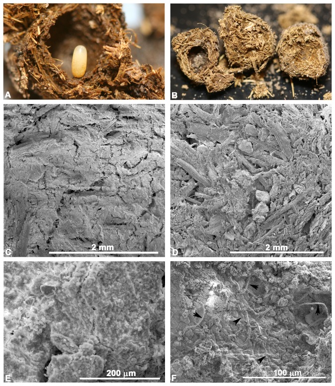
Figure 2. Brood ball chamber.
The brood ball chamber is a unique structure for the transmitted microbiome of dung beetles. (A) The innermost walls of the brood ball chamber with the egg are smooth as compared to the fibrous outer walls. (B) A brood ball is shown broken into 3 pieces. The hollow brood ball chamber is where the egg develops (left). The remainder of the brood ball is filled with cellulose-rich, fibrous dung (center and right). Images C-F are scanning electron micrographs of different portions of the brood ball. (C) Micrograph illustrating the smooth biofilm-like matrix coating the inner wall of the brood ball chamber. (D) Fibrous bits of grass from the cow dung found in the portion of the brood ball away from the chamber. (E) Higher magnification of the smooth biofilm-like matrix that coats the brood ball chamber walls. (F) When the smooth matrix is scraped away, rod shaped microbes in chains (arrows) are found underneath the smooth matrix of the brood ball chamber walls. Other rod-shaped structures in the background remain covered in the biofilm-like matrix (arrowheads). This matrix and microbes are only observed where the mother has prepared the brood ball chamber.
Based on the nutritionally incomplete diet dung provides, the nesting behavior of the female, and the larval feeding behavior, we hypothesize that a vertically transmitted microbiome is present that is transmitted through the brood ball in the dung beetle, Onthophagus taurus. Here we use a sterile rearing technique along with culture based and molecular methods to assess the potential for microbiome transmission and inheritance of the bull headed dung beetle, O. taurus, through the brood ball.
Methods
Beetle collection and sterile rearing of offspring
Adult female Onthophagus taurus were collected with permission from the land owner from fresh cow manure on the private organic dairy farms of Ms. Whitney Schlegel at Marble Hill Farm in Monroe County, IN, USA, and Mr. Bob Nutter from Maple View Farm in Wake County, NC, USA. Beetles collected in IN and NC were brought into the laboratory and reared as previously described [40], but with the following modifications to eliminate contamination with bacteria from human caretakers, manure, and the laboratory. All dung, sand, soil, and water were autoclaved at 15 PSI and 121 °C for 30 min to kill living bacteria. Sterile dung was stored at -80 °C until use. Non-autoclavable items (e.g., beetle colony rearing bins) were disinfected with 1% sodium hypochlorite and detergent and then rinsed with sterile water. Gloves were used at all times by human caretakers.
To obtain offspring, brood balls from each of two wild-collected females were harvested every 10 d and housed separately to avoid any transfer of microbiota among offspring. At least 15 brood balls were collected from each female so that offspring could be obtained at several developmental stages. Offspring were removed using sterilized forceps after the brood balls were opened with gloved hands. For the molecular based bacterial identification, the offspring were sacrificed in 95% ethanol for DNA extractions from 4 stages including 2 first instar larvae from IN, 1 second instar larva from IN, 2 second instar larvae from NC, 3 third instar larvae from IN, 3 third instar larvae from NC, and 2 pupae from IN. For culture-based work, 4 individual 3rd instar larvae from the NC population were collected and dissected to remove the entire digestive system. Original wild-caught female parent beetles from NC and IN were sacrificed in 95% ethanol after their final brood ball was collected. All samples were held in ethanol at 4 °C until DNA was extracted. In total, 17 offspring (9 from NC and 8 from IN) and two wild-caught females were harvested. From each of these individuals, multiple culturing and cloning efforts were conducted to characterize the beetles’ internal microbiota.
Scanning electron microscopy
We used scanning electron microscopy to survey brood balls for bacteria. We expected bacteria to be present on the brood ball chamber walls where the female deposited a matrix covering the walls. No bacteria were expected outside of the brood ball chamber in the remainder of the brood ball that is away from the chamber, which is composed only of dung, and lacks the matrix found in the brood ball chamber. Brood balls ~ 2-3 d old were collected from the rearing bins in the laboratory and immersed in a standard fixation solution of 2% paraformaldehyde, 2.5% gluteraldehyde and 0.1 mM cacodylate buffer containing 0.06% calcium chloride for 1 h. Samples were rinsed 3X for 15 min each rinse in the cacodylate buffer, post-fixed in 1% osmium tetroxide in cacodylate buffer, rinsed 3X in water, and subjected to an ethanol dehydration gradient ranging from 30% to 100%. Samples were held in 100% ethanol overnight at RT. Samples were then placed in a SamiDri-810 Critical Point Dryer (Tousimis) for 30 min, then broken open with gloved hands and mounted onto stubs for viewing in an FEI Quanta 200 scanning electron microscope.
Generation of 16S rRNA sequence data
Beetles were surface sterilized in 1% sodium hypochlorite and 0.1% Triton-X 100, followed by 2 rinses in sterilized water [21]. DNA was extracted from the egg, whole animal, or the cultured bacterial isolate with the PowerSoil Kit (MoBio, Carlsbad, CA), and ~1490 bp 16S rRNA amplicons were generated using universal primers, 10F and 1507R [41], which have been used for several studies of insect associated bacteria [42–45]. The primer analysis program TestProbe in Silva [46] showed that allowing for 2 mismatches, 10F matched ~ 26% (24725/95194) of the bacterial sequences present in the database while 1507R with 2 mismatches matched ~ 56% (63788/113979). The majority of 10F matches were to the Proteobacteria, especially γ-Proteobacteria. The reverse primer, 1507R, had matches similar to 10F, but also included Acidobacteria. As such, sequences obtained with these primers may be enriched for Proteobacteria, and more specifically γ-Proteobacteria.
As controls, PCR was run on various additional samples to ensure that sterile conditions were maintained. To ensure that surface sterilization removed all bacteria, 400 µl of the rinse water was subjected to a DNA extraction. DNA was also extracted from three samples of 0.25 g of fresh, frozen, or autoclaved dung and autoclaved soil. Wash water, dung, and soil DNA extractions were templates for 16S rRNA PCR as described for the beetle samples. DNA from the olive fruit fly, Bactrocera oleae, and its endosymbiont which amplifies with the primers 10F-1507R, were used as a positive control. Amplifications were conducted in a Mastercycler pro (Eppendorf, Hamburg, Germany) using a 65-55 °C touchdown program described previously [21]. PCR amplicons were purified using the Gene JET PCR purification kit (Fermentas, Glen Burnie, MD, USA). Purified PCR amplicons were cloned into the pJET1.2/blunt plasmid (Fermentas, Glen Burnie, MD, USA) and inserted into chemically competent 10G E. cloni® cells (Lucigen, Middleton, WI, USA). Transformed cells were grown overnight at 37 °C in LB media with 100 mg/ml ampicillin. Between 15 and 25 colonies/beetle individual were transferred to a new plate and grown overnight in liquid media to be preserved at -80 °C in a final concentration of 40% glycerol. Colonies cloned from 16S rRNA PCR product were prepared for sequencing using either GeneJet plasmid mini prep, (Fermentas, Glen Burnie, MD, USA) of the transformed cells or from PCR products using pJET1.2F and pJET1.2R sequencing primers (Fermentas, Glen Burnie, MD, USA). Cloned samples were sequenced using primers pJET1.2F and pJET1.2R on an ABI 3730xl by Functional Biosciences (Madison, WI, USA). Sequences were edited and contigs assembled using Phred/Phrap/Consed [47].
Culturing the microbiome
The entire digestive system of surface sterilized third instar larvae from NC female parent 2 (n=4) was dissected into 1 ml of sterile deionized water and homogenized using a plastic pestle in a 1.5 ml tube (USA Scientific, Orlando, FL). Serial dilutions of the homogenate were plated onto full strength and 1/10th strength LB agar plates. Plates were incubated at 37 °C for 24 h. Individual colonies were picked and streaked for single colonies. Cells were grown up into either full or 1/10th strength liquid LB, DNA was extracted as described for the beetle samples, and glycerol stocks were made. A total of 50 isolates were collected from 4 larval siblings of NC female parent 2. Cultured isolates were initially classified to family using 1425 bp of the 16S rRNA locus in CloVR as described below. Since 16S rRNA is known to have limited information for taxonomic resolution below the family level [48–50], the taxonomic identity of isolates that produced an amplicon with the lepA (~900 bp) and/or gyrB (~1300 bp) primers were resolved to genus or species using a phylogenomic analysis as described below.
Microbiome community diversity analysis using 16S rRNA
The CloVR pipeline with chimera check [51] was run on 275 sequences for a 1425 bp amplicon of the 16S rRNA gene. CloVR uses scripts from Qiime [52] to cluster reads into operational taxonomic units (OTUs), select representative sequences from each OTU, and use the Ribosomal Database naïve Bayesian classifier (0.5 confidence interval) to assign each OTU to a known taxon [53]. The nucleotide sequence identity within an OTU is 95%. OTU’s (n=22) that had less than an 80% confidence with RDP were not classified [54,55]. CloVR uses scripts from Mothur [56] to plot rarefaction curves of richness and diversity, which were calculated every five sequences [53,57].
To assess the relatedness of 16S rRNA sequences, sequences were aligned with MUSCLE v3.7 [58], visualized and trimmed with Bioedit v7.1.3.0 [59], and a dendrogram generated with RAxML v7.3.0 [60] using a rapid Bootstrap analysis and search for best-scoring ML tree in a single run, a GTRGAMMA substitution model, random seeds of 12345, and 100 bootstraps. The best tree as ascertained by RAxML is shown and is decorated with symbols denoting the key characteristics of the sample. Percent similarity was determined between all sequences (Table S3).
Cultured microbiome community diversity analysis using lepA/gyrB phylotyping
To compare the microbiome community composition of the cultured isolates to the microbiome community found using molecular methods, all cultured isolates were screened using previously described degenerate primer sets designed for 10 single copy loci. The lepA (lepABAUP2/lepABNDN1) and gyrB (gyrBBAUP2/gyrsBBNDN1) primer pairs [48] amplified the majority of the cultured isolates. Cultured isolates that amplified with the lepA or gyrB primers were classified to their most specific taxonomic ranking in a phylogenomic context using a modified version of the AMPHORA pipeline [61]. A maximum likelihood phylogenomic tree was constructed of 1095 complete bacterial genomes (Table S1) based on 264 protein-coding loci concatenated into a supermatrix (Dryad http://doi.org/10.5061/dryad.266qs), Table S2). This tree was then used as a constraint tree to place the lepA/gyrB amplicons sequenced from cultured bacteria isolated from the dung beetle larval digestive system. For the constrained analysis, the concatenated lepA/gyrB sequences from the complete and annotated genomes were analyzed along with the lepA/gyrB sequences from the dung beetle microbial isolates.
Nucleotide sequence accession numbers
The 296 sequences generated during this study were deposited in GenBank (Accession numbers: 16S rRNA from cultured isolates: KF193090-KF193120; 16S rRNA from cloned isolates: KF193120-KF193354; gyrB from cultured isolates: KF193355-KF193369; lepA from cultured isolates: KF193370-KF193385). Phylogenetic trees and amino acid alignments used for phylogenetic inference have been deposited in Dryad http://doi.org/10.5061/dryad.266qs.
Results
Sterile rearing of offspring
In order to assess the transmission pattern of the microbiome, dung beetles needed to be both surface sterilized and reared aseptically. Beetle offspring were surface sterilized by washing with dilute bleach and detergent followed by two water rinses. No 16S rRNA PCR amplicons were observed for DNA extracted from the rinse water from surface sterilized beetles (Figure S1, Lane RW). Under the same conditions, DNA extracted from Bactrocera oleae, the olive fruit fly, which is known to have bacterial symbionts that amplify with the primer pair 10F-1507R [21,62] yielded the ~1500 bp amplicon and served as a positive control (Figure S1, Lane +). The negative control with sterile water as the template did not produce an amplicon (Figure S1, Lane -). This experiment was repeated two additional times with the same results.
To ensure that bacteria were not living or that exogenous DNA could not be amplified from the dung, DNA was extracted from both frozen and autoclaved dung. The ~1500-bp 16S rRNA amplicon could not be obtained from either the frozen or autoclaved dung (Figure S1, Lane FD and AD). When the Bactrocera oleae positive control was added to these same negative control samples, a 1490 bp product was formed demonstrating that the lack of amplification was not due to the differential presence of inhibitors of the PCR reaction (Figure S1, Lane AD+ and AD++). No amplicons were formed from the DNA of the autoclaved water or autoclaved soil (Figure S1, Lanes AW and AS). Amplicons were produced in fresh, unautoclaved, unfrozen dung (Figure S1, Lane UG) and the DNA extracted from the homogenized digestive system of the 3rd larval instar (Figure S1, lane Gut). This experiment was repeated two additional times with the same results.
Scanning electron microscopy
The inner walls of the brood ball chamber were covered in a matrix making an extremely smooth lining of the chamber walls (Figure 2 A, B, C, and E), especially compared to the portions of the brood ball away from the brood ball chamber that were composed entirely of fibrous, cellulose-rich dung (Figure 2B and D). In regions where the matrix covering the brood ball chamber walls was scraped away, clusters of rod-shaped structures resembling large microbes were better observed (Figure 2F). Rod-shaped structures ranged in width from 1.79 to 2.83 µm (average of 2.10 µm; N=10) and lengths of individual rods ranging from 5.82 to 18.3 µm (average of 11.72 µm; N=10). Some rods formed chains (Figure 2F). These rod-shaped microbes are found sandwiched in a smooth matrix that covers the brood ball chamber walls and away from the fibrous component of the brood ball wall. Microbes and the smooth matrix were not found in our screening of the portions of the brood ball away from the brood ball chamber. Instead, only pieces of grass were seen (Figure 2B and D). No microbes were seen on the surface of the egg. Additionally, 11 eggs from a total of 8 different females did not produce amplicons using the 16S rRNA PCR primers.
Identification of the O. taurus microbiome using molecular methods.
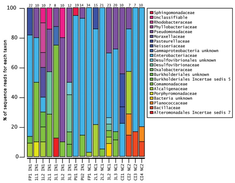
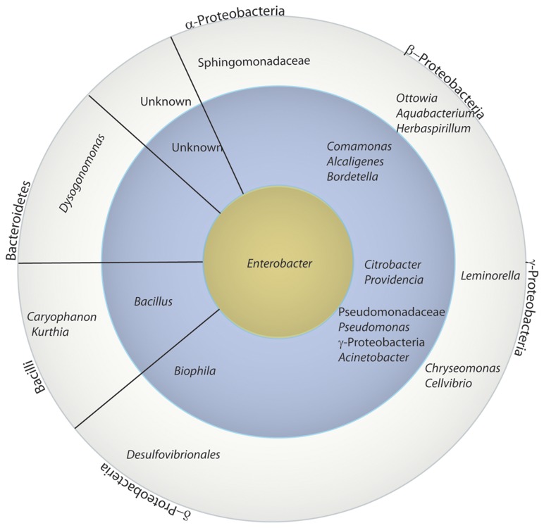
OTUs were assigned using the CloVR pipeline with a 95% threshold as described in the methods for the 275 16S rRNA sequences obtained. Predicted chimeric sequences (n=4) as well as sequences shorter than 470 bp (n=6) were discarded. From the 265 remaining sequences, 71 OTUs were identified across 19 individuals. Of those 71 OTUs, 49 OTUs were classified to the family level, 4 to the phylum level, and 4 could not be classified further than “root” (Figures 3, 4, and 5). The most abundant genus found across all samples was Enterobacter (Figures 3, 4, and 5).
Figure 3. Relative abundance of family level OTUs composing the dung beetle microbiome.
The relative abundance of each taxon is shown for each sample. Enterobacteriaceae (bright blue) were found in 95% of the samples, in all populations, and all life stages. Other major components include bacteria in the Comamonadaceae (green) and Pseudomonadaceae (bluish purple). Sample names are abbreviated as follows: 1L (1st instar larva), 2L (2nd instar larva), 3L (3rd instar larva), PU (pupa), CI (cultured isolates), FP (female parent), IN (Indiana population), and NC (North Carolina population). The number after the life stage indicates different individuals sampled.
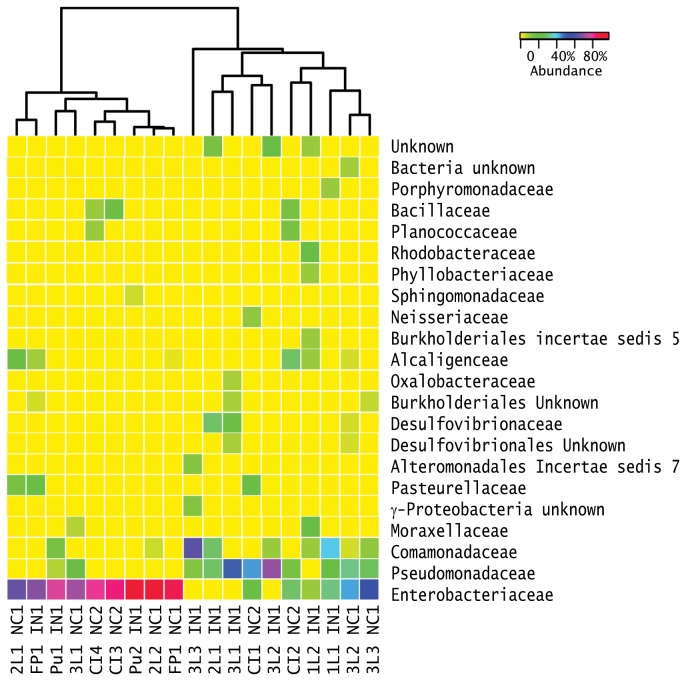
Figure 4. Clustering of samples based on microbiome composition.
The relative proportion of sequences in a specific OTU (color key in upper right corner) is illustrated on a heat map with samples clustered using a furthest neighbor clustering to determine similarity of both the composition and abundance of the microbiomes. Several OTUs were only seen once. Overall, samples did not cluster by population, parental female, life stage, or brood ball age except the cultured isolates (CI3NC1 and CI4NC1) from the digestive system of the 3rd instar larvae. Interestingly, the four cultured samples were oviposited at the same time; however, samples CI3NC1 and CI4NC1 that cluster were sampled 7 d later than CI1NC1 and CI2NC1. Thus, samples CI3NC1 and CI4NC1 were older and were transitioning from the 3rd larval instar to the pupal stages. Each sample is labeled by the source (CI=cultured isolate, 1L=1st instar, 3L=3rd instar, 2L=2nd instar, Pu=pupae, FP= female parent), the isolate or individual animal number, the US state from which the parental female was isolated (NC =North Carolina populations, USA, and IN=Indiana, USA), and the female parent number (NC1, NC2, IN1).
Figure 5. Abundance of lowest common ancestors across all microbiome samples.
Bacterial OTUs identified in Qiime are arranged in a bulls-eye configuration with the most commonly occurring group in the yellow center, those occurring in two or more individuals in the blue circle, and those genera occurring only one time in the outermost rim. The lowest common ancestor Qiime classification is given and may be at the genus, family, or class level and as such may overlap. Enterobacter was found in offspring of all parental females and in 68% of the individuals. Samples denoted with a star are those OTUs that were found using both culture-dependent and -independent techniques.
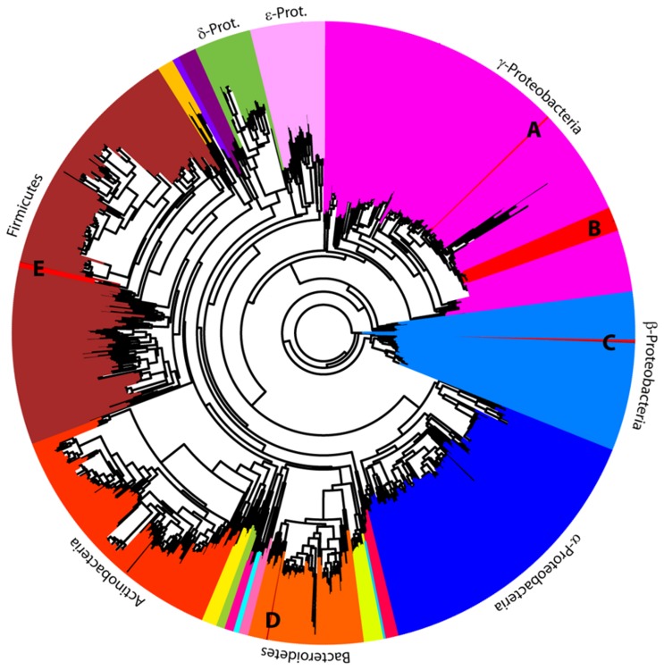
The 16S rRNA microbial profiles were clustered into two categories using a Euclidean distance metric and complete-linkage (furthest neighbor) clustering as implemented in CloVR (Figure 4). The first cluster was dominated by Enterobacteriaceae. We detected Enterobacteriaceae in ~74% of the individuals from all larvae and pupae, the two mothers, and from both populations. The gyrB and lepA loci used for the phylotyping indicated that the most abundant culturable bacterium from our samples was most sister to Enterobacter cloacae within the Enterobacteriaceae (Figures 6 and S2). The second cluster was more diverse with sequences from the Comamonadaceae, Pseudomonadaceae and Enterobacteriaceae families (Figure 4). A Pseudomonadaceae was abundant in only third larval instars (Figures 3 and 4). We detected it in 42% of individuals sampled at this life stage from both NC and IN populations from either whole animals or cultured gut isolates. An unknown γ-proteobacteria (n=5) was detected in 26% of the offspring from the IN population (Figure 4). An unknown Burkholderiales (n=4) was found in both third instar larvae and one of the parental females (Figure 4). Other OTUs that were found at least once in life stages in both populations included: Comamonas, Acinetobacter, and Bilophila (Figure 5).
Figure 6. hylotyping.
An unrooted phylogenomic tree based on 287 loci from 1095 sequenced bacterial genomes was made as a skeletal tree on which was placed the lepA and gyrB sequences from 16 bacterial cultures isolated from the digestive system of four different 3rd instar larvae from female parent 2 from the North Carolina population. This analysis placed the isolates sister to (A) Providencia stuartii, (B) Enterobacter cloacae, (C) Pusillimonas, (D) Pedobacter heparinus, and (E) Lysinibacillus sphaericus.
Several offspring and their female parents have bacteria with 100% identical 16S rRNA sequencing, suggesting vertical transmission through the brood ball and its contents (Figure S3 and Table S3). For example in the North Carolina samples, one sequence was recovered five times from the female parent, four times from the 2nd instar larva, and five times from the 3rd instar larvae (Table S3). Additionally, OTU’s classified as Alcaligenes and Pasteurella are similar between one of the female parents and the cultured isolates from a different female parent. Deeper sequencing of individuals may reveal more shared bacterial OTUs.
Rarefaction curves of the microbial community in several larvae and the IN parental female beetle suggests that additional sampling was needed to identify additional taxa in the microbiome (Figure S4). The rarefaction curves of the NC parental female beetle (35 sequenced clones) and of a second larval instar (20 sequenced clones) suggests that microbiome taxonomic sampling to be close to saturation (Figure S4). The highest bacterial taxonomic richness was isolated from the larval samples and the IN parental female, and lower richness was sampled in pupae and in the NC parental female. However, different primer sets and deeper sampling may amplify a wider diversity of bacterial taxa.
Identification of the O. taurus microbiome using culture based methods.
The nesting activities of the female parent and the feeding behaviors of the larva suggested that the microbiome may be vertically transmitted from the female parent via the brood ball chamber. Since the microbiome resides outside of its eukaryotic host during this time, we also expected the majority of the dung beetle microbiome could be cultured. The ability to culture members of the microbiome allowed for additional taxonomic resolution of some cultured isolates. Protein coding genes may contain more informative sites allowing resolution of taxonomic identity to species and strain level better than 16S rRNA [48–50]. The majority of the cultured isolates were Enterobacteriaceae based on both the CloVR pipeline analysis of 16S rRNA (Figures 3 and 4) from the cultured isolates and the phylotyping analysis using lepA/gyrB (Figure 6). Members of Bacillaceae and Alcaligenaceae were also identified by both methods. Planococcaceae and Pseudomonadaceae cultured isolates only amplified with the 16S rRNA primers (Figure 4). The lepA/gyrB phylotyping identified an isolate in the Sphingobacteriaceae, more specifically as sister to Pedobacter heparinus (Figures 6 and S2). The majority of isolates in the Enterobacteriaceae were placed as sister to Enterobacter cloaceae by the phylotyping analysis. A second enteric isolate was identified as Providencia by CloVR, whereas phylotyping resolved the identity most closely to Providencia stuartii. In the Bacillaceae, phylotyping identified two samples as sister to Lysinibacillus sphaericus. Both CloVR and phylotyping identified isolates in the Alcaligenaceae, with 16S rRNA identifying an Alcaligenes and lepA/gyrB identifying an isolate as sister to Pusillimonas (Figures 4, 6, and S2). However, a complete Alcaligenes genome was not available in GenBank at the time of sampling and thus was not included in the taxonomic sampling for the phylotyping. Overall, similar taxonomic composition is seen using both methods, and, as expected, the phylotyping method often provided more finely resolved taxonomic affiliations than the CloVR analyses. Several of the cultured isolates were classified using 16S rRNA as the same OTU as sequences from larvae or the adult beetles. These include Alcaligenes, Citrobacter, Pasteurella, Pseudomonadaceae, and Enterobacteriaceae (Figures 3 and 4).
Discussion
A potentially vertically transmitted digestive system microbiome
Our experimental design involved females being provided with autoclaved dung and soil so that offspring developed in a sterile environment. Two lines of evidence suggest that the microbiome is vertically transmitted in this system. First, microbial DNA was amplified from beetles reared in sterile conditions, but not from the sterilized dung in which the female parent beetles built their brood balls. We could not produce PCR amplicons from the water surface-sterilized beetles were rinsed in, from frozen or autoclaved dung, or from autoclaved soil. Thus, it is unlikely that the dung we provided the larvae was the source of their microbiome. Second, the brood ball is the only source for a microbiome. After an egg is deposited in the brood ball, the mother has no further contact with the developing offspring. Siblings are not a potential source of microbes, as they have no contact with each other until after they emerge from the brood balls as adults. In contrast, the female beetle constructs a brood ball through chewing, regurgitating, and defecating the cattle dung, so her digestive system microbiome could be present in the brood ball. Supporting maternal-offspring transmission, a female parent and several offspring from the same population have identical 16S rRNA sequences from the microbiome (Figure S3 and Table S3). Moreover, the Enterobacteriaceae were found in 95% of the samples, in all populations, and all life stages. The Comamonadaceae and Pseudomonadaceae were other dominant members. A complete overlap in the parental and offspring taxa was not seen. However, this is not unexpected given that the offspring physiology, anatomy, and diet may enrich for members of the microbiome that are not necessarily the dominant members in the female parent.
These initial results suggest that deeper sequencing of individuals may reveal more shared bacterial OTUs. The primers used are biased for amplifying γ-Proteobacteria, thus additional taxa may be identified using different primers. Lastly, some bacteria from the dung also may have a role in the microbiome of beetles in the wild. While, it is not a subject of this study such studies may be fruitful.
The behavior of the female parent and larval dung beetles suggests that the microbiome is vertically transmitted through structures in the brood ball or the egg itself. First, a single egg is laid on a pedestal of the female’s own excrement, or frass. The larva ecloses and immediately feeds on the pedestal. Second, the larvae then systematically alternate between feeding on the walls of the egg chamber and their own frass [31]. Many dung beetle researchers have considered this second stage of coprophagy (self-coprophagy) to be a method for further extracting the nutrients from their frass. However, we hypothesize that the larva may acquire their microbiome from the pedestal and brood ball walls. During self-coprophagy, the larva may be selecting for or concentrating the microbes that facilitate their digestion of the dung the female provides. Similarly, in other cellulose degrading taxa, such as wood roaches, juveniles do not survive unless they ingest frass from parents that have the microbiota needed for digestion [63]. No microbes or matrix were seen on the surface of the dung beetle egg. PCR amplicons were not produced when DNA from the egg was used as a template. Lack of amplification is never a definitive result because it can be explained in other ways such as lack of abundance. However, when paired with the SEM observations this suggests that egg smearing is not likely to be the mechanism of transmission.
How the microbiome survives and is maintained during metamorphosis remains unknown. The pupae sampled had less diversity of bacterial OTUs identified than other life stages (Figures 3 and 4), although that should not be confused with abundance, which was not assessed. A similar phenomenon is seen in some holometabolous insects [21,64,65], but not others [66]. However, in those systems, the bacteria that persist through metamorphosis are those that are thought to be essential to the symbiosis [66]. Alternatively, the microbiome may be reacquired from the puparium, meconium, or other structures the recently eclosed insect feeds upon post-emergence. Examining how symbionts are maintained during metamorphosis in holometabolous insects remains an intriguing focus of future research.
Scanning electron microscopy reveals rod-shaped structures embedded in the biofilm-like matrix of the brood ball chamber wall (Figure 2F), but lacking in the portions of the brood ball away from the chamber (Figure 2B and 2D). These rod-shaped structures were patchily distributed in the brood ball chamber walls (Figure 2F) and were best visualized when the matrix was scraped away from the chamber walls, supporting the idea that the larval self-coprophagy is necessary for increasing the microbiome population size or selecting for microbes that degrade the dung. Rod-shaped microbes could be seen within a biofilm-like matrix (Figure 2F). Although these microbes are large for many known bacterial cells, they are similar in size and form chains like Bacillus megaterium does. However, at this time, we cannot definitively determine if the microbes are fungal or bacterial. Whether the biofilm-like matrix was produced by the host insect, the bacteria, or both, also remains to be determined.
Collectively, the sterile rearing conditions, overlap of the microbiome between female parent and offspring, specialized dung processing behavior, and bacterial colonies found in the matrix on the brood ball chamber walls, but absent from other locations in the brood ball, suggest that the O. taurus microbiome is maternally transmitted via the brood ball.
The transmission from mother through the brood ball to offspring may be essential for provisioning specific beetle endosymbionts to the offspring since dung beetles develop in an environment rich with other microbes that were excreted from the digestive tract of the mammalian host, as well as bacteria from the soil. Thus, the brood ball provides offspring not only with a safe refuge and food, but it is likely that the female parent additionally provisions a microbiome alongside the egg. However, future experimental validation is needed to further test this hypothesis.
Vertical transmission via the brood ball may favor organisms that are culturable on standard microbiological media, as the bacteria must persist in an external environment prior to ingestion by the larvae. Our analyses based on separate loci for the cloning versus culture-based assays reveals shared bacterial taxa, suggesting a portion of the microbiome is culturable. This is in contrast to relative unculturability of the majority of vertically transmitted endosymbionts that have been studied thus far in insects.
Using almost the entire 16S rRNA amplicon for taxonomic identification allows for increased accuracy in taxonomic assignment [67]. Additionally, studies have shown that increasing sampling depth is not necessary when microbial communities are distinct [68]. Our taxon rarefaction curves (Figure S4) indicate that further sampling is required to fully characterize the dung beetle microbiota, despite that our sampling of the cultured isolates from the digestive system of the 3rd instar larvae captures much of the taxonomic variation seen using molecular approaches. However, on-going studies using next generation sequencing technologies to generate 16S rRNA profiles to increase sampling depth within individuals and additional culturing on a wider diversity of media and conditions will resolve this issue.
To our knowledge, only one other ground-dwelling insect has been described as provisioning offspring brood chambers with specific bacterial endosymbionts. Females in the beewolf genus, Philanthus, smear the larval brood chambers with the actinomycete, Candidatus Streptomyces philanthi, which produces antimicrobial compounds to protect the larvae and developing pupae from soil fungi and bacteria [24,69]. As in the bull-headed dung beetle, the beewolf offspring develop underground in individual brood cells the female creates and provisions with both food and endosymbionts. Upon hatching from the egg, the larva takes up the actinomycete endosymbiont. Prior to pupation, the larva smears the endosymbiont in the cocoon and takes it up after eclosion [24]. In the case of the beewolf, the endosymbionts are beneficial, protecting the developing offspring from fungal and bacterial pathogens [24,69]. Examining other ground and dung nesting insects may identify additional insect-bacterial mutualisms.
Taxonomic classification of the microbiome
The overall microbiome composition across the different locales, females, and life stages could be classified into two groups. Individuals either had microbiomes dominated by Enterobacteriaceae or microbiomes composed of bacteria from Enterobacteriaceae, Pseudomonadaceae, and Comamonadaceae. Additionally, unknown γ-proteobacteria and unknown Burkholderiales were present in several individuals. The function and importance of these different bacterial taxa to the microbiome community and insect host cannot be assessed with our current data and are the focus of future studies. Both culture based and molecular techniques to characterize the dung beetle microbiome provided similar results, though the phylogenomic analysis using lepA and gyrB classified isolates to a more specific taxonomic level.
The most abundant microbe identified was closely related to Enterobacter cloacae. Enterobacter are often found associated with insect digestive systems [21,70]. Different Enterobacter sp. have a diversity of functional capabilities in insect guts from digesting cellulose in Bombyx mori [71] to fixing nitrogen in tephritid fruit flies [70,72]. Another abundant microbe was an uncharacterized and uncultured γ-proteobacteria. Whether this bacterium is widespread in O. taurus or is present in other dung beetles remains to be seen. The next most abundant microbes were pseudomonads and comamonads. Pseudomonads are frequently found associated with insects where they assist in cellulose degradation. Pseudomonads have been found associating with detritivorous insects such as the rove beetle Paederus sp. (Coleoptera:Staphylinidae) [73] as well as plant-feeding beetles such as the Colorado potato beetle, Leptinotarsa decemlineata, (Coleoptera:Chrysomelidae) [74]. Cellulolytic pseudomonads dominated the microbiome of plant-feeding Holotrichia parallelea (Coleoptera:Scarabaeidae) [75]. Determining if the pseudomonads and other bacteria isolated from O. taurus have cellulose degrading properties is underway. If the endosymbionts of O. taurus have cellulose-degrading properties and are culturable, they may be bacterial isolates that could be used for processing cellulosic biofuel [75–77].
Conclusions
Our data suggest that the dung beetle, O. taurus, has endosymbionts that are vertically transmitted in a unique method – via a brood ball. Several of the endosymbionts are culturable. Due to the manipulability of the system, the dung beetle symbiosis may be an ideal system to understand how host and microbes co-operate, and perhaps co-evolve, to thrive on nutritionally unbalanced diets. Additionally, these data suggest that the essential ecosystem function of dung beetles, the degradation of cellulose-rich dung, may be due to communities of bacterial endosymbionts. Whether other dung feeding insects have a specific a vertically transmitted microbiome warrants future studies.
Supporting Information
Sterile rearing. DNA extractions and PCR amplification using the 10F and 1507R primers yielded an ~1500 bp product for fresh (unautoclaved) dung samples, homogenized dung beetle gut and a positive control (lanes UD, Gut,+). However, no such amplicon was produced from frozen or autoclaved dung samples, autoclaved sand and soil, and autoclaved water (lanes FD, AD, AS, AW). Lack of amplification was not due to an inhibitor present in the dung since adding either 0.5 µl or 1 µl of the positive control to the autoclaved products did produce an amplicon (Lane AD+ and AD++). Therefore, both freezing and autoclaving the dung and soil used for rearing the offspring degraded large fragments of bacterial DNA. Similarly, the rinse water of a surface sterilized beetle did not appear to inhibit amplicon production indicating that beetles can be effectively sterilized (Lane RW). The GeneRuler 1 kb ladder is used for reference (Lane Ld). Given these results, the microbiome profiles we examined are those that are inherited by the offspring from the female parent.
(TIF)
A more detailed version of Figure 6 that includes the species or strain names of the taxa used to build the tree.
(TIF)
Dendrogram illustrating sequence identity between 16S rRNA sequences relative to the samples. Abbreviations are the same as in Figure 4.
(TIF)
Rarefaction curves for selected individuals where the frequency with which diversity is calculated every five sequences.
(TIF)
Accession numbers and NCBI taxonomy of the bacterial genomes used for the phylotyping analysis.
(DOCX)
Abbreviations and full names of the loci used for the phylotyping analysis.
(DOCX)
Sequence identity matrix for 16S rRNA samples. Cells labeled ID are where the sequence is compared to itself. Cells labeled NA were those where the sequence was not long enough for comparison.
(XLSX)
Acknowledgments
We greatly appreciate Whitney Schlegel of the Marble Hill Farm near Bloomington, Indiana and Bob Nutter of Maple View Farm in Hillsborough, NC giving us permission to collect the wild female beetles that were used in this study. We thank Emily Zeller for maintaining the beetle colony. Matthew Hemm and Roland Roberts at Towson University graciously shared equipment necessary for this work. Discussions with David Riley and James White strengthened the microbiome data analysis. Comments from the Dunning Hotopp lab group, Vincent Bruno, Bernard Doube, Jacob Russell, and two anonymous reviewers strengthened the manuscript. We would like to thank the Plant and IT Groups at the J. Craig Venter Institute for their advice and support on the phylogenomic analysis. Mark Mazaitis made the graphical illustration for Figure 1. Ru-ching Hsia and John Strong of the University of Maryland School of Dentistry Core Imaging Facility assisted with the scanning electron microscope.
Funding Statement
This research was supported in part through funds from: the Towson University Fisher College Undergraduate Research Awards (KF, TA), a Towson University Undergraduate Research Award (KF), Orchid Society of Arizona funding (AME), NSF REU DBI0552654 (MF), NSF IOS 0445661 (AM), NSF IOS 0718522 (AM), and State of Maryland internal funds (JDH). The funders had no role in study design, data collection and analysis, decision to publish, or preparation of the manuscript.
References
- 1. Chin K, Gill BD (1996) Dinosaurs, dung beetles, and conifers: participants in a Cretaceous food web. Palaios 11: 280-285. doi: 10.2307/3515235. [DOI] [Google Scholar]
- 2. Krell F-T (2006) Fossil record and evolution of Scarabaeoidea (Coleoptera: Polyphaga). Coleopt Bull 60: 120-143. [Google Scholar]
- 3. Holter P, Scholtz CH (2013) Elongated hindguts in desert-living dung beetles (Scarabaeidae: Scarabaeinae) feeding on dry dung pellets or plant litter. J Morphol 274: 657-662. doi: 10.1002/jmor.20123. PubMed: 23450631. [DOI] [PubMed] [Google Scholar]
- 4. Holter P, Scholtz CH, Stenseng L (2009) Desert detritivory: Nutritional ecology of a dung beetle (Pachysoma glentoni) subsisting on plant litter in arid South African sand dunes. J Arid Environ 73: 1090-1094. doi: 10.1016/j.jaridenv.2009.04.009. [DOI] [Google Scholar]
- 5. Davis ALV, Scholtz CH, Philips TK (2002) Historical biogeography of scarabaeine dung beetles. J Biogeogr 29: 1217-1256. doi: 10.1046/j.1365-2699.2002.00776.x. [DOI] [Google Scholar]
- 6. Nichols E, Spector S, Louzada J, Larsen T, Amezquita S et al. (2008) Ecological functions and ecosystem services provided by Scarabaeinae dung beetles. Biol Conserv 141: 1461-1474. doi: 10.1016/j.biocon.2008.04.011. [DOI] [Google Scholar]
- 7. Philips TK (2011) The evolutionary history and diversification of dung beetles. Ecology and Evolution of Dung Beetles. John Wiley & Sons, Ltd. pp. 21-46. [Google Scholar]
- 8. Muller ZO (1980) Feed from animal wastes: State of knowledge. FAO Animal Product Health 18: 190. [Google Scholar]
- 9. Buchner P (1965) Endosymbiosis of animals with plant microorganisms. New York: Interscience Publishers. [Google Scholar]
- 10. Zientz E, Silva FJ, Gross R (2001) Genome interdependence in insect-bacterium symbioses. Genome Biol 2: 1-6. PubMed: 11790259. [DOI] [PMC free article] [PubMed] [Google Scholar]
- 11. Belda E, Moya A, Bentley S, Silva F (2010) Mobile genetic element proliferation and gene inactivation impact over the genome structure and metabolic capabilities of Sodalis glossinidius, the secondary endosymbiont of tsetse flies. BMC Genomics 11: 1-17. doi: 10.1186/1471-2164-11-S5-S1. PubMed: 20044946. [DOI] [PMC free article] [PubMed] [Google Scholar]
- 12. Macdonald SJ, Thomas GH, Douglas AE (2011) Genetic and metabolic determinants of nutritional phenotype in an insect–bacterial symbiosis. Mol Ecol 20: 2073-2084. doi: 10.1111/j.1365-294X.2011.05031.x. PubMed: 21392141. [DOI] [PubMed] [Google Scholar]
- 13. Baumann L, Baumann P, Moran NA, Sandström J, Thao ML (1999) Genetic characterization of plasmids containing genes encoding enzymes of leucine biosynthesis in endosymbionts (Buchnera) of aphids. J Mol Evol 48: 77-85. doi: 10.1007/PL00006447. PubMed: 9873079. [DOI] [PubMed] [Google Scholar]
- 14. Köhler T, Dietrich C, Scheffrahn RH, Brune A (2012) High-resolution analysis of gut environment and bacterial microbiota reveals functional compartmentation of the gut in wood-feeding higher termites (Nasutitermes spp.). Appl Environ Microbiol 78: 4691-4701. doi: 10.1128/AEM.00683-12. PubMed: 22544239. [DOI] [PMC free article] [PubMed] [Google Scholar]
- 15. Husseneder C, Ho HY, Blackwell M (2010) Comparison of the bacterial symbiont composition of the formosan subterranean termite from its native and introduced range. Open Microbiol J 4: 53-66. doi: 10.2174/1874285801004010053. PubMed: 21347207. [DOI] [PMC free article] [PubMed] [Google Scholar]
- 16. Bright M, Bulgheresi S (2010) A complex journey: transmission of microbial symbionts. Nat Rev Microbiol 8: 218-230. doi: 10.1038/nrmicro2262. PubMed: 20157340. [DOI] [PMC free article] [PubMed] [Google Scholar]
- 17. Noda S, Kitade O, Inoue T, Kawai M, Kanuka M et al. (2007) Cospeciation in the triplex symbiosis of termite gut protists (Pseudotrichonympha spp.), their hosts, and their bacterial endosymbionts. Mol Ecol 16: 1257-1266. doi: 10.1111/j.1365-294X.2006.03219.x. PubMed: 17391411. [DOI] [PubMed] [Google Scholar]
- 18. Attardo GM, Lohs C, Heddi A, Alam UH, Yildirim S et al. (2008) Analysis of milk gland structure and function in Glossina morsitans: Milk protein production, symbiont populations and fecundity. J Insect Physiol 54: 1236-1242. doi: 10.1016/j.jinsphys.2008.06.008. PubMed: 18647605. [DOI] [PMC free article] [PubMed] [Google Scholar]
- 19. Balmand S, Lohs C, Aksoy S, Heddi A (2013) Tissue distribution and transmission routes for the tsetse fly endosymbionts. J Invert Pathol, 112 Suppl: S116-S122. PubMed: 22537833. [DOI] [PMC free article] [PubMed] [Google Scholar]
- 20. Beard CB, Cordon-Rosales C, Durvasula RV (2002) Bacterial symbionts of the triatominae and their potential use in control of Chagas disease transmission. Annu Rev Entomol 47: 123-141. doi: 10.1146/annurev.ento.47.091201.145144. PubMed: 11729071. [DOI] [PubMed] [Google Scholar]
- 21. Estes AM, Hearn DJ, Bronstein JL, Pierson EA (2009) The Olive Fly endosymbiont, "Candidatus Erwinia dacicola," switches from an intracellular existence to an extracellular existence during host insect development. Appl Environ Microbiol 75: 7097-7106. doi: 10.1128/AEM.00778-09. PubMed: 19767463. [DOI] [PMC free article] [PubMed] [Google Scholar]
- 22. Rupert LLK (2003) Stadium-specific transmission of endosymbionts needed for pederin biosynthesis in three species of Paederus rove beetles. Entomol Exp Applicata 107: 115-124. doi: 10.1046/j.1570-7458.2003.00042.x. [DOI] [Google Scholar]
- 23. Fukatsu T, Hosokawa T (2002) Capsule-transmitted gut symbiotic bacterium of the Japanese common plataspid stinkbug, Megacopta punctatissima . Appl Environ Microbiol 68: 389-396. doi: 10.1128/AEM.68.1.389-396.2002. PubMed: 11772649. [DOI] [PMC free article] [PubMed] [Google Scholar]
- 24. Kaltenpoth M, Göttler W, Herzner G, Strohm E (2005) Symbiotic bacteria protect wasp larvae from fungal infestation. Curr Biol 15: 475-479. doi: 10.1016/j.cub.2004.12.084. PubMed: 15753044. [DOI] [PubMed] [Google Scholar]
- 25. Peccoud J, Simon J-C, McLaughlin HJ, Moran NA (2009) Post-Pleistocene radiation of the pea aphid complex revealed by rapidly evolving endosymbionts. Proc Natl Acad Sci USA 106: 16315-16320. doi: 10.1073/pnas.0905129106. PubMed: 19805299. [DOI] [PMC free article] [PubMed] [Google Scholar]
- 26. Pittman GW, Brumbley SM, Allsopp PG, O'Neill SL (2008) “Endomicrobia” and other bacteria associated with the hindgut of Dermolepida albohirtum larvae. Appl Environ Microbiol 74: 762-767. doi: 10.1128/AEM.01831-07. PubMed: 18083861. [DOI] [PMC free article] [PubMed] [Google Scholar]
- 27. Zhang H, Jackson TA (2008) Autochthonous bacterial flora indicated by PCR-DGGE of 16S rRNA gene fragments from the alimentary tract of Costelytra zealandica (Coleoptera: Scarabaeidae). J Appl Microbiol 105: 1277-1285. doi: 10.1111/j.1365-2672.2008.03867.x. PubMed: 18713286. [DOI] [PubMed] [Google Scholar]
- 28. Andert J, Marten A, Brandl R, Brune A (2010) Inter- and intraspecific comparison of the bacterial assemblages in the hindgut of humivorous scarab beetle larvae (Pachnoda spp.). FEMS Microbiol Ecol 74: 439-449. doi: 10.1111/j.1574-6941.2010.00950.x. PubMed: 20738398. [DOI] [PubMed] [Google Scholar]
- 29. Egert M, Wagner B, Lemke T, Brune A, Friedrich MW (2003) Microbial community structure in midgut and hindgut of the humus-feeding larva of Pachnoda ephippiata (Coleoptera: Scarabaeidae). Appl Environ Microbiol 69: 6659-6668. doi: 10.1128/AEM.69.11.6659-6668.2003. PubMed: 14602626. [DOI] [PMC free article] [PubMed] [Google Scholar]
- 30. Egert M, Stingl U, Bruun LD, Pommerenke B, Brune A et al. (2005) Structure and topology of microbial communities in the major gut compartments of Melolontha melolontha larvae (Coleoptera:Scarabaeidae). Appl Environ Microbiol 71: 4556-4566. doi: 10.1128/AEM.71.8.4556-4566.2005. PubMed: 16085849. [DOI] [PMC free article] [PubMed] [Google Scholar]
- 31. Goidanich A, Malan C (1964) Sulla nidificatione pedotrofica di alcune specie di Onthophagus europei e sulla microflora aerobica dell'apparato digerente delle larve di Onthophagus taurus Schreber (Coleoptera: Scarabaeidae). Ann Fac Sci Agr Univ Turino; 2: 213-378 [Google Scholar]
- 32. Howden HF, Cartwright OL (1963) Scarab beetles of the genus Onthophagus, Latreille north of Mexico (Coleoptera: Scarabaeidae). Proc United States Natl Museum 114: 1-135. doi: 10.5479/si.00963801.114-3467.1. [DOI] [Google Scholar]
- 33. Hoebeke ER, Beucke K (1997) Adventive Onthophagus (Coleoptera: Scarabaeidae) in North America: geographic ranges, diagnoses, and new distributional records Entomology. News 108: 345-361. [Google Scholar]
- 34. Hunter JS, Fincher GT (1985) Five new state records for the Afro-Asian dung beetle Onthophagus gazella (Coleoptera: Scarabaeidae). J Entomol Sci 20: 24-25. [Google Scholar]
- 35. Howden HF, Scholtz CH (1986) Changes in Texas dung beetle community between 1975 and 1985 (Coleoptera: Scarabaeidae: Scarabaeinae). Coleopt Bull 40: 313-316. [Google Scholar]
- 36. Fincher TG, Woodruff RE (1975) A European dung beetle, Onthophagus taurus Schreber, new to the US (Coleoptera: Scarabaeidae). Coleopt Bull 29: 349-350. [Google Scholar]
- 37. Price DL, Brenneman LM, Johnston RE (2012) Dung beetles (Coleoptera:Scarabaeidae and Geotrupidae) communities of Eastern Maryland 114 Entomological Society of Washington; pp. 142-151. [Google Scholar]
- 38. Simonnet F, Moczek AP (2011) Conservation and diversification of gene function during mouthpart development in Onthophagus beetles. Evol Dev 13: 280-289. doi: 10.1111/j.1525-142X.2011.00479.x. PubMed: 21535466. [DOI] [PubMed] [Google Scholar]
- 39. Halffter G, Edmonds WD (1982) The nesting behavior of dung beetles (Scarabaeinae):An ecological and evolutive approach. Publicaciones Instituto de Ecologia Mexico; pp. 1-176. [Google Scholar]
- 40. Moczek AP, Nagy LM (2005) Diverse developmental mechanisms contribute to different levels of diversity in horned beetles. Evol Dev 7: 175-185. doi: 10.1111/j.1525-142X.2005.05020.x. PubMed: 15876190. [DOI] [PubMed] [Google Scholar]
- 41. Lane DJ (1991) 16S and 23S rRNA sequencing In: Stackenbrandt E, Goodfellow M. Nucleic acid techniques in bacterial systematics. New York, NY: John Wiley and Sons; pp. 115–148. [Google Scholar]
- 42. Mateos M, Castrezana SJ, Nankivell BJ, Estes AM, Markow TA et al. (2006) Heritable endosymbionts of Drosophila . Genetics 174: 363-376. doi: 10.1534/genetics.106.058818. PubMed: 16783009. [DOI] [PMC free article] [PubMed] [Google Scholar]
- 43. Moran NA, Dale C, Dunbar H, Smith WA, Ochman H (2003) Intracellular symbionts of sharpshooters (Insecta : Hemiptera : Cicadellinae) form a distinct clade with a small genome. Environ Microbiol 5: 116-126. doi: 10.1046/j.1462-2920.2003.00391.x. PubMed: 12558594. [DOI] [PubMed] [Google Scholar]
- 44. Beninati T, Riegler M, Vilcins I-ME, Sacchi L, McFadyen R et al. (2009) Absence of the symbiont Candidatus Midichloria mitochondrii in the mitochondria of the tick Ixodes holocyclus . FEMS Microbiol Lett 299: 241-247. doi: 10.1111/j.1574-6968.2009.01757.x. PubMed: 19732154. [DOI] [PubMed] [Google Scholar]
- 45. Bressan A, Arneodo J, Simonato M, Haines WP, Boudon-Padieu E (2009) Characterization and evolution of two bacteriome-inhabiting symbionts in cixiid planthoppers (Hemiptera: Fulgoromorpha: Pentastirini). Environ Microbiol 11: 3265-3279. doi: 10.1111/j.1462-2920.2009.02055.x. PubMed: 19758348. [DOI] [PubMed] [Google Scholar]
- 46. Quast C, Pruesse E, Yilmaz P, Gerken J, Schweer T et al. (2013) The SILVA ribosomal RNA gene database project: improved data processing and web-based tools. Nucleic Acids Res 41: D590-D596. doi: 10.1093/nar/gks1219. PubMed: 23193283. [DOI] [PMC free article] [PubMed] [Google Scholar]
- 47. Gordon D, Abajian C, Green P (1998) Consed: a graphical tool for sequence finishing. Genome Res 8: 195-202. doi: 10.1101/gr.8.3.195. PubMed: 9521923. [DOI] [PubMed] [Google Scholar]
- 48. Santos SR, Ochman H (2004) Identification and phylogenetic sorting of bacterial lineages with universally conserved genes and proteins. Environ Microbiol 6: 754-759. doi: 10.1111/j.1462-2920.2004.00617.x. PubMed: 15186354. [DOI] [PubMed] [Google Scholar]
- 49. Fox GE, Wisotzkey JD, Jurtshuk P (1992) How close is close: 16S rRNA sequence identity may not be sufficient to guarantee species identity. Int J Syst Bacteriol 42: 166-170. doi: 10.1099/00207713-42-1-166. PubMed: 1371061. [DOI] [PubMed] [Google Scholar]
- 50. Roux S, Enault F, le Bronner G, Debroas D (2011) Comparison of 16S rRNA and protein-coding genes as molecular markers for assessing microbial diversity (Bacteria and Archaea) in ecosystems. FEMS Microbiol Ecol 78: 617-628. doi: 10.1111/j.1574-6941.2011.01190.x. PubMed: 22066608. [DOI] [PubMed] [Google Scholar]
- 51. Angiuoli SV, Matalka M, Gussman A, Galens K, Vangala M et al. (2011) CloVR: a virtual machine for automated and portable sequence analysis from the desktop using cloud computing. BMC Bioinformatics 12: 356. doi: 10.1186/1471-2105-12-356. PubMed: 21878105. [DOI] [PMC free article] [PubMed] [Google Scholar]
- 52. Kuczynski J, Stombaugh J, Walters WA, Gonzalez A, Caporaso JG et al. (2011) Using QIIME to analyze 16S rRNA gene sequences from microbial communities. Current Protoc Bioinforma Chapter 10: Unit 10 17. PubMed: 2318459222161565 [DOI] [PMC free article] [PubMed] [Google Scholar]
- 53. White JR, Arze C, Matalka M the CloVR team, White O, et al. (2011) CloVR-16S: Phylogenetic microbial community composition analysis based on 16S ribosomal RNA amplicon sequencing – standard operating procedure,version 2.0. Nature Precedings. [Google Scholar]
- 54. Cole JR, Chai B, Farris RJ, Wang Q, Kulam SA et al. (2005) The Ribosomal Database Project (RDP-II): sequences and tools for high-throughput rRNA analysis. Nucleic Acids Res 33: D294-D296. PubMed: 15608200. [DOI] [PMC free article] [PubMed] [Google Scholar]
- 55. Vilo C, Dong Q (2012) Evaluation of the RDP classifier accuracy using 16S rRNA gene variable regions. Metagenomics 1: 1-5. doi: 10.4303/mg/235571. [DOI] [Google Scholar]
- 56. Schloss PD, Westcott SL, Ryabin T, Hall JR, Hartmann M et al. (2009) Introducing mothur: open-source, platform-independent, community-supported software for describing and comparing microbial communities. Appl Environ Microbiol 75: 7537 - 7541. doi: 10.1128/AEM.01541-09. PubMed: 19801464. [DOI] [PMC free article] [PubMed] [Google Scholar]
- 57. White J, Arze C, Matalka M, Team TC, White. O, et al. (2011) CloVR-16S: Phylogenetic microbial community composition analysis based on 16S ribosomal RNA amplicon sequencing; ——standard operating procedure, version 1.0. Nature Precedings. [Google Scholar]
- 58. Edgar RC (2004) MUSCLE: multiple sequence alignment with high accuracy and high throughput. Nucleic Acids Res 32: 1792-1797. doi: 10.1093/nar/gkh340. PubMed: 15034147. [DOI] [PMC free article] [PubMed] [Google Scholar]
- 59. Hall TA (1999) BioEdit: a user-friendly biological sequence alignment editor and analysis program for Windows 95/98/NT. Nucleic Acids Symp S 41: 95-98. [Google Scholar]
- 60. Stamatakis A, Ludwig T, Meier H (2005) RAxML-III: a fast program for maximum likelihood-based inference of large phylogenetic trees. Bioinformatics 21: 456-463. doi: 10.1093/bioinformatics/bti191. PubMed: 15608047. [DOI] [PubMed] [Google Scholar]
- 61. Wu M, Eisen JA (2008) A simple, fast, and accurate method of phylogenomic inference. Genome Biol 9: R151. doi: 10.1186/gb-2008-9-10-r151. PubMed: 18851752. [DOI] [PMC free article] [PubMed] [Google Scholar]
- 62. Estes AM, Hearn DJ, Burrack HJ, Rempoulakis P, Pierson EA (2012) Prevalence of Candidatus Erwinia dacicola in wild and laboratory Olive Fruit Fly populations and across developmental stages. Environ Entomol 41: 265-274. doi: 10.1603/EN11245. PubMed: 22506998. [DOI] [PubMed] [Google Scholar]
- 63. Nalepa C (1984) Colony composition, protozoan transfer and some life history characteristics of the woodroach Cryptocercus punctulatus Scudder (Dictyoptera: Cryptocercidae). Behav Ecol Sociobiol 14: 273-279. doi: 10.1007/BF00299498. [DOI] [Google Scholar]
- 64. Delalibera I, Vasanthakumar A, Burwitz BJ, Schloss PD, Klepzig KD et al. (2007) Composition of the bacterial community in the gut of the pine engraver, Ips pini (Say) (Coleoptera) colonizing red pine. Symbiosis 43: 97-104. [Google Scholar]
- 65. Greenberg B (1959) Persistence of bacteria in the developmental stages of the housefly. III. Quantitative distribution in prepupae and pupae. Am J Trop Med Hyg 8: 613-617. PubMed: 13851548. [DOI] [PubMed] [Google Scholar]
- 66. Stoll S, Feldhaar H, Fraunholz MJ, Gross R (2010) Bacteriocyte dynamics during development of a holometabolous insect, the carpenter ant Camponotus floridanus. BMC Microbiol 10: 308. doi: 10.1186/1471-2180-10-308. PubMed: 21122115. [DOI] [PMC free article] [PubMed] [Google Scholar]
- 67. Schloss PD (2010) The effects of alignment quality, distance calculation method, sequence filtering, and region on the analysis of 16S rRNA gene-based studies. PLOS Comput Biol 6: e1000844 PubMed: 20628621. [DOI] [PMC free article] [PubMed] [Google Scholar]
- 68. Kuczynski J, Costello EK, Nemergut DR, Zaneveld J, Lauber CL et al. (2010) Direct sequencing of the human microbiome readily reveals community differences. Genome Biol 11: 210. doi: 10.1186/gb-2010-11-5-210. PubMed: 20441597. [DOI] [PMC free article] [PubMed] [Google Scholar]
- 69. Kroiss J, Kaltenpoth M, Schneider B, Schwinger M-G, Hertweck C et al. (2010) Symbiotic streptomycetes provide antibiotic combination prophylaxis for wasp offspring. Nat Chem Biol 6: 261-263. doi: 10.1038/nchembio.331. PubMed: 20190763. [DOI] [PubMed] [Google Scholar]
- 70. Martinez-Sañudo I, Savio C, Mazzon L, Girolami V, Ciolfi S et al. (2011) Green fluorescent protein (GFP)-labeling of enterobacteria associated with fruit flies (Diptera: Tephritidae) and persistence in their natural host Rhagoletis completa Cresson. Can J Microbiol 57: 969-973. doi: 10.1139/w11-057. PubMed: 22004066. [DOI] [PubMed] [Google Scholar]
- 71. Anand AA, Vennison SJ, Sankar SG, Prabhu DI, Vasan PT et al. (2010) Isolation and characterization of bacteria from the gut of Bombyx mori that degrade cellulose, xylan, pectin and starch and their impact on digestion. J Insect Sci 10: 107 PubMed: 20874394. [DOI] [PMC free article] [PubMed] [Google Scholar]
- 72. Ben-Yosef M, Jurkevitch E, Yuval B (2008) Effect of bacteria on nutritional status and reproductive success of the Mediterranean fruit fly Ceratitis capitata . Physiol Entomol 33: 145-154. doi: 10.1111/j.1365-3032.2008.00617.x. [DOI] [Google Scholar]
- 73. Kellner RLL (2002) Interspecific transmission of Paederus endosymbionts: relationship to the genetic divergence among the bacteria associated with pederin biosynthesis. Chemoecology 12: 133-138. doi: 10.1007/s00012-002-8338-1. [DOI] [Google Scholar]
- 74. Castrillo LA, Lee RE Jr., Lee MR, Rutherford ST (2000) Identification of ice-nucleating active Pseudomonas fluorescens strains for biological control of overwintering Colorado potato beetles (Coleoptera: Chrysomelidae). J Econ Entomol 93: 226-233. doi: 10.1603/0022-0493-93.2.226. PubMed: 10826166. [DOI] [PubMed] [Google Scholar]
- 75. Huang S, Sheng P, Zhang H (2012) Isolation and identification of cellulolytic bacteria from the gut of Holotrichia parallela larvae (Coleoptera: Scarabaeidae). Int J Mol Sci 13: 2563-2577. doi: 10.3390/ijms13032563. PubMed: 22489111. [DOI] [PMC free article] [PubMed] [Google Scholar]
- 76. Huang S-W, Zhang H-Y, Marshall S, Jackson TA (2010) The scarab gut: A potential bioreactor for bio-fuel production. J Insect Sci 17: 175-183. doi: 10.1111/j.1744-7917.2010.01320.x. [DOI] [Google Scholar]
- 77. Shi W, Xie S, Chen X, Sun S, Zhou X et al. (2013) Comparative genomic analysis of the endosymbionts of herbivorous insects reveals eco-environmental adaptations: Biotechnology applications. PLOS Genet 9: e1003131. [DOI] [PMC free article] [PubMed] [Google Scholar]
Associated Data
This section collects any data citations, data availability statements, or supplementary materials included in this article.
Supplementary Materials
Sterile rearing. DNA extractions and PCR amplification using the 10F and 1507R primers yielded an ~1500 bp product for fresh (unautoclaved) dung samples, homogenized dung beetle gut and a positive control (lanes UD, Gut,+). However, no such amplicon was produced from frozen or autoclaved dung samples, autoclaved sand and soil, and autoclaved water (lanes FD, AD, AS, AW). Lack of amplification was not due to an inhibitor present in the dung since adding either 0.5 µl or 1 µl of the positive control to the autoclaved products did produce an amplicon (Lane AD+ and AD++). Therefore, both freezing and autoclaving the dung and soil used for rearing the offspring degraded large fragments of bacterial DNA. Similarly, the rinse water of a surface sterilized beetle did not appear to inhibit amplicon production indicating that beetles can be effectively sterilized (Lane RW). The GeneRuler 1 kb ladder is used for reference (Lane Ld). Given these results, the microbiome profiles we examined are those that are inherited by the offspring from the female parent.
(TIF)
A more detailed version of Figure 6 that includes the species or strain names of the taxa used to build the tree.
(TIF)
Dendrogram illustrating sequence identity between 16S rRNA sequences relative to the samples. Abbreviations are the same as in Figure 4.
(TIF)
Rarefaction curves for selected individuals where the frequency with which diversity is calculated every five sequences.
(TIF)
Accession numbers and NCBI taxonomy of the bacterial genomes used for the phylotyping analysis.
(DOCX)
Abbreviations and full names of the loci used for the phylotyping analysis.
(DOCX)
Sequence identity matrix for 16S rRNA samples. Cells labeled ID are where the sequence is compared to itself. Cells labeled NA were those where the sequence was not long enough for comparison.
(XLSX)


