
FIGURE 9.
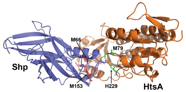
A docking complex with the juxtaposed heme pockets of Shp and lowest energy. Structure elements: Blue, Shp protein; orange, HtsA protein; light pink, heme; red, the M66 and M153 axial residues in Shp; and green, the M79 and H229 axial residues.