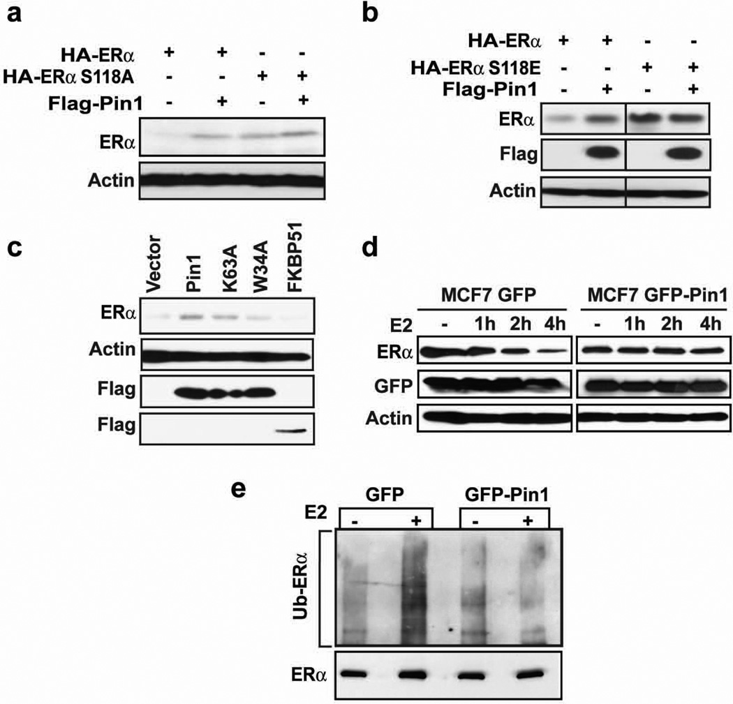
Figure 2. Pin1 stabilizes ERα in an S118-dependent manner and blocks its ubiquitination.
a and b) MEF Pin1−/− were co-transfected with 0.1 µg Flag-Pin1 and 0.3 µg of HA-ERα or HA-ERα S118A (a) or HA-ERα S118E (b) for 24 h and treated with 10 nM E2 for another 24 h. Western blot analysis was performed to assess the level of ERα by using anti-HA antibody and Pin1 by anti-Flag antibody. The actin band represents the loading control.
c) MEF Pin1−/− cells were co-transfected with HA-ERα and Flag vector, Flag-Pin1, Flag-Pin1 K63A, Flag-Pin1 W34A, or Flag-FKBP51. 24 h post-transfection, cells were treated with 10 nM E2 for 24 h and Western blot was performed for ERα, Flag, and actin.
d) MCF-7 cells stably expressing GFP or GFP-Pin1 were treated with and without 10 nM E2 for the indicated length of time and Western blot was performed for ERα, GFP, and actin.
e) MCF-7 cells overexpressing GFP or GFP Pin1 were treated with 10 µM MG132 for 30 min followed by 4 h treatment with 10 nM E2 (+) or EtOH (-). ERα was immunoprecipitated using anti-ERα antibody and the level of ubiquitination was evaluated by Western blot using anti-ubiquitin antibody (Ub).

